

Beyond lipofilling: adipose-derived stem cells in plastic surgery
 0
0 Graphical Abstract
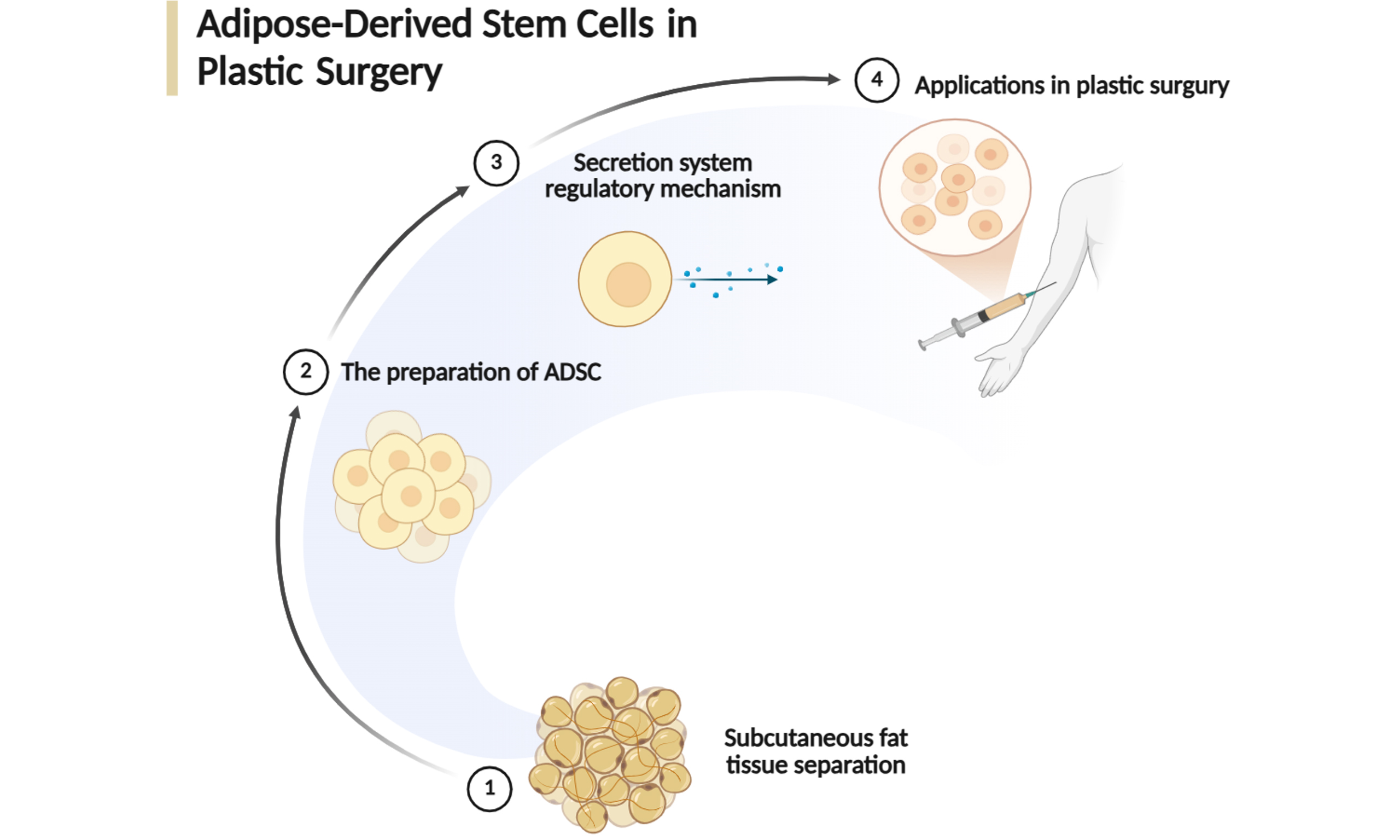
Abstract
Adipose-derived stem cells (ADSCs) have emerged as a pivotal tool in the paradigm shift of plastic and reconstructive surgery from traditional tissue transposition toward regenerative medicine, owing to their abundance, minimally invasive harvesting, and potent regenerative capabilities. Beyond their multilineage differentiation potential, the therapeutic efficacy of ADSCs is increasingly attributed to their sophisticated paracrine activity and exosome-mediated signaling. This review provides a comprehensive synthesis of the biological characteristics of ADSCs, their underlying molecular mechanisms, and their diverse clinical applications in plastic surgery. We discuss the orchestrated roles of the ADSC secretome in promoting neoangiogenesis, modulating the immune microenvironment, and regulating extracellular matrix remodeling. Clinically, we highlight the latest evidence for cell-assisted lipotransfer (CAL) in fat grafting, the acceleration of chronic wound healing, anti-fibrotic strategies for scar management, and the emerging role of ADSCs in facial rejuvenation and anti-aging. While issues regarding standardization and long-term oncological safety remain, the integration of ADSCs with biofabrication and cell-free therapies represents a promising frontier. Ultimately, ADSCs hold the potential to achieve functional and scarless tissue regeneration and may redefine the future of plastic and reconstructive surgery.
Keywords


 Download PDF
INTRODUCTION
Plastic and aesthetic surgery has traditionally relied on the transposition of autologous tissues, such as skin grafts and vascularized flaps, to repair defects and restore function. However, the limitations of these conventional approaches include donor-site morbidity, limited tissue availability, and unpredictable long-term outcomes, which have catalyzed a shift toward regenerative medicine. At the forefront of this evolution are adipose-derived stem cells (ADSCs), a subset of mesenchymal stem cells (MSCs) that have redefined the landscape of tissue engineering and aesthetic surgery[1].
First characterized in the early 2000s, ADSCs are typically isolated from the stromal vascular fraction (SVF) of adipose tissue[2]. Compared to bone marrow-derived MSCs, ADSCs offer several distinct clinical advantages: they are more abundant, can be harvested through minimally invasive liposuction with lower donor-site morbidity, and exhibit a higher proliferative capacity in vitro[3-5]. Beyond their multi-lineage differentiation potential into adipogenic, osteogenic, and chondrogenic lineages, the therapeutic efficacy of ADSCs is increasingly attributed to their potent paracrine activity. By secreting a diverse array of growth factors, cytokines, and extracellular vesicles like exosomes[6], ADSCs effectively modulate the inflammatory microenvironment, promote neoangiogenesis, and accelerate tissue remodeling[7-9].
Despite the proliferation of preclinical studies demonstrating the regenerative potential of ADSCs in fat grafting, wound healing, and scar management, the transition from “bench to bedside” remains complex[10]. Challenges such as the standardization of isolation protocols, the long-term oncological safety of cell-assisted lipotransfer (CAL), and the regulatory hurdles for clinical translation continue to be subjects of intense debate.
Therefore, this review aims to provide a comprehensive overview of the biological characteristics and underlying regenerative mechanisms of ADSCs. Furthermore, we summarize the latest clinical advancements in plastic surgery, aiming to offer insights into future research directions and the potential for personalized regenerative treatments.
BIOLOGY AND CHARACTERIZATION OF ADSCs
The clinical ascendancy of ADSCs is traditionally predicated on their surgical accessibility and expansive potential[5,10-12]. However, contemporary regenerative medicine has moved beyond these logistical advantages, unveiling a biological landscape defined by intrinsic heterogeneity and context-dependent plasticity. Understanding ADSC biology now requires a shift from viewing them as a monolithic cell population to recognizing them as a dynamic, responsive therapeutic system[13] [Figure 1].
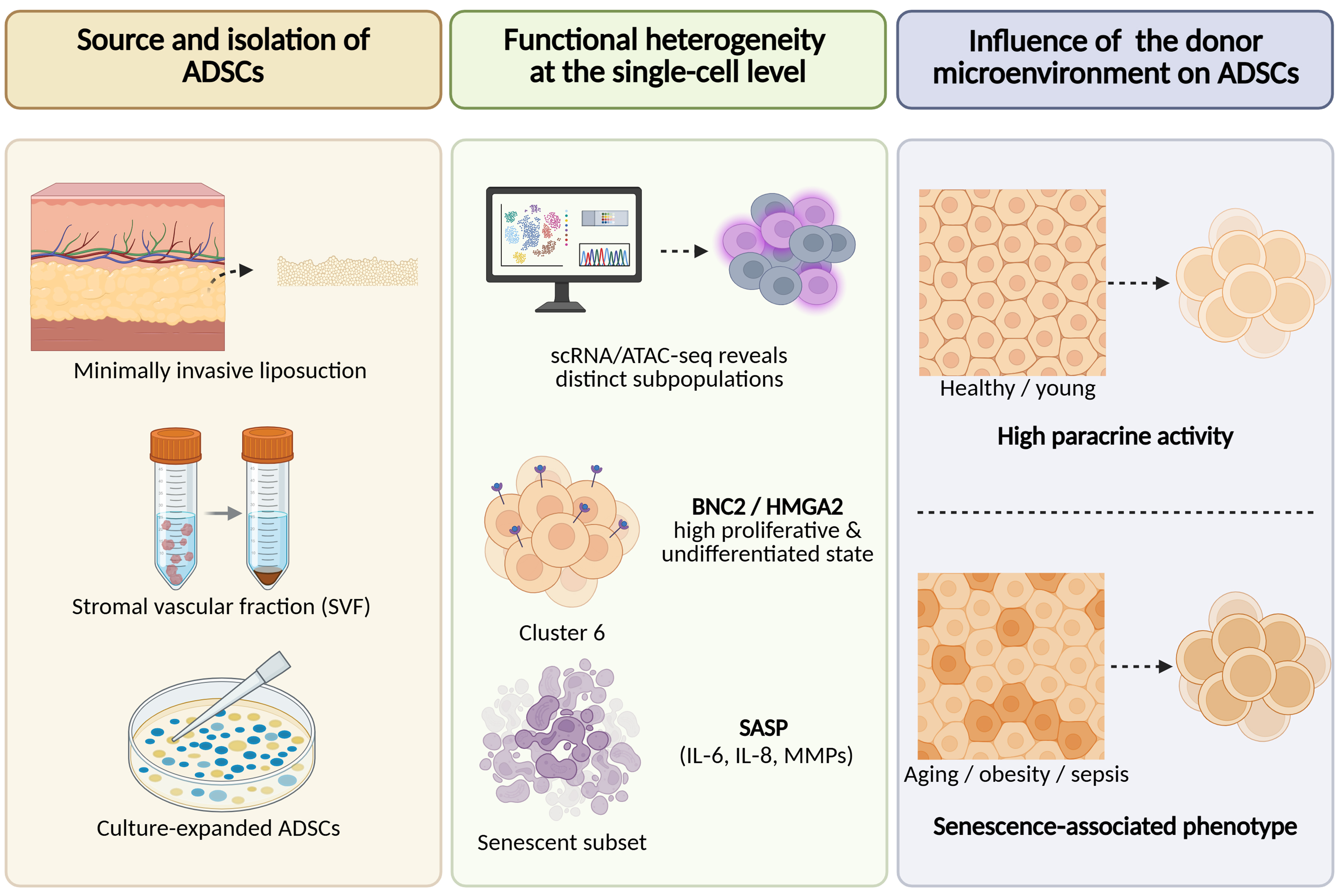
Figure 1. Biological characteristics and functional heterogeneity of ADSCs. ADSCs are isolated from adipose tissue via minimally invasive liposuction, followed by enzymatic digestion and culture expansion (Left). scRNA/ATAC-seq reveals functional subpopulations within cultured ADSCs, including a highly proliferative cluster (Cluster 6) marked by BNC2 and HMGA2, and a senescent subset exhibiting SASP (Middle). The regenerative capacity of ADSCs is heavily influenced by donor-specific factors; young/healthy donors yield cells with potent paracrine activity, whereas aging, obesity, or sepsis drive ADSCs toward a pro-inflammatory SASP, compromising their therapeutic potential (Right). Created in BioRender. (2026) https://BioRender.com/ru1r8t8. ADSCs: Adipose-derived stem cells; BNC2: basonuclin 2; HMGA2: high mobility group AT-hook 2; IL-6: interleukin-6; IL-8: interleukin-8; MMPs: matrix metalloproteinases; SASP: senescence-associated secretory phenotype; scRNA-seq/ATAC-seq: single-cell RNA and assay for transposase-accessible chromatin sequencing; SVF: stromal vascular fraction.
Phenotypic baseline and functional heterogeneity
While the International Society for Cell & Gene Therapy (ISCT) provides a necessary phenotypic baseline (CD73+/90+/105+ and CD34-/45-)[11,12], these minimal criteria fail to capture the functional diversity inherent in ADSCs. Recent single-cell RNA and assay for transposase-accessible chromatin sequencing (scRNA-seq/ATAC-seq) studies have unmasked this biological noise, revealing that cultured ADSCs are a mosaic of proliferative, functional, and senescent subsets[13]. The identification of specific clusters like Cluster 6 and biomarkers like basonuclin 2 (BNC2) and high mobility group AT-hook 2 (HMGA2) suggests that future clinical success will depend less on bulk cell quantity and more on the enrichment of high-potency, non-senescent sub-lineages. However, the intrinsic molecular traits governing heightened regenerative capacity often overlap with oncogenic hallmarks. Specifically, the core transcription factors BNC2 and HMGA2, while essential for maintaining the undifferentiated state and proliferative vigor of ADSCs, are frequently implicated in malignant transformation and epithelial-mesenchymal transition. HMGA2, for instance, can promote cancer cells to acquire stem cell-like characteristics by regulating chromatin structure, and enhance their invasive and migratory abilities[14]. Therefore, aggressive selection for these super-subpopulations warrants rigorous scrutiny to ensure that therapeutic potency does not come at the cost of genomic stability or an increased risk of long-term oncogenicity.
Secretome-mediated therapeutic framework
While traditional research emphasized the multi-lineage differentiation potential of ADSCs[15], contemporary focus has pivoted toward a paracrine-driven model. In this framework, the secretome serves as the primary vehicle for therapeutic delivery[16,17]. This paracrine dominance is particularly pronounced within wound microenvironments; here, ADSCs modulate the M1-to-M2 macrophage phenotypic transition and facilitate neoangiogenesis through the regulated secretion of vascular endothelial growth factor (VEGF), transforming growth factor-β (TGF-β), and interleukin-6 (IL-6)[18,19]. Furthermore, ADSC-derived exosomes (ADSC-exos) - specifically those harboring bioactive cargo such as miR-378 - provide a robust cell-free mechanism to attenuate oxidative stress and enhance keratinocyte viability[20]. Such advancements underscore a significant transition toward standardized, off-the-shelf precision regenerative medicine.
Environmental modulation and bioengineering of ADSC function
Notably, the functional efficacy of ADSCs is not an inherent constitutive attribute but a context-dependent state dictated by the host microenvironment. This phenomenon, characterized by Wang et al. as “reciprocal regulation”, entails the induction of an immunosuppressive phenotypic shift in response to inflammatory cues [e.g., interferon-γ (IFN-γ) and tumor necrosis factor-α (TNF-α)] via indoleamine 2,3-dioxygenase (IDO) or inducible nitric oxide synthase (iNOS) pathways[21]. Conversely, deleterious environments - including donor senescence, obesity, or systemic sepsis - can “de-license” these cells, precipitating a transition toward a pro-inflammatory senescence-associated secretory phenotype (SASP)[22-25]. To circumvent these environmental constraints, emerging bioengineering strategies, such as mitochondrial transplantation for metabolic reprogramming[26] and the development of smart chimeric antigen receptor (CAR)-modified immunosuppression platforms[27], aim to transition ADSCs from inert cellular grafts into autonomous, responsive modulators of tissue regeneration.
MECHANISMS OF ACTION
Extracellular vesicles as functional mediators of paracrine signaling
The clinical application of ADSCs has undergone a strategic shift, moving from direct cellular replacement toward a paracrine-centered model[9] [Figure 2]. Within this framework, extracellular vesicles (EVs), particularly exosomes, serve as bioactive nanovesicles that mirror the regenerative potential of their parental cells while offering enhanced safety profiles and superior scalability[28-32]. Distinct from passive biological carriers, ADSC-EVs function as dynamic regulatory units, delivering functional non-coding RNAs (ncRNAs) and proteomic payloads that reprogram the recipient microenvironment through the sophisticated modulation of autophagy. Specifically, ADSC-EVs exhibit context-specific pleiotropy: they can either attenuate myofibroblast transformation by inhibiting TGF-β/Smad2-mediated autophagic flux[33] or mitigate fibrotic pathology by activating phosphoinositide 3-kinase (PI3K)/protein kinase B (AKT)/mammalian target of rapamycin (mTOR)-dependent mitophagy[34], with the functional outcome being contingent upon the specific pathological cues of the wound environment.
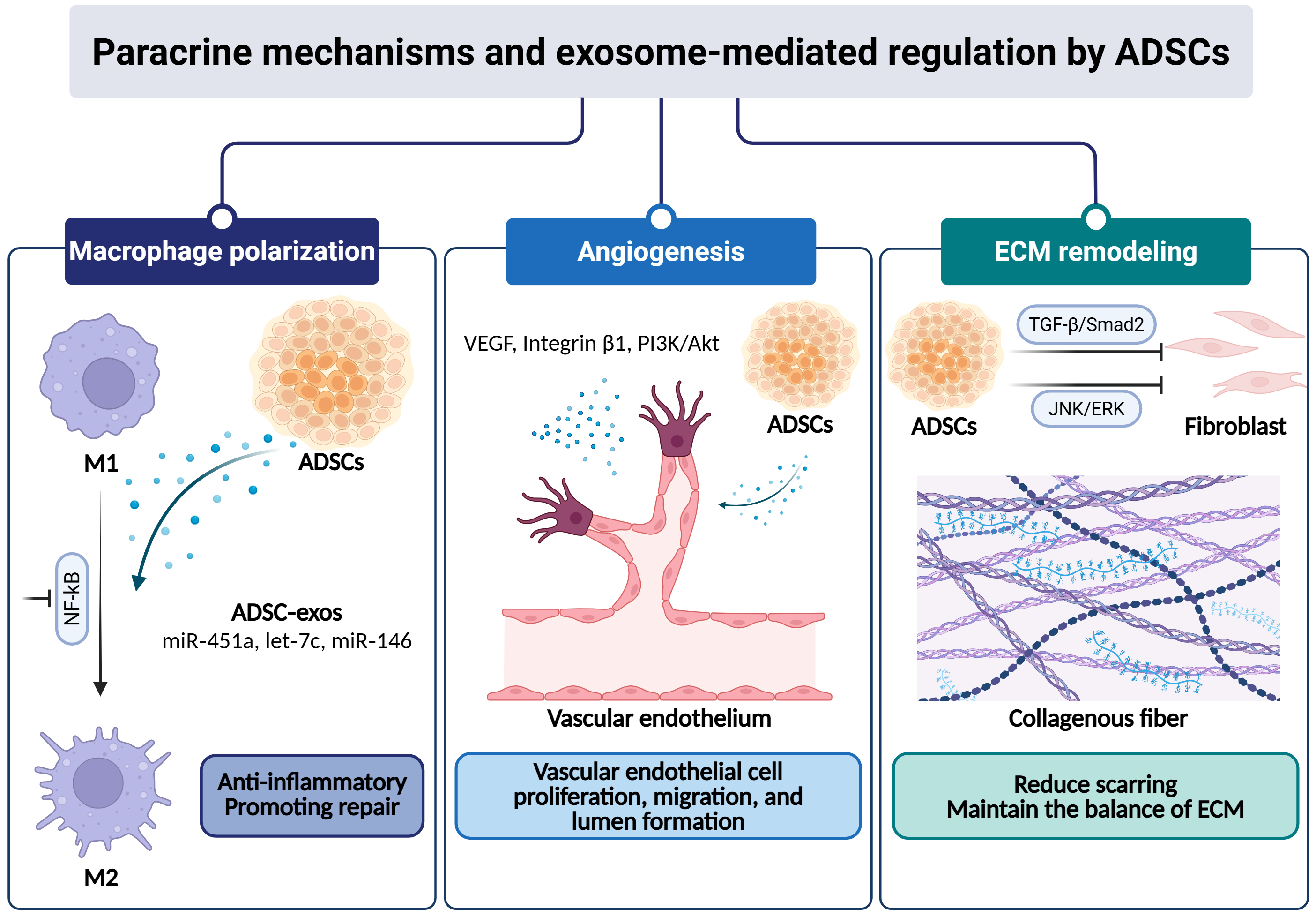
Figure 2. Paracrine mechanisms and exosome-mediated regulation by ADSCs. ADSCs secrete a diverse array of EVs/exosomes, miRNAs, and long non-coding RNAs. These bioactive factors orchestrate three key processes in tissue repair: (1) Macrophage polarization: ADSC-exos deliver miR-451a, let-7c, and miR-146 to suppress NF-κB signaling, driving the transition from pro-inflammatory M1 to reparative M2 macrophages; (2) Angiogenesis: ADSCs promote neovascularization via direct endothelial differentiation and paracrine activation of the integrin β1/PI3K/AKT pathway, supported by VEGF and other growth factors; (3) ECM remodeling and anti-fibrosis: ADSC-derived factors exert anti-fibrotic effects through distinct signaling mechanisms, including inhibition of the JNK/ERK pathway and modulation of TGF-β/Smad2 signaling, thereby suppressing myofibroblast activation and pathological scar formation. Created in BioRender. (2026) https://BioRender.com/1teq1ae. ADSC-exos: Adipose-derived stem cell-derived exosomes; ADSCs: adipose-derived stem cells; AKT: protein kinase B; ECM: extracellular matrix; ERK: extracellular signal-regulated kinase; EVs: extracellular vesicles; JNK: c-Jun N-terminal kinase; miRNAs: microRNAs; NF-κB: nuclear factor kappa-light-chain-enhancer of activated B cells; PI3K: phosphoinositide 3-kinase; TGF-β: transforming growth factor-β; VEGF: vascular endothelial growth factor.
The competitive endogenous RNA (ceRNA) regulatory axis and networks
The functional versatility of ADSC-EVs is sustained by a complex repertoire of ncRNAs operating via ceRNA networks. Long non-coding RNAs (lncRNAs), such as H19 and Neat1, function as molecular sponges that sequester microRNAs (miRNAs) to derepress regenerative pathways, including the Wnt/β-catenin and Ulk1-mediated autophagic axes[35,36]. Consistently, the miRNA profile of ADSC-EVs - further optimized by hypoxic preconditioning - functions as a coordinated toolkit for reversing ischemic stagnation. Upregulated miR-21, miR-126, and miR-31 facilitate high-quality healing by activating the PI3K/AKT signaling cascade, thereby promoting targeted vascularization and keratinocyte migration[37-39]. Recent identification of circular RNAs adds an additional layer of epigenetic control, modulating the miR-144/signal transducer and activator of transcription 3 (STAT3) axis to resolve chronic inflammation[29].
Inflammatory priming and macrophage plasticity
Inflammatory priming represents a key mechanism through which the context-dependent properties of ADSCs translate into immunomodulatory effects[21,29,31,32]. ADSC-EVs further reinforce this response by delivering miRNAs and lncRNAs that attenuate nuclear factor kappa-light-chain-enhancer of activated B cells (NF-κB) signaling. Moreover, unbiased omics analyses have elucidated pivotal molecular checkpoints, such as epiregulin (EREG) and cystatin A (CSTA). The targeted modulation of these factors by ADSCs correlates with enhanced M2 macrophage infiltration and accelerated re-epithelialization in diabetic foot ulcers (DFUs)[40].
Spatiotemporal coordination of angiogenesis and extracellular matrix homeostasis
Optimal tissue restoration necessitates the precise integration of chemotaxis, revascularization, and extracellular matrix (ECM) reorganization. The recruitment of ADSCs is primarily mediated via the stromal cell-derived factor-1α (SDF-1α)/C-X-C motif chemokine receptor 4 (CXCR4)/CXCR7 signaling axis, a pathway amenable to targeted manipulation for enhanced site-specific homing[41]. Following recruitment, integrin β1/PI3K/AKT signaling contributes to the angiogenic response[39,42]. Concurrently, ADSC-EVs modulate fibroproliferative dynamics by fine-tuning collagen deposition and regulating matrix metalloproteinases (MMPs)[28,43]. Specifically, miR-141-3p-mediated suppression of c-Jun N-terminal kinase (JNK)/extracellular signal-regulated kinase (ERK) signaling inhibits myofibroblast differentiation, thereby favoring functional tissue regeneration over hypertrophic scar formation[44]. This multifaceted regulatory network is further reinforced by the broader ADSC secretome, which preserves ECM structural integrity and attenuates environmental senescence[43,45].
CLINICAL APPLICATIONS IN PLASTIC SURGERY
The translation of ADSC biology into clinical practice has found its most fertile ground in plastic and reconstructive surgery. The mechanistic pillars detailed previously provide the biological scaffolding for a wide clinical spectrum, ranging from enhancing the fidelity of autologous fat grafting to the management of recalcitrant wounds and pathological scars[16,46,47] [Figure 3].
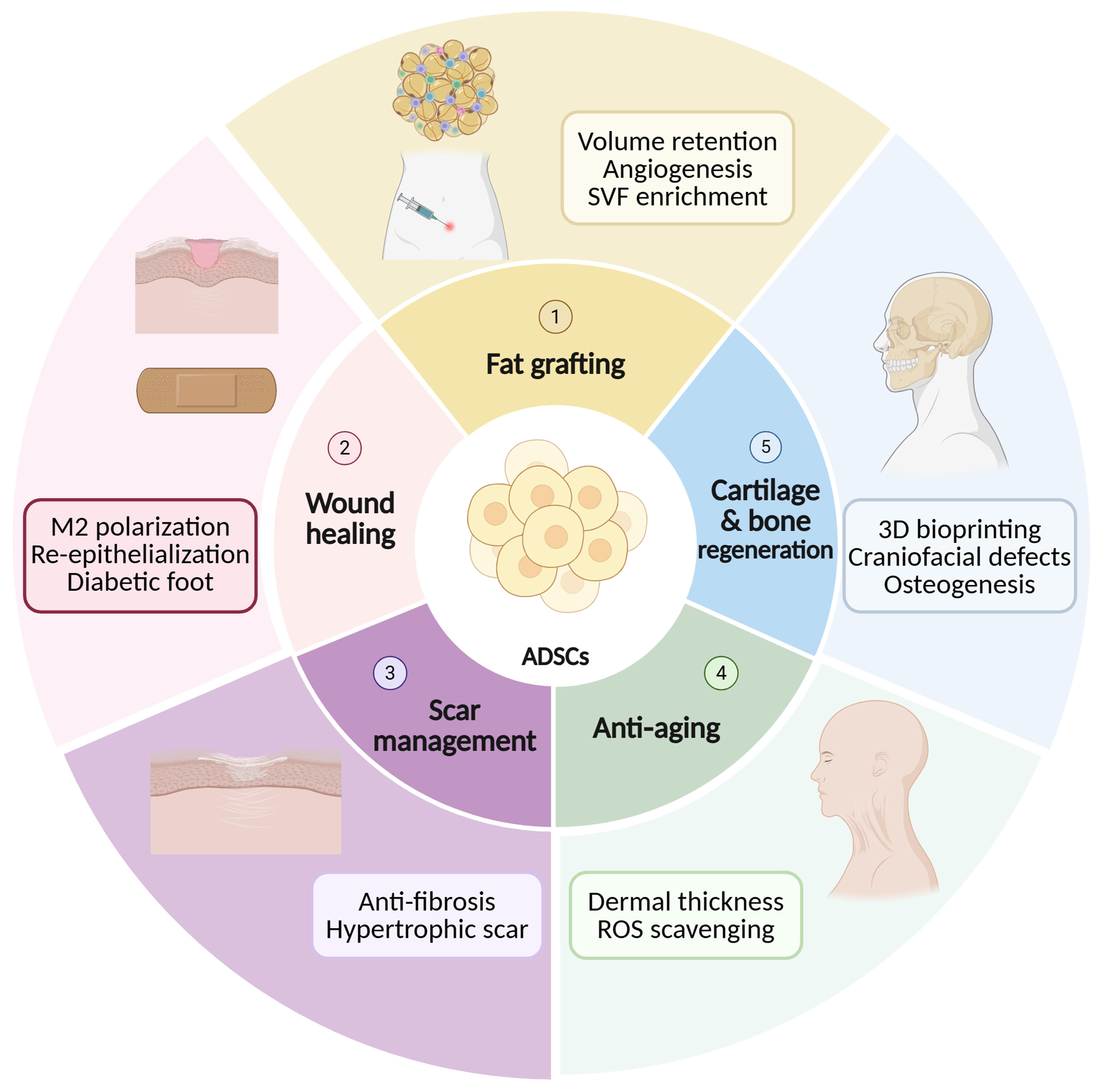
Figure 3. Major clinical applications of ADSCs in plastic and reconstructive surgery. Surrounding sectors illustrate five key areas where ADSCs have demonstrated therapeutic potential: (1) CAL for fat grafting; (2) chronic wound healing (e.g., diabetic foot ulcers); (3) scar management and anti-fibrosis; (4) facial rejuvenation and anti-aging; and (5) cartilage and bone regeneration for craniofacial reconstruction. Each sector highlights representative mechanisms or key molecules involved in ADSC-mediated tissue repair. Created in BioRender. (2026) https://BioRender.com/mdkvzrj. ADSCs: Adipose-derived stem cells; CAL: cell-assisted lipotransfer; M2: type 2 (alternatively activated) macrophage; ROS: reactive oxygen species; SVF: stromal vascular fraction.
Fat grafting and CAL
Autologous fat grafting (AFG) remains the standard for soft tissue restoration, yet it is plagued by a volumetric lottery, with unpredictable resorption rates that range from 10% to 90%[48]. This phenomenon is primarily driven by delayed neovascularization, which subjects the central graft to lethal ischemic stress[49].
The identification of ADSCs within the SVF offered a transformative solution[50]. This catalyzed the advent of CAL, a strategy where lipoaspirates are enriched with either freshly isolated SVF or ex vivo culture-expanded ADSCs[51]. In this context, ADSCs function as “biologic factories”, secreting a potent cocktail of pro-angiogenic and immunomodulatory factors that mitigate early inflammatory insult and facilitate host-graft integration[52]. The clinical applications of CAL have been increasingly explored. A landmark randomized controlled trial by Kølle et al. reported a remarkable 80.9% volume retention on day 121 with ADSC enrichment, compared to a mere 16.3% in controls [an absolute difference of 64.6 percentage points (pp)][53,54]. Although promising results have been reported, the clinical superiority of CAL over conventional fat grafting has not been consistently reproduced across all studies[51]. Differences in cell preparation methods, enrichment strategies, and outcome assessment criteria may contribute to the observed variability. This distinction underscores the importance of cell potency and dosage, though the ADSCs still face stringent regulatory and logistical hurdles[4,55-57].
Beyond volume, ADSCs are being instructed through innovative co-culture or functional enhancement strategies. For instance, co-culturing ADSCs with vascular regenerative cells (RE-01 cells) has been shown to achieve superior graft retention even with a tenfold reduction in cell dosage, signaling a shift toward precision cell therapy[58]. Furthermore, the field is pivoting toward a cell-free protocol, utilizing ADSC-exos. These nanovesicles recapitulate the parent cell’s benefits while offering a superior safety profile, easier storage, and reduced risks of immunogenicity or unwanted differentiation[59-61].
Wound healing and chronic ulcers
Managing recalcitrant wounds, such as DFUs and major burns, remains a formidable challenge where ADSCs offer significant therapeutic leverage[62]. The pathophysiology of chronic wounds - often characterized by a stalled M1 inflammatory phase - is directly countered by the ADSCs’ ability to drive M2 macrophage polarization, thereby re-initiating the regenerative cascade[47,63]. The key mechanism by which ADSCs promote the healing of DFUs is closely related to their powerful paracrine effect. Studies have shown that ADSC-exos are rich in various functional miRNAs, such as miR-451a, let-7c, miR-146, etc.[63,64]. These miRNAs target key molecules such as macrophage migration inhibitory factor (MIF), interferon regulatory factor 1 (IRF1), and CCAAT/enhancer-binding protein delta (C/EBP-δ), and jointly regulate the polarization of macrophages to the M2 anti-inflammatory phenotype, thereby breaking the pathological state of the chronic wound where the M1 inflammatory phase is stagnant. This creates a favorable immune microenvironment for tissue regeneration. Additionally, the growth factors secreted by ADSCs activate signaling pathways, promoting the proliferation and migration of vascular endothelial cells, protecting fibroblasts from apoptosis and accelerating re-epithelialization and collagen deposition. Based on these mechanisms, to enhance the therapeutic efficacy of ADSC-exos, researchers have adopted various engineering strategies. Among them, hypoxic preconditioning has been proven to effectively enrich the functional miRNAs in exosomes, which target inflammatory-related genes and activate signaling pathways such as PI3K/AKT, jointly regulating the polarization of macrophages to the M2 type, promoting the proliferation and migration of fibroblasts, and enhancing the function of vascular endothelial cells, thereby accelerating wound healing through multiple targets[46,47,61]. For deep-tissue defects, 3D-bioprinted hydrogel scaffolds co-delivering ADSCs and nitric oxide (NO) donors have demonstrated synergistic effects, promoting rapid re-epithelialization and organized collagen deposition[52,65,66]. Despite these encouraging findings, the clinical evidence for ADSC-based treatment of chronic wounds remains less mature than the preclinical evidence. Most available studies are experimental or translational, with substantial variation in cell source, delivery strategy, wound models, and outcome assessment. Therefore, the reproducibility and clinical relevance of these findings remain to be established in adequately powered controlled clinical trials.
Scar management and anti-fibrosis
ADSCs are equally pivotal in modulating the late stages of tissue repair to prevent pathological scarring. Their anti-fibrotic efficacy is primarily mediated through the inhibition of TGF-β1/Smad signaling, which prevents the transition of fibroblasts into hyperactive myofibroblasts[47]. Additionally, ADSCs help restore the ECM balance by upregulating MMPs while suppressing their inhibitors [tissue inhibitors of metalloproteinases (TIMPs)][67]. Clinical evidence, including a systematic review of 665 patients, suggests potential benefits of ADSCs or SVF in improving both the functional and aesthetic outcomes of scars[68]. Emerging data highlight the role of ADSC-exos in alleviating the progression of various fibrotic diseases via mitochondrial restoration and targeted miRNA delivery[47,60,63,68]. In aesthetic practice, the efficacy of local ADSC-exos or direct ADSC injection for acne scars has been confirmed to be comparable to that of fractional CO2 laser treatment, indicating their potential as independent regenerative therapies. A randomized controlled study by Abou Eitta et al. demonstrated that a single ADSC injection was as effective as three sessions of fractional CO2 laser in improving acne scars[69]. Additionally, ADSC-exos can serve as an adjunct to fractional CO2 laser treatment, significantly enhancing efficacy, reducing erythema, and shortening recovery time[59]. These findings collectively support ADSCs and their exosomes as effective strategies for acne scar treatment, with their non-invasive and repeatable nature offering unique application value in aesthetic practice.
However, the strength of this evidence remains limited by heterogeneity in scar type, cell preparation, treatment protocols, comparator interventions, and outcome measures. Although some controlled studies suggest efficacy comparable to established treatments, the available evidence does not yet establish ADSC-based therapy as a standardized or superior treatment for scar management.
Facial rejuvenation and anti-aging
In the realm of aesthetic medicine, ADSCs have garnered attention for their ability to combat both intrinsic and extrinsic aging[70,71]. By neutralizing reactive oxygen species (ROS) and stimulating resident dermal fibroblasts, ADSCs can increase dermal thickness and restore elastic fiber architecture[72,73]. While clinical data suggest improvements in skin elasticity and hydration, meta-analyses have highlighted substantial heterogeneity in aesthetic outcomes, with some studies reporting limited or non-significant effects on wrinkle reduction[70]. This variability may reflect differences in administration routes, treatment protocols, and patient characteristics. Nevertheless, high-dose intradermal ADSC injections have demonstrated sustained facial rejuvenation for over a year, potentially through the activation of underlying facial musculature via exosomal signaling[73]. The use of ADSC-conditioned medium (ADSC-CM) also represents a growing trend, offering a potent, non-invasive secretome cocktail that protects against ultraviolet (UV)-induced senescence[28].
Cartilage and bone regeneration
The chondrogenic and osteogenic potential of ADSCs is being harnessed for complex craniofacial reconstruction[74]. Most clinical applications involve combining ADSCs with osteoconductive scaffolds like β-tricalcium phosphate (β-TCP) for mandibular or cranial defects[61]. In rhinoplasty, the convergence of ADSC biology and 3D bioprinting allows for the creation of patient-specific, stable cartilage constructs using bio-inks like gelatin methacryloyl (GelMA)[75]. Innovative injectable systems using ADSC-incorporated microspheres now offer a minimally invasive alternative for minor nasal contouring, bypassing the morbidity associated with traditional rib cartilage harvesting[76]. The evidence for craniofacial cartilage and bone regeneration remains predominantly preclinical or early translational. Outcomes are highly dependent on scaffold composition, cell source, differentiation conditions, and defect characteristics, which limits direct comparison between studies. Further long-term clinical studies are needed to determine the durability, safety, and functional relevance of these approaches.
A summary of key clinical studies on ADSCs in plastic surgery is provided in Table 1.
Summary of key clinical studies on ADSCs in plastic surgery
| Study (Year) | Design | n | Cell type/Dose | Follow-up | Primary outcome | Key findings | Evidence level/limitations |
| CAL and fat grafting | |||||||
| Kølle et al., 2013[54] (Lancet) | RCT (Triple-blind, placebo-controlled) | 10 | Ex vivo-expanded autologous ASCs 20 × 106 cells/mL fat 30 mL fat graft enriched vs. 30 mL control (contralateral arm) | 121 days | Residual graft volume by MRI | ASC-enriched grafts: 80.9% retention vs. 16.3% control (P < 0.0001); Absolute difference = 64.6 pp. No serious AEs | High-level clinical evidence; small sample size and short follow-up |
| Li and Chen, 2021[51] (Aesthetic Plast Surg) | Meta-analysis | 353 (6 studies) | CAL (ex vivo ADSC or SVF enrichment); dose heterogeneous across studies | Variable | Fat survival rate; Complication rate | CAL superior to conventional lipotransfer for fat survival (P = 0.02). No significant difference in complications (OR = 1.34, P = 0.43) | Moderate-to-high level evidence; limited by heterogeneity of included studies, cell preparations, and outcome measures |
| Hasiba-Pappas et al., 2025[77] (Aesthetic Surg J) | Systematic review (12 clinical studies, 15 animal studies) | 12 clinical studies (case numbers variable) | SVF and/or ADSC enrichment; also PRP, Vitamin D3, botulinum toxin A, etc. | Variable | Graft survival; enhancement strategies | SVF/ADSC and PRP most promising for graft enhancement; marked protocol heterogeneity limits comparability. No gold standard established | Moderate-level evidence; substantial protocol heterogeneity and no established gold standard |
| Gao et al., 2022[57] (J Cosmet Dermatol) | In vivo preclinical (nude mouse model; clinical ADSC harvest) | 15 PHA patients vs. 15 healthy donors (fat grafts in nude mice) | ADSC-assisted fat graft; exosome-assisted fat graft; non-cell-assisted (control) | 2, 4, 8, 12 weeks | Volume & weight retention; Histology (HE, CD31, CD68, perilipin) | PHA-ADSC group: lower retention than NORM-ADSC but superior to non-cell group. Exosome-assisted also improved fat survival. Supports CAL for hemifacial atrophy | Preclinical evidence; limited direct clinical generalizability |
| Wound healing & chronic ulcers | |||||||
| Cao et al., 2025[40] (J Inflammation Res) | Translational (Bioinformatics + ML + in vivo/in vitro validation) | GEO datasets: GSE134431, GSE80178; in vivo: DFU mouse model | ADSCs applied to DFU wound model | Not specified (wound closure endpoint) | AUC of 2-gene prognostic model; M2 macrophage polarization; wound healing rate | EREG & CSTA identified as key MA-DEGs. AUC > 0.944 in training and validation. ADSCs downregulate EREG/CSTA → promote M2 polarization → accelerate DFU healing | Translational/preclinical evidence; supported by bioinformatics and experimental validation but lacks direct human clinical evidence |
| Kohlhauser et al., 2024[67] (Cell Mol Biol Lett) | Systematic Review (21 in vivo studies; rodent + 1 porcine model) | 21 studies (all preclinical; no human RCTs included) | Heterogeneous across studies | Variable | Immunomodulation; neovascularization; granulation tissue; re-epithelialization; remodeling | ADSCs promoted wound healing in all 21 in vivo studies via immunomodulation, neovascularization, granulation tissue formation, re-epithelialization, and remodeling. The majority of studies reported positive outcomes across multiple healing phases. Clinical translation remains at an early experimental stage | Preclinical evidence; systematic review of animal studies without human RCTs, limiting clinical translation |
| Skin rejuvenation & anti-aging | |||||||
| Ichihashi et al., 2023[73] (J Pers Med) | Case Series (single-arm, open-label) | 8 patients | 1 × 10⁸ autologous ADSCs (cultured from abdominal subcutaneous fat) Single intradermal injection to face | 12 months (1, 3, 6, 12 months) | Photographic assessment of wrinkles (glabella, crow’s feet, nasolabial), pore size, double eyelid | All 8 cases: wrinkle shallowing/disappearance from 1-several months, lasting > 1 year. Double eyelid sharpening and pore reduction. No adverse events reported | Moderate-level clinical evidence; single-arm case series with only 8 patients and no control group |
| Chon et al., 2025[70] (Aesthetic Surg J Open Forum) | Systematic review (17 studies; SVF, ADSC, AD-MSC) | 17 studies (sample sizes per included study) | SVF, ADSCs, or conditioned medium; heterogeneous protocols | Variable; heterogeneity limits pooling | Rhytid severity; skin elasticity; pigmentation (melanin index); texture | SVF and ADSC-CM reduced rhytid severity, especially periorbital and nasolabial. Meta-analysis of 2 studies: rhytid reduction non-significant (P = 0.12). Combination therapy (SVF+laser/PRP) outperformed SVF alone | Moderate-level evidence; substantial heterogeneity and limited statistical power for pooled clinical outcomes |
| Surowiecka and Strużyna, 2022[78] (J Pers Med) | Narrative review | Multiple clinical studies summarized | ADSCs or SVF injected intradermally; no standard dose | Variable | Skin density; appearance; hydration; capillary density | ADSC intradermal injection improved skin density, overall appearance, hydration, and capillary vessel number. Graft survival main limitation. No standardized protocol; longer follow-up needed | Moderate quality evidence; heterogeneous interventions and outcome measures |
| Scar treatment | |||||||
| Stachura et al., 2021[68] (J Clin Med) | Systematic review (19 clinical studies) | 665 total patients (19 included studies) | ADSCs or SVF (enzymatic/mechanical/nanofat); heterogeneous across studies | Variable | Microscopic, functional, and aesthetic scar outcomes; Comparison to PRP/CO2 laser | Low-to-average quality evidence for beneficial effects. Some studies: ADSC interventions non-inferior to PRP or fractional CO2 laser. No gold standard established | Moderate quality evidence; heterogeneous interventions and outcome measures, with no established gold-standard treatment |
| Vascular & other applications | |||||||
| Shikanai et al., 2025[58] (Regen Ther) | Translational (in vitro co-culture + in vivo nude mouse fat graft model) | In vitro: human PBMC donors; in vivo: nude mice | ADSCs co-cultured with RE-01 cells (ex vivo peripheral blood MNCs); reduced ADSC number vs. ADSC-alone | Fat graft engraftment endpoint | Fat engraftment rate; blood vessel density; adipose tissue quality | ADSC+RE-01 combination significantly improved fat engraftment rate, blood vessel number, and fat quality vs. ADSC alone in vivo. RE-01 reduces ADSC requirement for breast reconstruction fat grafting | Translational/preclinical evidence; promising in vitro and animal findings but limited direct evidence of clinical efficacy |
CHALLENGES AND SAFETY CONSIDERATIONS
Standardization and regulatory hurdles
Despite the promising clinical outcomes, the lack of standardized protocols for ADSC isolation, expansion, and administration remains a primary bottleneck[79]. The biological properties of ADSCs are significantly influenced by donor characteristics [e.g., body mass index (BMI), age, and anatomical harvest site] and processing techniques (enzymatic vs. mechanical)[12,22]. To move toward large-scale clinical application, it is imperative to establish Good Manufacturing Practice (GMP) standards and universal characterization criteria[80]. Furthermore, varying international regulatory classifications - ranging from minimally manipulated tissue to advanced therapy medicinal products - pose significant hurdles for global clinical translation and multicenter trials[11,32]. At present, the lack of standardized separation protocols and the significant impact of donor characteristics on cell quality make it extremely difficult to establish a unified risk monitoring system. Therefore, future research should not be limited to the observation of short-term complications, but should focus on building an early warning model based on single-cell omics, to achieve a leap from empirical application to precise regenerative medicine.
Oncological safety and long-term monitoring
A critical concern in plastic surgery, particularly in breast reconstruction following malignancy, is the potential for ADSCs to promote tumor recurrence or metastasis via their pro-angiogenic and anti-apoptotic signaling[81]. While available clinical evidence has not demonstrated a clear increase in oncological risk associated with CAL, the pro-angiogenic secretome of ADSCs, including VEGF and hepatocyte growth factor (HGF), warrants continued vigilance in post-oncologic reconstruction, particularly given the potential for interactions with residual or dormant tumor cells[82-84]. The powerful paracrine effect of ADSCs, through the secretion of VEGF, HGF and various cytokines, promotes the vascularization of transplanted fat but may also influence the local tumor microenvironment, highlighting the need for continued oncologic surveillance. In addition, the transcriptional heterogeneity of ADSCs further complicates risk assessment. Studies have shown that the drift of cell subpopulations during in vitro culture may increase the risk of embolism in the host[79]. Rigorous, large-scale prospective randomized controlled trials with extended follow-up periods are essential to definitively establish the safety profile of ADSC-based therapies. Clinically, these potential risks support careful patient selection and individualized timing of CAL rather than a universal waiting period. CAL should preferably be considered after completion of oncological treatment and confirmation of no active disease, with the timing determined according to tumor characteristics and recurrence risk; an interval of approximately 6-12 months may be considered in selected post-radiotherapy patients[85].
Future directions: biofabrication and cell-free therapies
The future of ADSCs in plastic surgery lies at the intersection of bioengineering and molecular medicine. 3D Bioprinting technology enables the creation of customized, ADSC-laden scaffolds that mimic the complex architecture of craniofacial bones or soft tissue defects, offering a path toward precision reconstructive surgery[86]. Simultaneously, the shift toward cell-free therapies using ADSC-derived exosomes represents a transformative trend[87]. Exosome-based products may retain some of the regenerative properties associated with ADSCs while offering potential advantages in terms of stability, immunogenicity, and translational feasibility, potentially serving as biological agents for wound repair and aesthetic enhancements[88]. Beyond these emerging strategies, further investigation is also warranted to clarify and optimize the potential applications of ADSCs in breast remodeling and reconstruction, facial soft-tissue restoration, and protection against UV-related skin damage and photoaging[89]. Future studies should focus on improving the consistency, safety, and long-term efficacy of these approaches and on translating promising preclinical findings into well-controlled clinical applications.
CONCLUSION
In conclusion, ADSCs represent a transformative pillar in the field of plastic and reconstructive surgery, bridging the gap between traditional surgical techniques and advanced regenerative medicine. Their abundance, ease of isolation, and multifaceted mechanisms - ranging from direct differentiation to potent paracrine and exosome-mediated signaling - have redefined our approach to tissue repair and aesthetic enhancement. While CAL and chronic wound therapies have already shown encouraging preclinical efficacy, the emergence of cell-free strategies and biofabrication technologies marks the next frontier. Despite the remaining challenges in standardization and long-term regulatory oversight, ADSCs hold the potential to realize the goal of personalized, scarless, and functional tissue regeneration. As research continues to unravel the molecular intricacies of these cells, ADSCs will undoubtedly remain at the heart of the next generation of plastic surgery.
DECLARATIONS
Acknowledgments
The Graphical Abstract and figures were created using BioRender. Publication licenses were obtained for the BioRender content used in this manuscript. (2026) https://BioRender.com/smn4uzm.
Authors’ contributions
Made substantial contributions to the conception and design of the review, and performed literature search and analysis: Sun C
Provided critical feedback on the structure and intellectual content, and revised the manuscript: Jin M
Supervised the study, acquired funding, and revised the manuscript for important intellectual content: An Y
All authors read and approved the final manuscript.
Availability of data and materials
Not applicable.
AI and AI-assisted tools statement
During the preparation of this manuscript, the AI tool ChatGPT (GPT-5.6 Luna, released 2026-07-09) was used solely for language polishing and proofreading to improve readability and grammatical clarity. The tool did not influence the study design, data collection, analysis, interpretation, or the scientific content of the work. All authors take full responsibility for the accuracy, integrity, and final content of the manuscript.
Financial support and sponsorship
This work was supported by the Grant Beijing Natural Science Foundation (Nos. L244086 and 7264349).
Conflicts of interest
All authors declared that there are no conflicts of interest.
Ethical approval and consent to participate
Not applicable.
Consent for publication
Not applicable.
Copyright
© The Author(s) 2026.
REFERENCES
1. Zhou C, Zhang B, Yang Y, et al. Stem cell-derived exosomes: emerging therapeutic opportunities for wound healing. Stem Cell Res Ther. 2023;14:107.
2. Condé-Green A, Marano AA, Lee ES, et al. Fat grafting and adipose-derived regenerative cells in burn wound healing and scarring: a systematic review of the literature. Plast Reconstr Surg. 2016;137:302-12.
3. Wang L, Jiang X, Zhao F, Duan P, Li Z, Luo Y. A review of adipose-derived mesenchymal stem cells’ impacts and challenges: metabolic regulation, tumor modulation, immunomodulation, regenerative medicine and genetic engineering therapies. Front Endocrinol. 2025;16:1606847.
4. Shimizu Y, Inoue Y, Sowa Y, et al. Adipose-derived stem cell-enhanced versus conventional fat grafting for breast reconstruction: a systematic review and meta-analysis. Plast Reconstr Surg. 2026;157:338e-49.
5. Anjum A, Vijakumaran U, Hussain HB, Khalid S, Ghosh M. Stem cell-based nerve regenerative therapies: the role of adipose-derived stem cells (ADSCs) in preventing nerve adhesions and promoting axonal nerve repair. Stem Cell Rev Rep. 2026;22:1309-24.
6. Shukla L, Yuan Y, Shayan R, Greening DW, Karnezis T. Fat therapeutics: the clinical capacity of adipose-derived stem cells and exosomes for human disease and tissue regeneration. Front Pharmacol. 2020;11:158.
7. Yi Y, Hu W, Zhao C, et al. Deciphering the emerging roles of adipocytes and adipose-derived stem cells in fat transplantation. Cell Transplant. 2021;30:963689721997799.
8. Plangger A, Haslik W, Rath B, Neumayer C, Hamilton G. Interactions of BRCA1-mutated breast cancer cell lines with adipose-derived stromal cells (ADSCs). J Mammary Gland Biol Neoplasia. 2021;26:235-45.
9. Qin Y, Ge G, Yang P, et al. An update on adipose-derived stem cells for regenerative medicine: where challenge meets opportunity. Adv Sci. 2023;10:e2207334.
10. Dong L, Li X, Leng W, et al. Adipose stem cells in tissue regeneration and repair: From bench to bedside. Regen Ther. 2023;24:547-60.
11. Mazini L, Ezzoubi M, Malka G. Overview of current adipose-derived stem cell (ADSCs) processing involved in therapeutic advancements: flow chart and regulation updates before and after COVID-19. Stem Cell Res Ther. 2021;12:1.
12. Khazaei S, Keshavarz G, Bozorgi A, Nazari H, Khazaei M. Adipose tissue-derived stem cells: a comparative review on isolation, culture, and differentiation methods. Cell Tissue Bank. 2022;23:1-16.
13. Long Q, Yuan Y, Liu Z, Zhang P, Yuan X. Single-cell RNA and ATAC sequencing reveals cell subsets characteristics in adipose-derived stem cells. Biochim Biophys Acta Mol Cell Biol Lipids. 2026;1871:159734.
14. Kong D, Zha L, Yao Y, et al. Effects of HMGA2 on the biological characteristics and stemness acquisition of gastric cancer cells. Arab J Gastroenterol. 2024;25:135-42.
15. Park JH, Choi Y, Shin JM, Yang HW, Jeong SH, Park IH. Ultrasound cavitation: a reliable non-enzymatic method for adipose-derived mesenchymal stem cell (ADSC) isolation. Stem Cell Res Ther. 2023;14:153.
16. Yuan X, Li L, Liu H, et al. Strategies for improving adipose-derived stem cells for tissue regeneration. Burns Trauma. 2022;10:tkac028.
17. Mazini L, Rochette L, Admou B, Amal S, Malka G. Hopes and limits of adipose-derived stem cells (ADSCs) and mesenchymal stem cells (MSCs) in wound healing. Int J Mol Sci. 2020;21:1306.
18. Hao Z, Qi W, Sun J, Zhou M, Guo N. Review: research progress of adipose-derived stem cells in the treatment of chronic wounds. Front Chem. 2023;11:1094693.
19. Gandolfi S, Sanouj A, Chaput B, Coste A, Sallerin B, Varin A. The role of adipose tissue-derived stromal cells, macrophages and bioscaffolds in cutaneous wound repair. Biol Direct. 2024;19:85.
20. Zheng T, Shao W, Tian J. Exosomes derived from ADSCs containing miR-378 promotes wound healing by targeting caspase-3. J Biochem Mol Toxicol. 2021;35:e22881.
21. Wang Y, Fang J, Liu B, Shao C, Shi Y. Reciprocal regulation of mesenchymal stem cells and immune responses. Cell Stem Cell. 2022;29:1515-30.
22. Foti R, Storti G, Palmesano M, et al. Senescence in adipose-derived stem cells: biological mechanisms and therapeutic challenges. Int J Mol Sci. 2024;25:8390.
23. Li K, Shi G, Lei X, et al. Age-related alteration in characteristics, function, and transcription features of ADSCs. Stem Cell Res Ther. 2021;12:473.
24. Fang J, Lu R, Lin Y, et al. Effects of sepsis serum on the fate of adipose tissue-derived stem cells. Front Biosci. 2023;28:72.
25. Navarro-Perez J, Carobbio S. Adipose tissue-derived stem cells, in vivo and in vitro models for metabolic diseases. Biochem Pharmacol. 2024;222:116108.
26. Tang J, Han Y, Ren P, et al. Mitochondria transplanted adipose-derived stem cells/decellularized adipose tissue hydrogel for adipose tissue regeneration. Mater Today Bio. 2025;34:102193.
27. Sirpilla O, Sakemura RL, Hefazi M, et al. Mesenchymal stromal cells with chimaeric antigen receptors for enhanced immunosuppression. Nat Biomed Eng. 2024;8:443-60.
28. Wang Y, Cheng L, Zhao H, et al. The therapeutic role of ADSC-EVs in skin regeneration. Front Med. 2022;9:858824.
29. Jia Q, Zhao H, Wang Y, Cen Y, Zhang Z. Mechanisms and applications of adipose-derived stem cell-extracellular vesicles in the inflammation of wound healing. Front Immunol. 2023;14:1214757.
30. Chen L, Amraee F, Sadegh-Nejadi S, et al. Molecular mechanisms linking adipose tissue-derived small extracellular vesicles/exosomes to the development or amelioration of obesity, insulin resistance, and diabetes-related complications. Eur J Med Res. 2025;30:1049.
31. Zhou Z, Tao Y, Zhao H, Wang Q. Adipose extracellular vesicles: messengers from and to macrophages in regulating immunometabolic homeostasis or disorders. Front Immunol. 2021;12:666344.
32. Zhang J, Liu Y, Chen Y, et al. Adipose-derived stem cells: current applications and future directions in the regeneration of multiple tissues. Stem Cells Int. 2020;2020:8810813.
33. Xu J, Wang Y, Shao Z, et al. Adipose-derived stem cell exosomes attenuates myofibroblast transformation via inhibiting autophagy through TGF-β/Smad2 axis in oral submucosal fibrosis. J Nanobiotechnology. 2024;22:780.
34. Liu C, Khairullina L, Qin Y, Zhang Y, Xiao Z. Adipose stem cell exosomes promote mitochondrial autophagy through the PI3K/AKT/mTOR pathway to alleviate keloids. Stem Cell Res Ther. 2024;15:305.
35. Qian L, Pi L, Fang BR, Meng XX. Adipose mesenchymal stem cell-derived exosomes accelerate skin wound healing via the lncRNA H19/miR-19b/SOX9 axis. Lab Invest. 2021;101:1254-66.
36. An Y, Huang F, Tan X, et al. Exosomes of adipose tissue-derived stem cells promote wound healing by sponging miR-17-5p and inducing autophagy protein Ulk1. Plast Reconstr Surg. 2023;151:1016-28.
37. Wang J, Wu H, Peng Y, et al. Hypoxia adipose stem cell-derived exosomes promote high-quality healing of diabetic wound involves activation of PI3K/Akt pathways. J Nanobiotechnology. 2021;19:202.
38. An Y, Zhao J, Nie F, et al. Exosomes from adipose-derived stem cells (ADSCs) overexpressing miR-21 promote vascularization of endothelial cells. Sci Rep. 2019;9:12861.
39. Biniazan F, Stoian A, Haykal S. Adipose-derived stem cells: angiogenetic potential and utility in tissue engineering. Int J Mol Sci. 2024;25:2356.
40. Cao J, Zhang X, Li Z, et al. Adipose-derived mesenchymal stem cells accelerate diabetic foot ulcer healing by promoting macrophage M2 polarization through downregulation of EREG and CSTA. J Inflamm Res. 2025;18:7749-68.
41. Ji F, Wang Y, Yuan J, Wu Q, Wang J, Liu D. The potential role of stromal cell-derived factor-1α/CXCR4/CXCR7 axis in adipose-derived mesenchymal stem cells. J Cell Physiol. 2020;235:3548-57.
42. Wang Q, Zhang N, Hu L, Xi Y, Mi W, Ma Y. Integrin β1 in adipose-derived stem cells accelerates wound healing via activating PI3K/AKT pathway. Tissue Eng Regen Med. 2020;17:183-92.
43. Guo S, Wang T, Zhang S, et al. Adipose-derived stem cell-conditioned medium protects fibroblasts at different senescent degrees from UVB irradiation damages. Mol Cell Biochem. 2020;463:67-78.
44. Zhang Y, Wang X, Wang Z, et al. The efficacy of miR-141-3p to facilitate the healing of wounds and prevent scarring in mice by blocking the JNK/ERK pathway via HDAC6 silencing. Mol Biol Rep. 2025;52:237.
45. An YH, Kim DH, Lee EJ, et al. High-efficient production of adipose-derived stem cell (ADSC) secretome through maturation process and its non-scarring wound healing applications. Front Bioeng Biotechnol. 2021;9:681501.
46. Zou ML, Liu SY, Sun ZL, et al. Insights into the role of adipose-derived stem cells: Wound healing and clinical regenerative potential. J Cell Physiol. 2021;236:2290-7.
47. Wang M, Zhao J, Li J, Meng M, Zhu M. Insights into the role of adipose-derived stem cells and secretome: potential biology and clinical applications in hypertrophic scarring. Stem Cell Res Ther. 2024;15:137.
48. Ansari Lari S, Zumot MS, Nemrish S, Fredericks S. Role of mesenchymal cells in enhancing cosmetic outcomes for autologous augmented fat transfers for facial rejuvenation and reconstructive surgery. Front Med. 2024;11:1466939.
49. Lynch EB, Anderson WM, DeCoster RC, et al. Update on the basic science concepts and applications of adipose-derived stem cells in hand and craniofacial surgery. Plast Reconstr Surg. 2021;148:475e-86.
50. Suchanecka M, Grzelak J, Farzaneh M, et al. Adipose derived stem cells - sources, differentiation capacity and a new target for reconstructive and regenerative medicine. Biomed Pharmacother. 2025;186:118036.
51. Li M, Chen C. The efficacy of cell-assisted lipotransfer versus conventional lipotransfer in breast augmentation: a systematic review and meta-analysis. Aesthetic Plast Surg. 2021;45:1478-86.
52. Trotzier C, Sequeira I, Auxenfans C, Mojallal AA. Fat graft retention: adipose tissue, adipose-derived stem cells, and aging. Plast Reconstr Surg. 2023;151:420e-31.
53. Li TH, Li ZM, Qin XH, Yu NZ, Huang JZ, Long X. Global analyses and latest research hot spots of adipose-derived stem cells in fat grafting: a bibliometric and visualized review. Aesthetic Plast Surg. 2023;47:1192-204.
54. Kølle SF, Fischer-Nielsen A, Mathiasen AB, et al. Enrichment of autologous fat grafts with ex-vivo expanded adipose tissue-derived stem cells for graft survival: a randomised placebo-controlled trial. Lancet. 2013;382:1113-20.
55. Glass GE, Ferretti P. Adipose-derived stem cells in aesthetic surgery. Aesthet Surg J. 2019;39:423-38.
56. Meretsky CR, Polychronis A, Schiuma AT. A comparative analysis of the advances in stem cell therapy in plastic surgery: a systematic review of current applications and future directions. Cureus. 2024;16:e67067.
57. Gao Q, Qi Z, Jin X, et al. Biological characteristics of adipose-derived stem cells from patients with progressive hemifacial atrophy: an in vivo study. J Cosmet Dermatol. 2022;21:6134-44.
58. Shikanai A, Furukawa S, Jiang S, et al. Novel breast reconstruction technique using ex vivo mononuclear (RE-01) cells and adipose-derived mesenchymal stem cells. Regen Ther. 2025;29:271-81.
59. McGraw IT, Wilson EE, Behfar A, Paradise CR, Rohrich RJ, Wyles SP. Evolving role of exosomes in plastic and reconstructive surgery and dermatology. Plast Reconstr Surg Glob Open. 2024;12:e6061.
60. Tang H, He Y, Liang Z, Li J, Dong Z, Liao Y. The therapeutic effect of adipose-derived stem cells on soft tissue injury after radiotherapy and their value for breast reconstruction. Stem Cell Res Ther. 2022;13:493.
61. Lau CS, Park SY, Ethiraj LP, et al. Role of adipose-derived mesenchymal stem cells in bone regeneration. Int J Mol Sci. 2024;25:6805.
62. Schneider I, Calcagni M, Buschmann J. Adipose-derived stem cells applied in skin diseases, wound healing and skin defects: a review. Cytotherapy. 2023;25:105-19.
63. Dong Z, Fu Y, Cai Z, Dai H, He Y. Recent advances in adipose-derived mesenchymal stem cell-derived exosomes for regulating macrophage polarization. Front Immunol. 2025;16:1525466.
64. An Y, Lin S, Tan X, et al. Exosomes from adipose-derived stem cells and application to skin wound healing. Cell Prolif. 2021;54:e12993.
65. Wu Y, Liang T, Hu Y, et al. 3D bioprinting of integral ADSCs-NO hydrogel scaffolds to promote severe burn wound healing. Regen Biomater. 2021;8:rbab014.
66. Fu H, Zhang D, Zeng J, et al. Application of 3D-printed tissue-engineered skin substitute using innovative biomaterial loaded with human adipose-derived stem cells in wound healing. Int J Bioprint. 2023;9:674.
67. Kohlhauser M, Tuca A, Kamolz LP. The efficacy of adipose-derived stem cells in burn injuries: a systematic review. Cell Mol Biol Lett. 2024;29:10.
68. Stachura A, Paskal W, Pawlik W, Mazurek MJ, Jaworowski J. The use of adipose-derived stem cells (ADSCs) and stromal vascular fraction (SVF) in skin scar treatment-a systematic review of clinical studies. J Clin Med. 2021;10:3637.
69. Abou Eitta RS, Ismail AA, Abdelmaksoud RA, Ghezlan NA, Mehanna RA. Evaluation of autologous adipose-derived stem cells vs. fractional carbon dioxide laser in the treatment of post acne scars: a split-face study. Int J Dermatol. 2019;58:1212-22.
70. Chon J, Randall SE, Schumann TA, et al. A systematic review of adipose-derived cell therapies on skin quality. Aesthet Surg J Open Forum. 2025;7:ojaf098.
71. Czop JK, Jałowska M. Stem cells in plastic surgery and aesthetic medicine. Postepy Dermatol Alergol. 2023;40:504-9.
72. Li C, Wei S, Xu Q, Sun Y, Ning X, Wang Z. Application of ADSCs and their exosomes in scar prevention. Stem Cell Rev Rep. 2022;18:952-67.
73. Ichihashi M, Tanaka M, Iizuka T, et al. A single intradermal injection of autologous adipose-tissue-derived stem cells rejuvenates aged skin and sharpens double eyelids. J Pers Med. 2023;13:1162.
74. Zhang L, Yu Z, Liu S, et al. Advanced progress of adipose-derived stem cells-related biomaterials in maxillofacial regeneration. Stem Cell Res Ther. 2025;16:110.
75. Farahani PK. Application of tissue engineering and biomaterials in nose surgery. JPRAS Open. 2024;40:262-72.
76. Gao C, Yuan W, Wang D, Zhang X, Zhang T, Zhou Z. Adipose-derived mesenchymal stem cell-incorporated PLLA porous microspheres for cartilage regeneration. Animal Model Exp Med. 2024;7:685-95.
77. Hasiba-Pappas S, Opriessnig E, Nischwitz SP, et al. Optimizing autologous fat grafting: a systematic review of enhancement strategies and graft survival. Aesthet Surg J. 2025:sjaf242.
78. Surowiecka A, Strużyna J. Adipose-derived stem cells for facial rejuvenation. J Pers Med. 2022;12:117.
79. Yan K, Zhang J, Yin W, et al. Transcriptomic heterogeneity of cultured ADSCs corresponds to embolic risk in the host. iScience. 2022;25:104822.
80. Gong Y, Ma H, Zheng Z, Wang X, Zhang J, Zhao X. Adipose-derived stem cell exosomes: emerging roles and therapeutic application. Front Pharmacol. 2025;16:1637342.
81. Yuan Z, Zhu Z, Zhu F, et al. Impact of human adipose tissue-derived stem cells on dermatofibrosarcoma protuberans cells in an indirect co-culture: an in vitro study. Stem Cell Res Ther. 2021;12:440.
82. Strong AL, Syrjamaki JD, Kamdar N, Wilkins EG, Sears ED. Oncological safety of autologous fat grafting for breast reconstruction. Ann Plast Surg. 2024;92:21-7.
83. Wu Q, Chen S, Peng W, Chen D. Current perspectives on cell-assisted lipotransfer for breast cancer patients after radiotherapy. World J Surg Oncol. 2023;21:133.
84. Gentile P, Cervelli V. Systematic review: oncological safety of reconstruction with fat grafting in breast cancer outcomes. J Plast Reconstr Aesthet Surg. 2022;75:4160-8.
85. Weber WP, Shaw J, Pusic A, et al. Oncoplastic breast consortium recommendations for mastectomy and whole breast reconstruction in the setting of post-mastectomy radiation therapy. Breast. 2022;63:123-39.
86. Airuddin SS, Halim AS, Wan Sulaiman WA, Kadir R, Nasir NAM. Adipose-derived stem cell: “Treat or Trick”. Biomedicines. 2021;9:1624.
87. Alonso-Alonso ML, García-Posadas L, Diebold Y. Extracellular vesicles from human adipose-derived mesenchymal stem cells: a review of common cargos. Stem Cell Rev Rep. 2022;18:854-901.
88. Papadopoulos KS, Piperi C, Korkolopoulou P. Clinical applications of adipose-derived stem cell (ADSC) exosomes in tissue regeneration. Int J Mol Sci. 2024;25:5916.
Cite This Article
 Open Access
How to Cite
Sun C, Jin M, An Y. Beyond lipofilling: adipose-derived stem cells in plastic surgery. Plast Aesthet Res. 2026;13:28. https://dx.doi.org/10.20517/2347-9264.2026.70
Download Citation
If you have the appropriate software installed, you can download article citation data to the citation manager of your choice. Simply select your manager software from the list below and click on download.
Export Citation File
Type of Import
Tips on Downloading Citation
Citation Manager File Format
Type of Import
Direct Import: When the Direct Import option is selected (the default state), a dialogue box will give you the option to Save or Open the downloaded citation data. Choosing Open will either launch your citation manager or give you a choice of applications with which to use the metadata. The Save option saves the file locally for later use.
Indirect Import: When the Indirect Import option is selected, the metadata is displayed and may be copied and pasted as needed.
About This Article
Special Topic
Copyright

Data & Comments
Data
0









Comments
Comments must be written in English. Spam, offensive content, impersonation, and private information will not be permitted. If any comment is reported and identified as inappropriate content by OAE staff, the comment will be removed without notice. If you have any queries or need any help, please contact us at [email protected].