

Microvascular replantation of extensive craniofacial amputations: case series
 0
0 
Abstract
Facial replantation is the preferred treatment for traumatic injuries because native tissue best preserves the unique contours and textures of the face. Successful outcomes depend heavily on the condition of the amputated tissue and the identification of viable vessels for anastomosis. This case series reviews the technical challenges and surgical techniques involved in two complex cases of multi-subunit facial replantation. In the first case, a near-total facial amputation involving the maxilla was salvaged through retrograde perfusion of the facial vein using a temporary arteriovenous inflow shunt to provide early perfusion. The second case involved a total scalp, bilateral ear, and eyelid avulsion that was replanted using bilateral superficial temporal artery and vein anastomoses. Both cases resulted in successful salvage with only partial necrosis, demonstrating that diminutive or unconventional vascular connections can still lead to viable outcomes. These cases reflect four decades of microsurgical advancement and demonstrate that innovative anastomotic strategies can overcome significant vascular limitations in extensive facial trauma.
Keywords


 Download PDF
INTRODUCTION
Craniofacial amputations are rare, severe traumatic injuries most often caused by tangential forces[1]. These injuries involve loss or detachment of skull, facial, or scalp components and present complex reconstructive challenges. Successful reconstruction requires specialized microsurgical techniques and often multiple staged procedures[2]. Replantation of extensive craniofacial amputations remains difficult even for experienced microvascular surgeons. Although successful scalp, nasal, and auricular replantations were first reported decades ago, the fundamental surgical principles guiding these cases have remained largely unchanged despite advances in free tissue transfer[3].
Core principles include meticulous preparation of amputated segment, identification of viable flap and recipient vessels, prioritization of arterial revascularization to minimize ischemia time, and close postoperative monitoring of perfusion and drainage. High-energy trauma frequently compromises potential anastomotic sites, limiting standard artery-to-artery and vein-to-vein reconstruction[4]. Given that these injuries often occur in the setting of life-threatening trauma, definitive microsurgical repair may be delayed, requiring alternative strategies for tissue vascularization.
Over the past four decades, numerous methods have been described to address venous congestion, including milking, pinpricking, leech therapy, and artery-to-vein shunts[5]. When suitable recipient vessels are unavailable within the injury zone, long interposition grafts from proximal sites may be required. In the absence of adequate venous outflow, retrograde perfusion, through a second artery anastomosed to a recipient vein or arteriovenous flow-through techniques, has been successfully used to salvage amputated tissue of the lip, scalp, and ear[6-9].
Traumatic amputations involving multiple facial subunits are particularly uncommon and differ substantially from partial or single-subunit injuries. Outcome data for these cases remain limited. In this report, we describe two complex craniofacial amputations managed with advanced microsurgical techniques, including the use of a temporary arteriovenous inflow shunt as a salvage strategy.
CASE REPORTS
Case 1
A 20-year-old male suffered a severe facial segment amputation in a motor vehicle accident involving a metal pole, resulting in extensive tissue loss encompassing the nose, most of the right cheek, part of the upper lip, and most of the hard palate [Figure 1A]. The avulsed tissue, measuring 12 cm × 11 cm × 3 cm, was preserved and brought to the emergency department [Figure 1B]. Additionally, the patient had open extremity fractures and cervical spine lesions requiring immediate attention. Following examination and tracheotomy, successful revascularization was completed after seven hours of ischemia time. The flap-side did not have any viable arterial vessels for artery-to-artery anastomosis after thorough examination on the back table. The flap was ischemic for 7 h. Therefore, to vascularize the flap, an arteriovenous (AV) anastomosis was performed from the patient-side facial artery to the flap-side facial vein, relying on the valveless nature of the facial venous system to allow retrograde perfusion to the flap. This AV anastomosis was performed using 9-0 nylon suture in an end-to-end fashion. This created an AV flow-through with perfusion of oxygenated blood from the patient-side facial artery to the flap-side superior facial vein to perfuse the flap . The flap-side inferior facial vein stump was anastomosed to a branch of the patient-side facial vein with a 2.5 mm coupler to bring venous blood out of the flap back to the patient’s circulation. This created an AV flow-through with perfusion from the patient-side facial artery to the flap-side facial vein leading to retrograde perfusion, and a standard veno-venous anastomosis to relieve venous congestion.
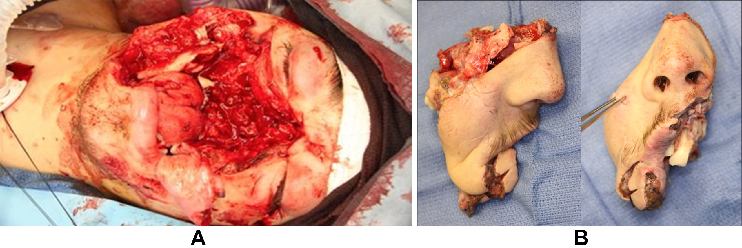
Figure 1. Injury at the time of incident. (A) 20-year-old male transferred to the emergency department after a metal pole amputated the majority of his mid-face during a motor vehicle accident; (B) The avulsed tissue after cleaning and thorough dissection. No suitable arterial anastomosis was identified on the flap for revascularization.
While the flap was perfusing, inset was performed. The posterior septum was first identified on the patient and the flap tissue; Doyle splints were secured to the remaining bony septum. These splints were passed retrograde through the patient’s nasal passage to secure the nose to the face at a known landmark while maintaining nasal patency. The radix and left cheek tissue were then secured using absorbable suture.
At the right side of the defect, there was a fractured piece of mandible without periosteum lying underneath the newly anastomosed facial vessels. Risk of infection or migration of this bone leading to damage to the anastomosis prompted removal of the mandible without a reconstructive bar or bone graft. The mucosal and cutaneous tissues were then closed.
Post-procedure, the patient showed immediate improvement in facial distention, indicating successful perfusion [Figure 2]. After insetting, the patient was transferred to the intensive care unit with a heparin drip to prevent flap site thrombosis.
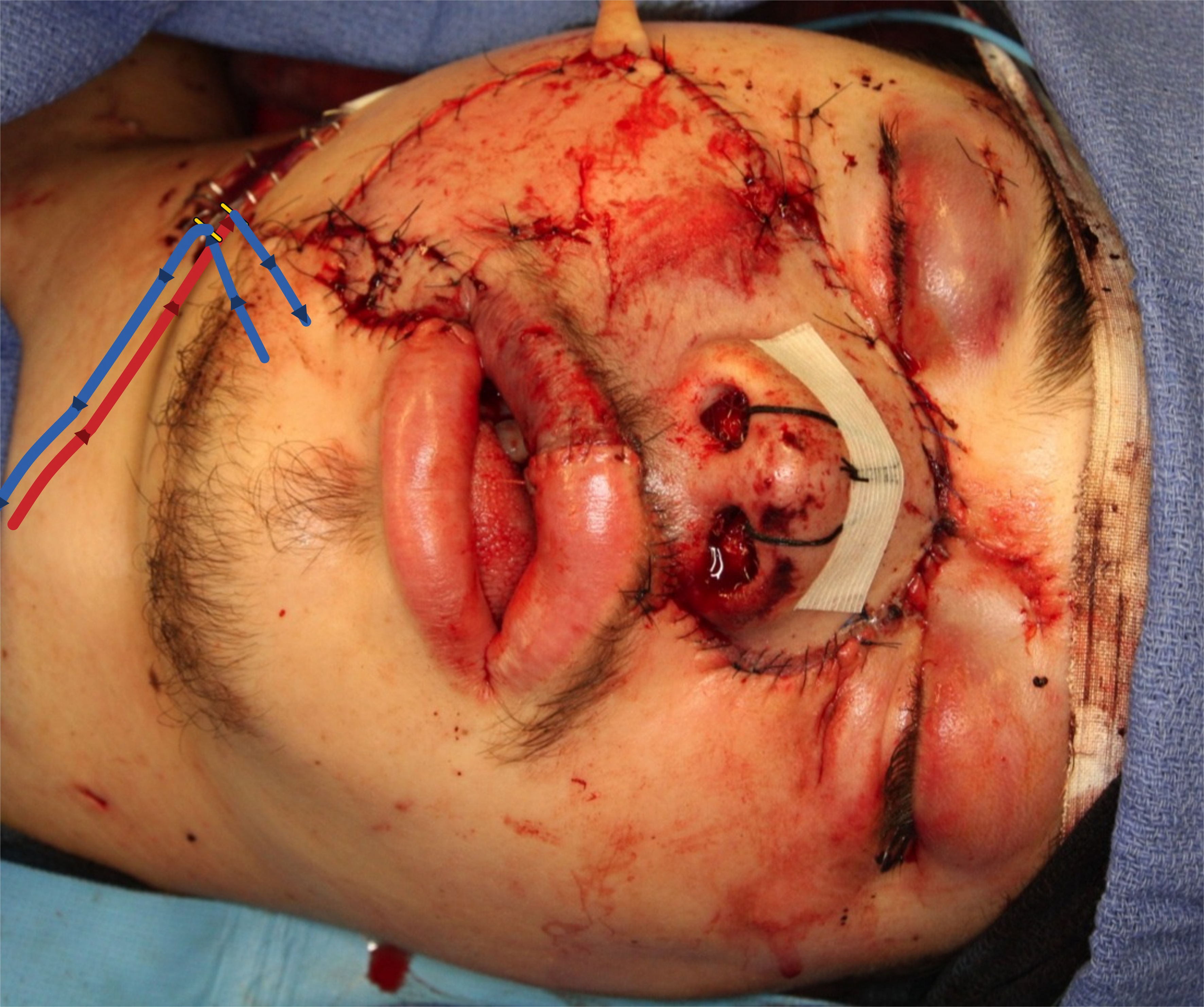
Figure 2. Facial appearance at the time of anastomosis. After creation of the AV anastomosis with venous outflow the flap exhibited good perfusion. Overlayed vessel schematic showing the flow of blood from the patient’s facial artery to the flap-side facial vein to provide retrograde perfusion to the flap (red-to-blue lines). Outflow of the flap runs through a separately identified facial vein to the patient-side facial vein (solid blue lines). Arrows denoting direction of blood flow. AV: Arteriovenous.
On post-operative day 6, the flap developed venous congestion, necessitating drainage of a hematoma and removal of the AV anastomosis. Exploration of the flap was performed and a suitable artery on the flap was identified which was not encountered on the initial exploration. An artery-to-artery anastomosis was performed using 10-0 Nylon, connecting a 1 mm branch of the previously identified patient side facial artery to the newly identified flap side facial branch to provide anterograde perfusion to the flap. Tension was noted on the venous anastomosis so a portion of the parotid tail was removed, the retromandibular vein (RMV) was dissected cephalad then clipped and mobilized anteriorly [Figure 3]. The superior vein was clipped and the inferior vein carefully trimmed before anastomosing it to the RMV. Leech therapy was temporarily initiated to address venous congestion, and on post-operative day 42, the patient was discharged with a well-perfused flap. Over three months, partial flap necrosis led to a 2 cm × 1 cm oronasal fistula, treated with an osteocutaneous free fibula flap using proximal superficial temporal vessels for anastomosis. Six months later, exposed maxillary hardware was debrided and arterial anastomosis was performed. Subsequent procedures involved scar revisions, orbit and nasal reconstruction with split calvarial grafts, and rib grafting overlay for dorsum elevation. Four years post-injury, concerns include right lower eyelid malposition, facial asymmetry, and right facial nerve paralysis, with planned medial canthopexy and temporalis transfer; however, the patient has not pursued further surgical treatments. Final reparative outcomes are shown in Figure 4.
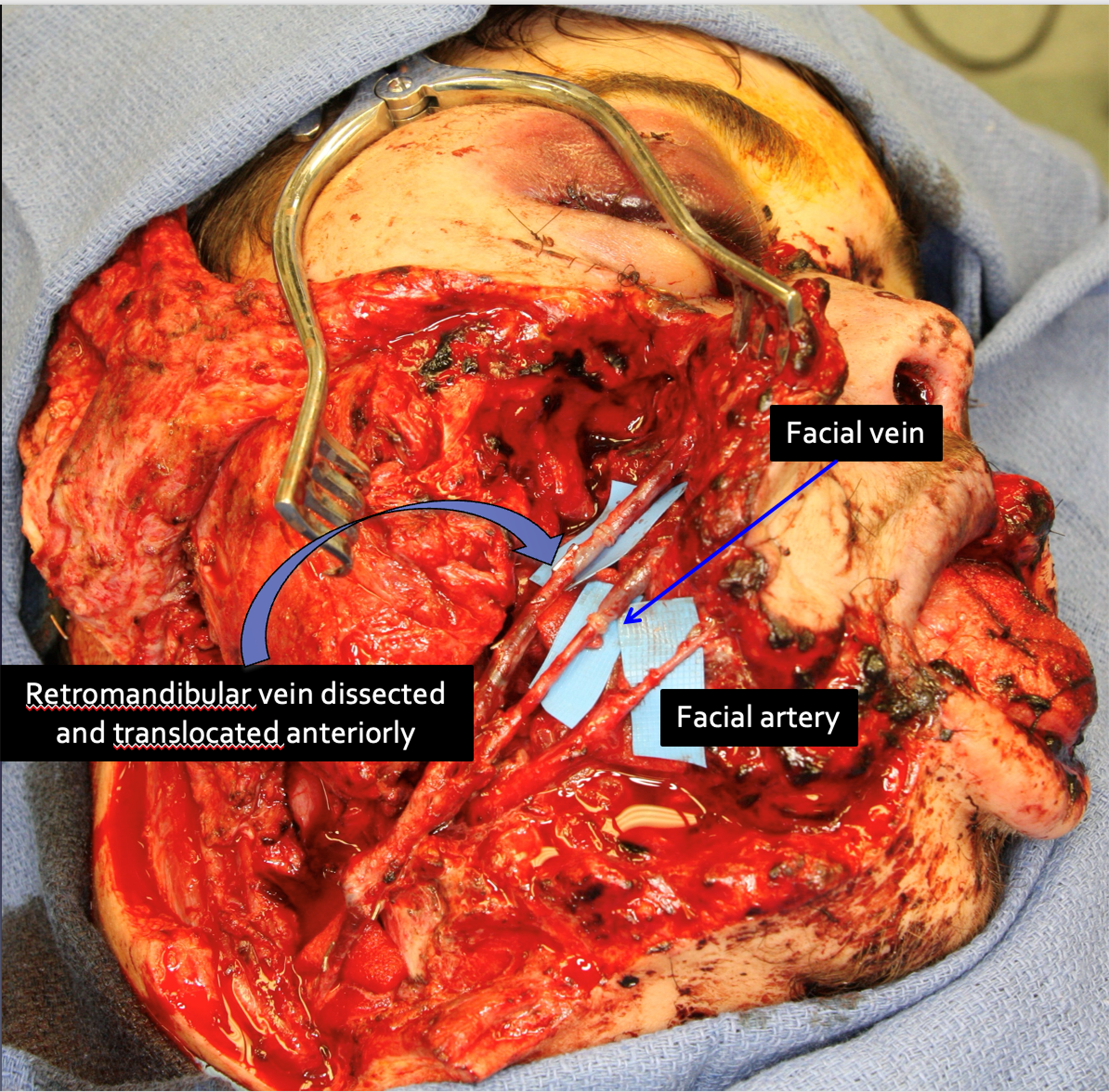
Figure 3. Second intraoperative repair. After returning to the OR for venous congestion and hematoma of the buccal space, the AV anastomosis was taken down. After exploration a suitable artery on the flap (facial artery stump) was identified and anastomosed to the donor facial artery - previously anastomosed to the facial vein. To avoid further venous congestion two facial veins were isolated on the flap and used to drain to the recipient facial vein and a mobilized retromandibular vein. AV: Arteriovenous; OR: operating room.
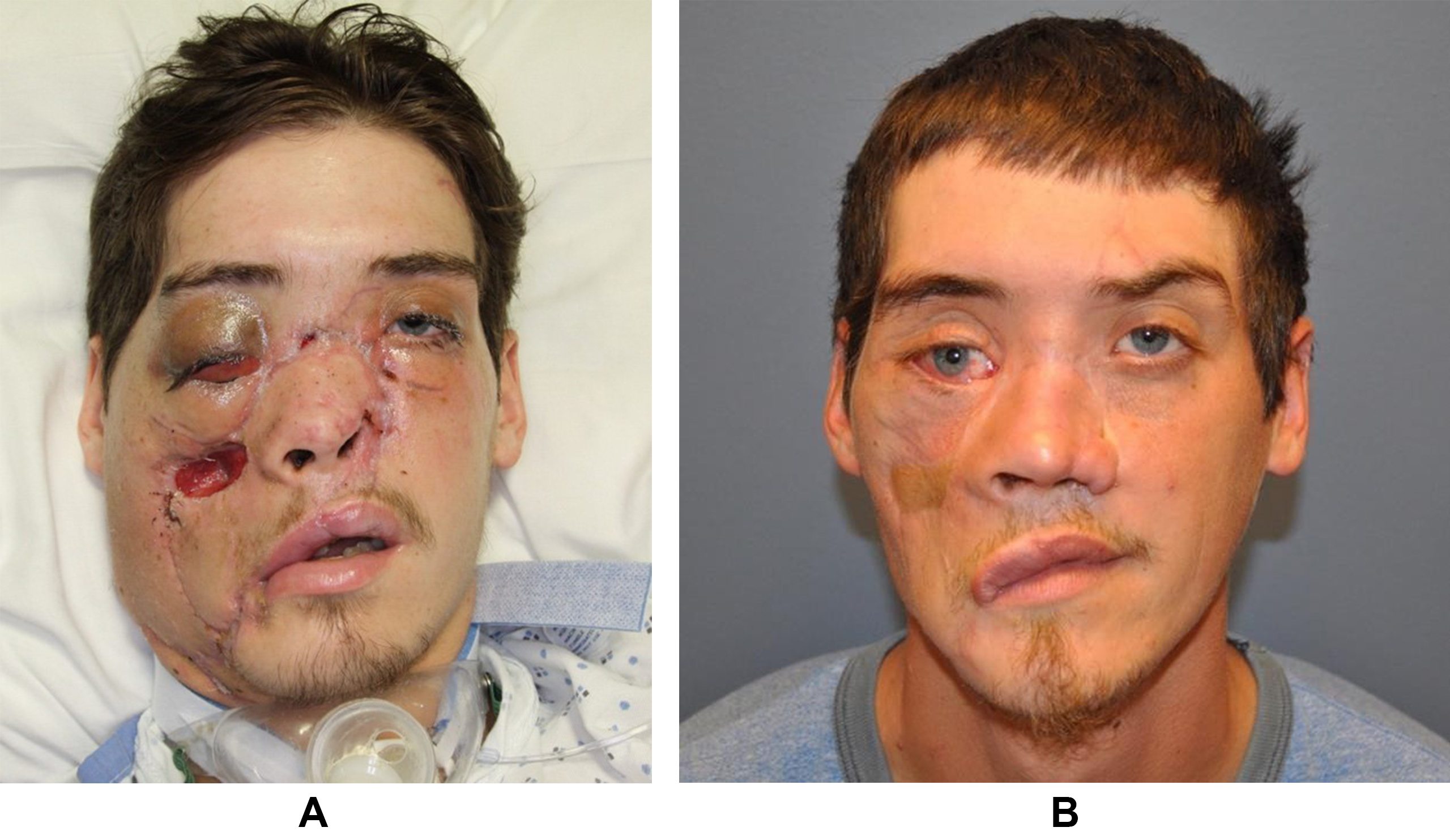
Figure 4. Post-reparative outcomes. (A) Forty-two days after replantation, prior to discharge from the hospital. There was partial necrosis of the left ala, a 3 cm × 3 cm defect of the right cheek, partial loss of palatal tissue and septal cartilage (not pictured) which required debridement; (B) Four years after facial replantation. There is a right malar eminence from the free flap reconstruction and hypopigmented region of the right cheek from free flap skin paddle. In the right orbit, there is dystopia, vertical hypoglobus, medial canthal displacement, excess scleral show and ectropion.
Case 2
A 21-year-old female caught her hair in an industrial machine, completely degloving her entire scalp, forehead, both eyebrows and eyelids, and both ears superior to the external auditory canals [Figure 5A-C]. The amputated tissue was preserved and brought to the emergency department without associated cervical or intracranial injuries. Before exploration, the scalp was shaved and irrigated to check for concealed lacerations. Tension-free arterial anastomosis with a 1 mm branch of the superficial temporal artery (STA) was followed by venous anastomosis with the superficial temporal vein (STV) using a 2.0 mm coupler [Figure 6A and B]. Bleeding from the donor side was observed before clamping the artery. The same procedure was performed on the left side. Arterial anastomosis was completed first using a one-to-one match with 10-0 Nylon, followed by venous anastomosis using a 1.5 mm coupler.
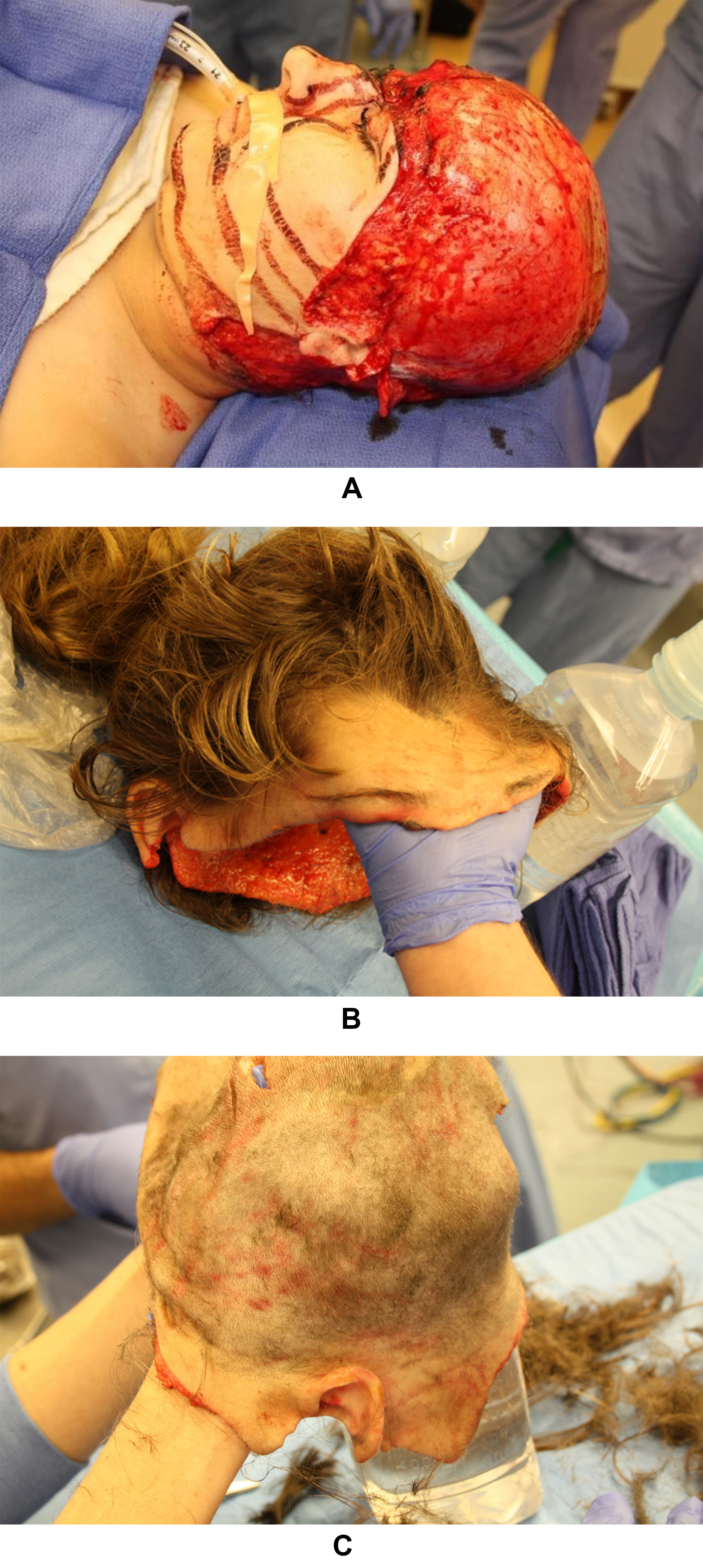
Figure 5. Pre-operative evaluation and preparation. (A) Lateral view of a 21-year-old female who caught her hair in an industrial machine; (B) The degloved tissue was preserved on ice and transported with the patient to the emergency department; (C) The degloved tissue after thorough cleaning and shaving.
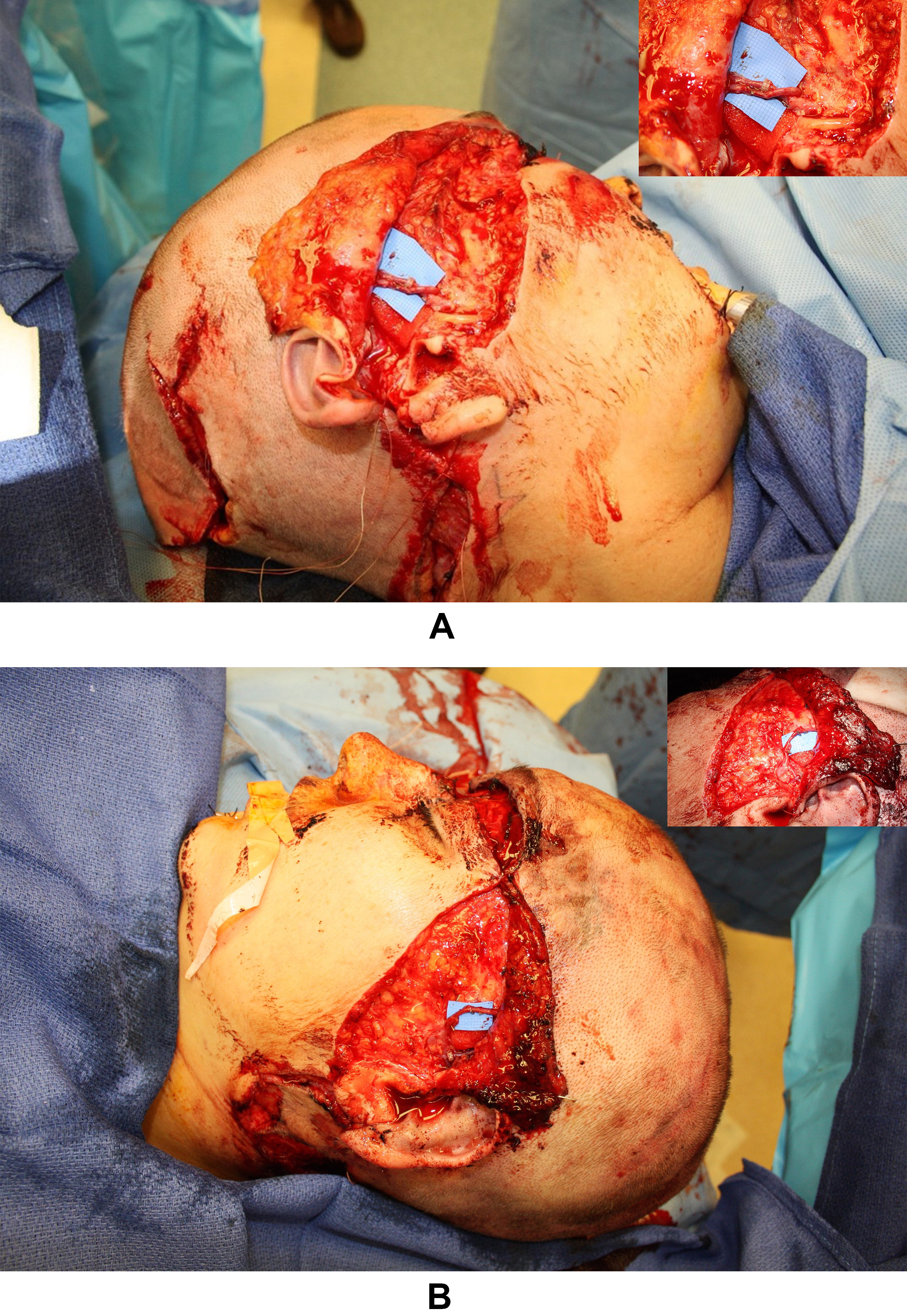
Figure 6. Intra-operative photography. (A) Right sided microanastomoses at the superficial temporal vascular bundle; (B) Left sided microanastomosis at the superficial temporal vascular bundle.
The patient’s eyebrows were realigned and secured to the frontal bone periosteum. Nasal bone reconstruction involved re-suspension from the periosteum. Eyelids were reconstructed with 5-0 fast-absorbing sutures. Ears were reconstructed using 3-0 chromic and 5-0 fast absorbing gut sutures, and auricular cartilage was approximated with chromic suture. Doppler signals were strong bilaterally, with good scalp capillary refill. Total ischemia time was around 5 h, and total operative time was 8 h. Two months post-surgery, the entire scalp remained viable other than a 1.5 cm border along the occiput. Necrotic tissue was debrided and closed with local tissue arrangement [Figure 7A]. One year following the injury the patient had full hair growth from the scalp with thin scarring along the repair [Figure 7B].
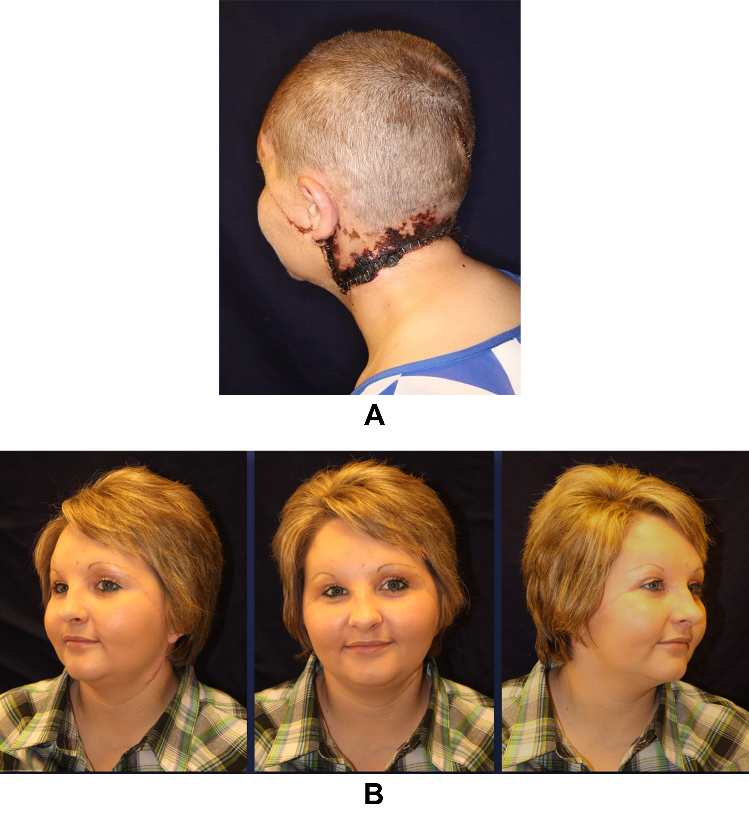
Figure 7. Post-replantation outcomes. (A) Two months after the accident, the entirety of the scalp remained viable, except for a 1-2 cm margin along the occiput. The necrotic tissue was subsequently debrided and closed with local tissue rearrangement; (B) One year after replantation. The patient experienced a complete recovery of hair growth with no long-term adverse sequelae.
DISCUSSION
AV inflow anastomosis
Confusion often exists because the nomenclature “AV shunt” does not differentiate whether the inflow is from the arterial or venous side of the capillaries (Case 1). In 1992, Morris et al. successfully anastomosed the STA to the STV in a partially amputated scalp[8]. Seven years later, Hallock described another replantation of a partial scalp using the frontal branch of the STA anastomosed to the unnamed scalp vein and achieved excellent aesthetic results[6]. In 2020, Lee et al. used a right posterior tibial vein as a venous graft to connect the STA to STV of the amputated scalp[10]. Hendrick and Tiwari achieved the first successful lip replantation by connecting the superior labial artery to a facial branch at the nasolabial fold. They suggested the absence of venous valves at this level might have enabled successful replantation, or insufficient valve function under arterial perfusion[11].
The viability risk of venous-perfused tissues depends on anastomotic site, flow direction, and venous valve density[6]. Valve scarcity within the scalp and ear replantations with valve trimming may contribute to their success[9]. The midface does have a lower density of valves compared to other anatomical regions of the body and face, which may partly explain why the vast majority of the amputated midface in Case 1 survived in our report[11]. Studies show severed rabbit auricles can survive through arterialization of the venous system[9,12]. Some argue nutrient flow occurs via AV shunts, while others suggest retrograde perfusion from artery to vein, reaching capillaries through arterialized venules[8]. From there, blood returns centrally through venules of the low-pressure system. Artery-to-vein anastomosis is a valid technique for replanting non-salvageable maxillofacial tissue, supported by clinical and basic science experiments. Our report’s first case uniquely employs AV inflow anastomosis to salvage osteocutaneous tissue in a facial and maxillary artery watershed region.
Lip, nose, and palate
The lip receives consistent arterial inflow but variable venous outflow, making venous anastomoses challenging. Lip replantations have a high success rate (approximately 95%), with common complications including diminutive venous vasculature and fragility of small lip veins. Venous drainage occurs through plexuses, with over 50% of replantations lacking venous anastomoses[13]. In a review of sizable lip defects (n = 13; mean defect 10.6 cm2), the most important factor for survival was re-establishment of arterial (typically labial) flow[7].
Nose replantations are less frequent than lip ones but are vital for facial aesthetics. Like lips, over half of these cases do not involve venous anastomosis, with various techniques used for venous congestion relief, such as open venous drainage, nasal tip abrasion, pin-pricking, stab incisions, medicinal leeches, AV outflow fistulas, intra-replant subcutaneous heparin, and heparin sponges[14]. Arterial anastomosis in nose replantations involves various arteries, including the labial, facial, supratrochlear, angular, and anterior ethmoidal arteries[5]. To our knowledge, there have been no reported cases where artery-to-artery anastomosis was not performed in the initial replantation attempt.
Scalp, ears, and eyebrows
Scalp amputations often result from hair getting caught in industrial machines[5]. The scalp played a crucial role both cosmetically and physiologically, influencing appearance, social standing, and regulating body temperature[15]. One-stage scalp replantation has a 90%-95% success rate in large-scale reviews. While the entire scalp can survive on a single artery and vein, two sets are typically considered safer. The STA is commonly used in such procedures, as in the second case cited[5]. The occipital artery is an option but may be problematic with cervical spine injuries[3]. If artery-to-artery anastomosis is not possible, AV inflow anastomosis is reported[6,8]. Independent of anastomosis technique, partial necrosis is relatively common and may occur in as high as a third of cases[5].
Because of their common muscular connections to the galea aponeurotica, scalp avulsions often involve the ears and eyebrows. The eyebrows and associated frontalis muscle usually survive, allowing for gradual brow elevation. Revascularization of the ears is less predictable, with partial or complete loss possible, mainly due to venous insufficiency. Patient satisfaction with ear position and angulation can pose challenges, as can correcting issues such as right lagophthalmos post-replantation[4].
CONCLUSION
Facial replantation studies over four decades show the maxillofacial region’s resilience, yielding excellent functional and aesthetic outcomes. This series presents diverse facial trauma cases, highlighting evolving microsurgical techniques. A unique case involving near-total facial replantation with maxillary involvement saved by a temporary AV inflow shunt to maintain early perfusion is featured. Further research is crucial for understanding the procedure’s physiology and its adaptation based on facial angiosome relevance.
DECLARATIONS
Authors’ contributions
Made substantial contributions to the conception and design of the study and performed data analysis and interpretation: Nguyen K, Boyette JR, Sunde J, Vural E, Moreno MA
Performed data acquisition and provided administrative, technical, and material support: Nguyen K, Hairston HC, Farsi S
Drafted the manuscript or provided critical revision of the text for important content: Nguyen K, Hairston HC, Farsi S
Conceived and supervised the study and provided final approval of the version to be published: Boyette JR, Sunde J, Vural E, King D, Moreno MA
Availability of data and materials
Not applicable.
AI and AI-assisted tools statement
Not applicable.
Financial support and sponsorship
None.
Conflicts of interest
All authors declared that there are no conflicts of interest.
Ethical approval and consent to participate
After review by the Institutional Review Board (IRB) of UAMS, this project was determined not to constitute human subjects research as defined by applicable federal regulations or IRB policies; therefore, IRB review was not required. Informed consent was obtained from the patients.
Consent for publication
Informed consent for the publication of the patient's photographs has been obtained.
Copyright
© The Author(s) 2026.
REFERENCES
1. Singh K, Aggarwal K. Total scalp replantation after traumatic avulsion. Indian J Plast Surg. 2020;53:311-2.
2. Cho DY, Willborg BE, Lu GN. Management of traumatic soft tissue injuries of the face. Semin Plast Surg. 2021;35:229-37.
3. Miller GD, Anstee EJ, Snell JA. Successful replantation of an avulsed scalp by microvascular anastomoses. Plast Reconstr Surg. 1976;58:133-6.
4. Kayikçioğlu A, Karamürsel S, Keçik A. Replantation of nearly total nose amputation without venous anastomosis. Plast Reconstr Surg. 2001;108:702-4.
5. Yin JW, Matsuo JM, Hsieh CH, Yeh MC, Liao WC, Jeng SF. Replantation of total avulsed scalp with microsurgery: experience of eight cases and literature review. J Trauma. 2008;64:796-802.
6. Hallock GG. Secondary expansion of a replanted scalp salvaged by an intrinsic arteriovenous shunt. Plast Reconstr Surg. 1999;103:1957-60.
7. Walton RL, Beahm EK, Brown RE, et al. Microsurgical replantation of the lip: a multi-institutional experience. Plast Reconstr Surg. 1998;102:358-68.
8. Morris SF, MacGill KA, Taylor GI. Scalp replantation by arterialised venous network flow-through. Br J Plast Surg. 1992;45:187-92.
9. Zamboni WA, Lozano DD, Vitkus K, et al. Single-vessel arteriovenous revascularization of the amputated ear. J Reconstr Microsurg. 1999;15:9-13.
10. Lee JH, Kim DG, Choi KY, Chung HY, Lee JS. Successful microsurgical replantation of scalp amputation using medicinal and chemical leech therapy: a case report. Arch Hand Microsurg. 2020;25:320-5. (in Korean).
11. Hendrick RG Jr, Tiwari P. Successful replantation of upper lip avulsion injury using an arterialized venous anastomosis. Plast Reconstr Surg. 2012;130:628e-30.
12. Farrior EH, Baker SR. Use of arterialization of the venous system in reattachment of the avulsed auricle. Arch Otolaryngol Head Neck Surg. 1988;114:1385-8.
13. Daraei P, Calligas JP, Katz E, Etra JW, Sethna AB. Reconstruction of upper lip avulsion after dog bite: case report and review of literature. Am J Otolaryngol. 2014;35:219-25.
14. Kim S, Jeong H, Choi TH, Kim JS. Successful re-plantation of an amputated nasal segment by supermicrosurgery: a case report and review of the literature. J Plast Reconstr Aesthet Surg. 2010;63:e380-3.
Cite This Article
 Open Access
How to Cite
Nguyen K, Hairston HC, Farsi S, Boyette JR, Sunde J, Vural E, King D, Moreno MA. Microvascular replantation of extensive craniofacial amputations: case series. Plast Aesthet Res. 2026;13:27. https://dx.doi.org/10.20517/2347-9264.2026.30
Download Citation
If you have the appropriate software installed, you can download article citation data to the citation manager of your choice. Simply select your manager software from the list below and click on download.
Export Citation File
Type of Import
Tips on Downloading Citation
Citation Manager File Format
Type of Import
Direct Import: When the Direct Import option is selected (the default state), a dialogue box will give you the option to Save or Open the downloaded citation data. Choosing Open will either launch your citation manager or give you a choice of applications with which to use the metadata. The Save option saves the file locally for later use.
Indirect Import: When the Indirect Import option is selected, the metadata is displayed and may be copied and pasted as needed.
About This Article
Copyright

Data & Comments
Data
0









Comments
Comments must be written in English. Spam, offensive content, impersonation, and private information will not be permitted. If any comment is reported and identified as inappropriate content by OAE staff, the comment will be removed without notice. If you have any queries or need any help, please contact us at [email protected].