
fig6
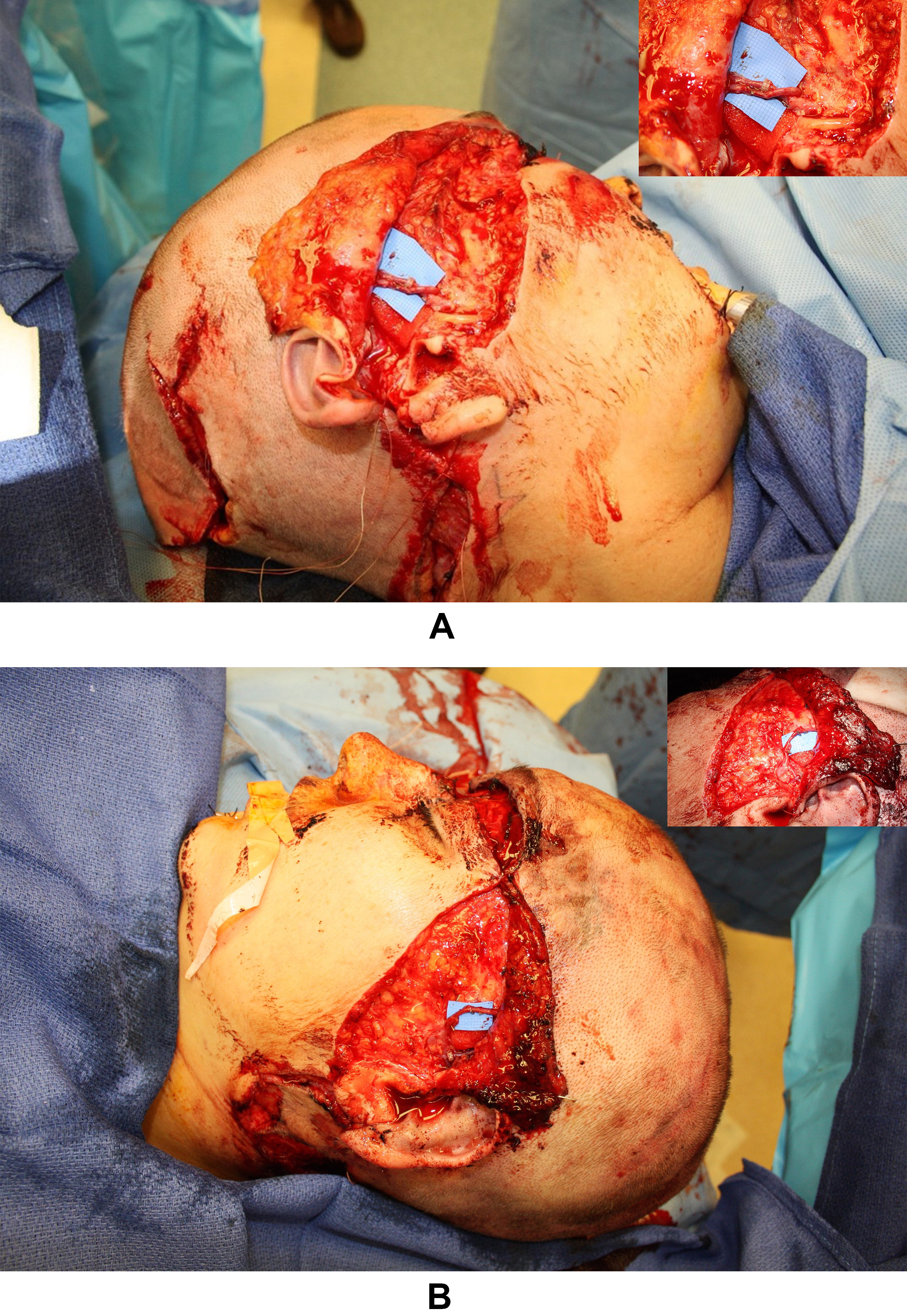
Figure 6. Intra-operative photography. (A) Right sided microanastomoses at the superficial temporal vascular bundle; (B) Left sided microanastomosis at the superficial temporal vascular bundle.
Figure 6. Intra-operative photography. (A) Right sided microanastomoses at the superficial temporal vascular bundle; (B) Left sided microanastomosis at the superficial temporal vascular bundle.


All published articles are preserved here permanently:
https://www.portico.org/publishers/oae/