
fig4
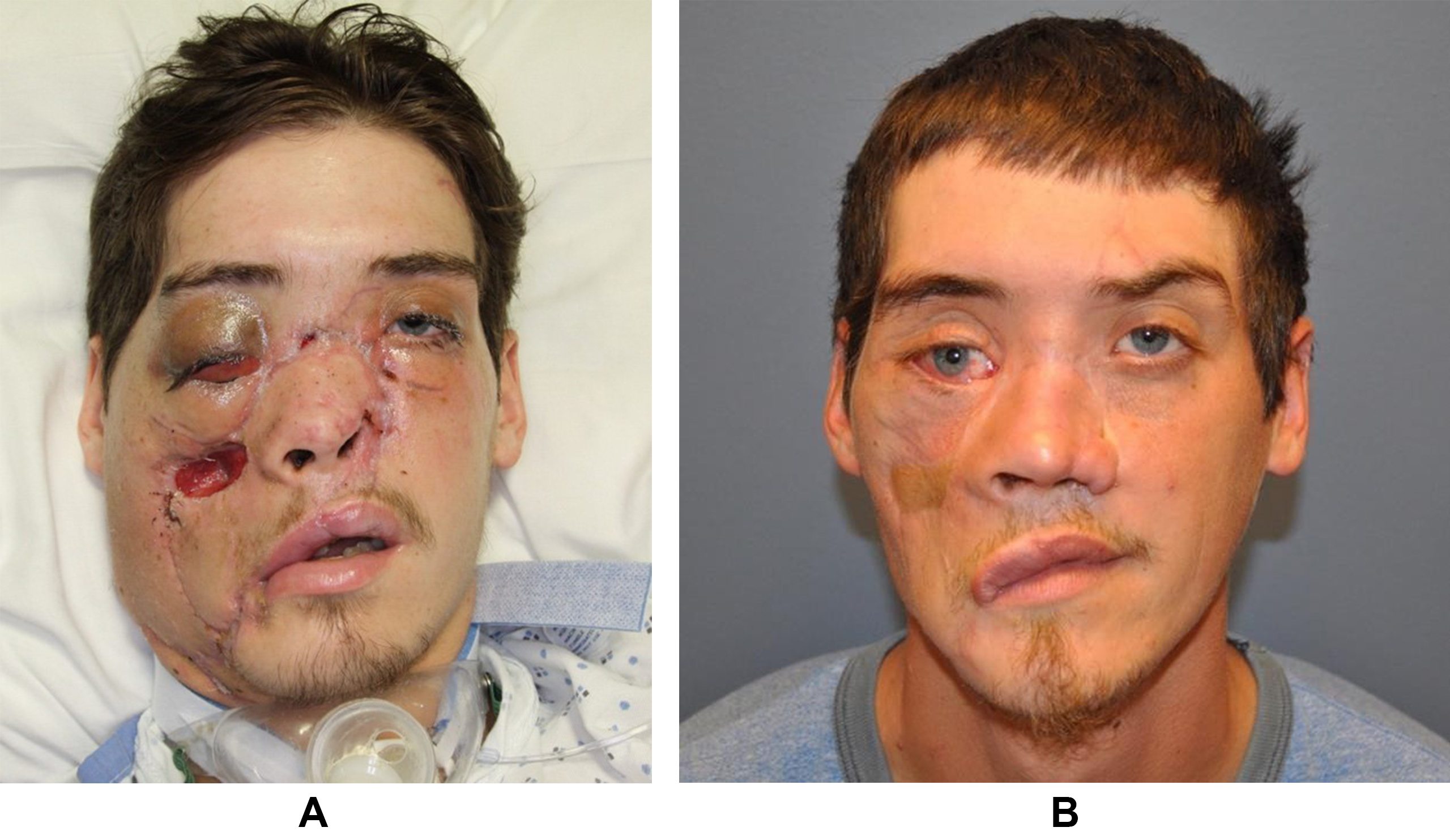
Figure 4. Post-reparative outcomes. (A) Forty-two days after replantation, prior to discharge from the hospital. There was partial necrosis of the left ala, a 3 cm × 3 cm defect of the right cheek, partial loss of palatal tissue and septal cartilage (not pictured) which required debridement; (B) Four years after facial replantation. There is a right malar eminence from the free flap reconstruction and hypopigmented region of the right cheek from free flap skin paddle. In the right orbit, there is dystopia, vertical hypoglobus, medial canthal displacement, excess scleral show and ectropion.