

Inflammation-targeted nanomedicines for atherosclerotic cardiovascular disease: from atherosclerosis regression to post-myocardial infarction management
 0

Graphical Abstract
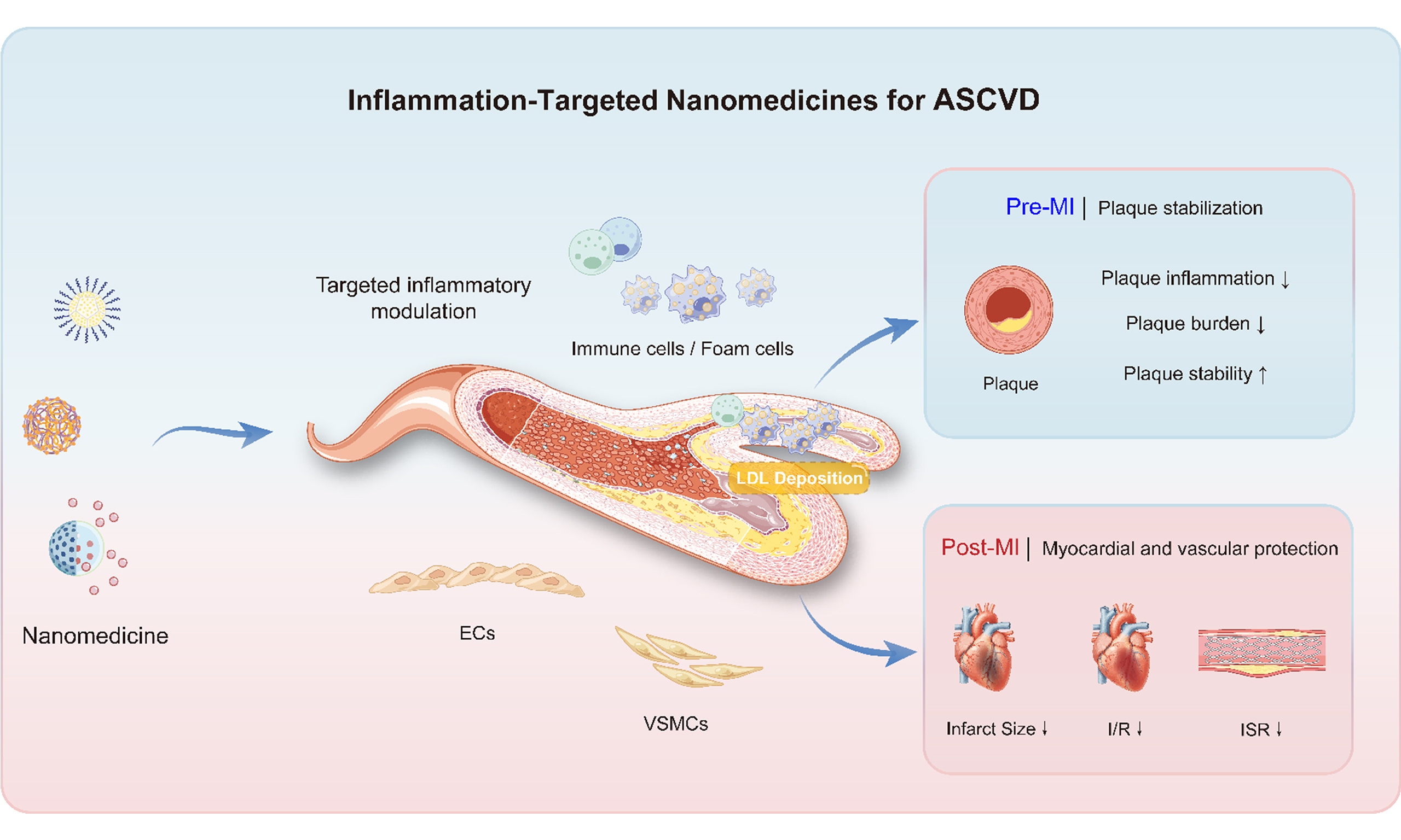
Abstract
Cardiovascular diseases (CVDs) remain the leading cause of death and disability worldwide, with atherosclerotic cardiovascular disease (ASCVD) accounting for the largest proportion. Vascular inflammation is a major driver of ASCVD, contributing to plaque formation, progression, and rupture and influencing myocardial repair after infarction. Conventional anti-inflammatory therapies are limited by systemic toxicity and insufficient drug accumulation at lesion sites, restricting their capacity for precise intervention. Nanomedicines can deliver therapeutics to lesion sites and release them in response to local stimuli, potentially reducing systemic toxicity and offering a promising approach to inflammation-targeted treatment of ASCVD. Existing studies have largely cataloged nanocarrier types or evaluated efficacy in isolated disease models but have not provided an integrated perspective on inflammation management across the full ASCVD continuum. This review discusses the role of nanomedicines in preventive plaque stabilization and post-interventional management, including recovery after myocardial infarction and prevention of in-stent restenosis. Preclinical studies indicate that nanomedicines can reduce atherosclerotic plaque size and infarct size and mitigate ischemia-reperfusion injury and stent intimal hyperplasia, potentially improving cardiovascular outcomes. Future work should prioritize mechanistic validation in clinically relevant models and artificial intelligence (AI)-assisted precision medicine to accelerate the safe clinical adoption of inflammation-targeted nanomedicines for CVDs.
Keywords


 Download PDF
INTRODUCTION
Cardiovascular diseases (CVDs) are among the leading causes of cardiovascular-related death and disability worldwide[1,2], with atherosclerotic cardiovascular disease (ASCVD) accounting for the largest proportion. The central pathological features of ASCVD are subendothelial deposition of low-density lipoprotein (LDL) and sustained vascular inflammation[3,4]. For decades, the treatment of ASCVD has focused on lipid accumulation and the thrombotic events that follow. The success of statins in controlling low-density lipoprotein cholesterol (LDL-C) has supported the lipid hypothesis[5]. However, large-scale clinical trials such as CANTOS have demonstrated that vascular inflammation is not merely a secondary consequence of lipid deposition but an active driver of disease progression from its earliest stages[6,7]. Targeting inflammatory processes with precision has therefore emerged as a central research priority in ASCVD prevention and management[8].
This review outlines key inflammatory pathways involved in the immunology of atherosclerosis that are relevant to the design of inflammation-targeted nanomedicines[4,9]. Building on previous reviews of nanomedicine design and targeting methods, this review presents an integrated framework covering the full ASCVD continuum - from early endothelial dysfunction and stable plaque to plaque rupture and acute myocardial infarction (MI), followed by post-interventional complications such as myocardial ischemia-reperfusion injury (MIRI) and in-stent restenosis (ISR). Several reviews have cataloged nanocarrier classes, cell-specific atherosclerosis targeting, or ISR management[8,10-12]. The present review adds a stage-by-stage framework that links changing inflammatory states to therapeutic targets, delivery timing, nano-bio interface behavior, measurable efficacy endpoints, and translational maturity. This review provides a systematic framework for designing inflammation-targeted nanomedicines to stabilize plaques and protect the myocardium and vasculature after intervention [Figure 1].
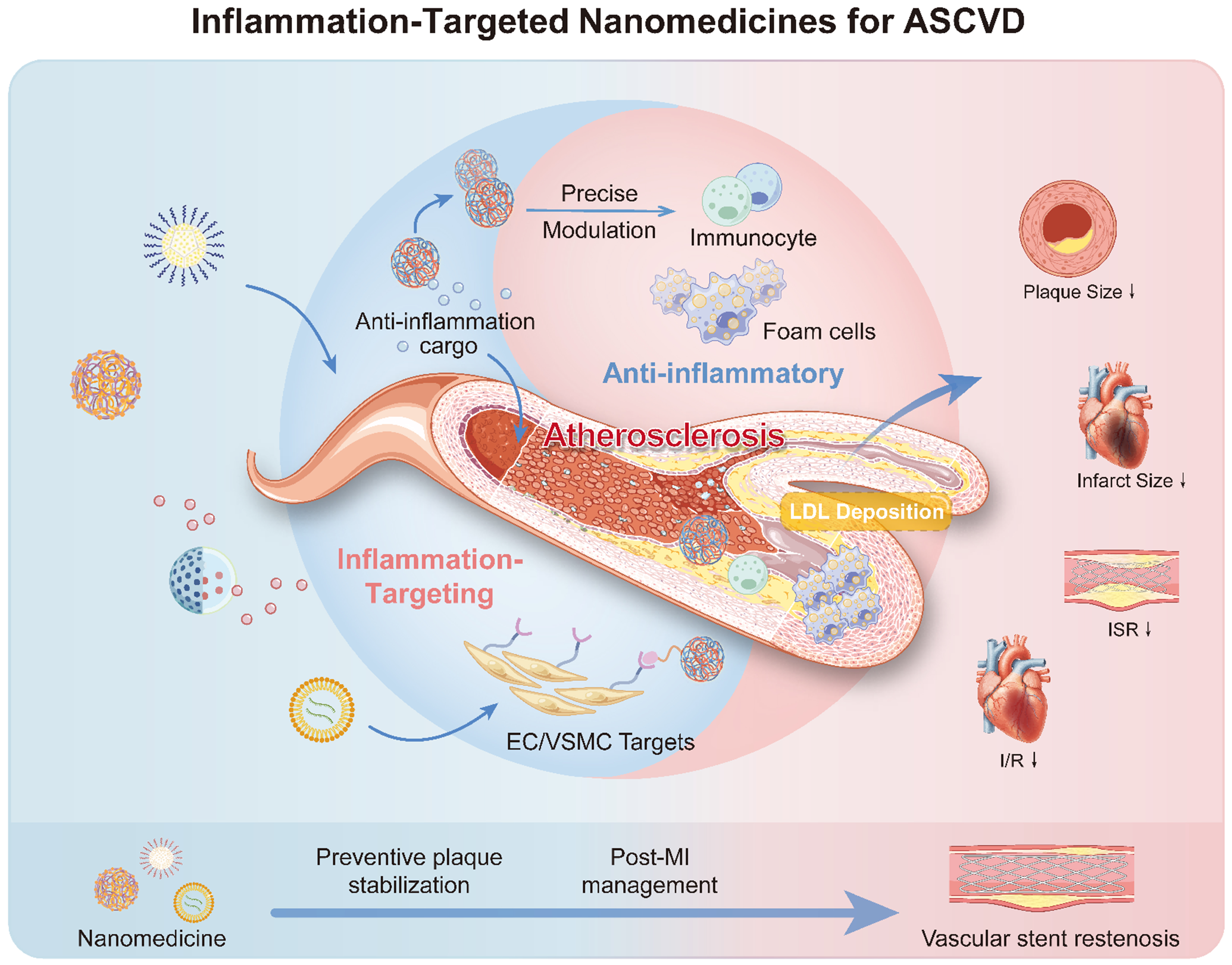
Figure 1. Schematic illustration of inflammation-targeted nanomedicines for ASCVD. ASCVD: Atherosclerotic cardiovascular disease; EC: Endothelial cell; VSMC: vascular smooth muscle cell; ISR: in-stent restenosis; MI: myocardial infarction.
LITERATURE SEARCH AND STUDY SELECTION
We conducted a structured narrative search of PubMed, Web of Science, and Scopus for English-language articles published from January 2010 through August 2026, supplemented by earlier landmark trials and mechanistic studies. Search terms combined atherosclerosis, ASCVD, MI, ischemia-reperfusion injury, percutaneous coronary intervention (PCI), and ISR with inflammation, nanomedicine, nanoparticles, nanozymes, liposomes, polymeric nanoparticles, biomimetic membranes, exosomes, responsive delivery, interfacial binding, protein corona, and shear stress. We prioritized randomized human trials, primary translational studies, and in vivo studies that reported lesion targeting, biodistribution, therapeutic efficacy, or safety. Studies focused on non-atherosclerotic kidney injury, pulmonary embolism, isolated stroke, cancer, or non-cardiovascular inflammatory disease were excluded unless they established a general nano-bio interface principle directly relevant to intravascular delivery. This is a structured narrative review rather than a systematic review or meta-analysis; therefore, no pooled effect estimate was calculated.
LIMITATIONS OF TRADITIONAL ANTI-INFLAMMATORY THERAPY
Inflammation can drive vulnerable plaque formation, coronary artery occlusion, and MI[4,13]. Clinical trial results are pathway and context-dependent. Canakinumab reduced recurrent cardiovascular events in CANTOS without lowering lipids, whereas colchicine reduced events in COLCOT and LoDoCo2[6,14,15]. In contrast, low-dose methotrexate in CIRT did not lower interleukin-1β (IL-1β), interleukin-6 (IL-6), high-sensitivity C-reactive protein, or cardiovascular events, and colchicine initiated after acute MI did not reduce the primary cardiovascular outcome in CLEAR SYNERGY[16,17]. These contrasting findings support selective modulation of validated inflammatory pathways while underscoring the need to match the target, patient inflammatory phenotype, and treatment window. Most current anti-inflammatory agents for ASCVD are administered systemically and do not selectively accumulate in atherosclerotic plaques. Consequently, suppressing harmful inflammation within plaques may also compromise normal host immune defenses. Long-term anti-inflammatory therapy may increase susceptibility to infection, highlighting the need to balance anti-inflammatory efficacy against potential impairment of host immune defense[18]. This concern is particularly relevant to therapies targeting key innate immune pathways, such as IL-1β signaling. Meanwhile, the intrinsic toxicities of anti-inflammatory agents further restrict their tolerability. For example, although colchicine is among the anti-inflammatory agents with the most extensive clinical evidence in ASCVD, it frequently causes gastrointestinal adverse effects, such as diarrhea, which may reduce patient adherence[17]. In addition, concomitant use of certain cytochrome P450 3A4 (CYP3A4) and/or P-glycoprotein inhibitors can increase colchicine exposure, highlighting the need to assess drug interactions and dosing regimens carefully[19]. Although most studies of inflammation-targeted nanomedicines have reported some efficacy in ASCVD, developing formulations that avoid systemic immunosuppression remains a critical unmet clinical need.
INFLAMMATORY MECHANISMS AND PRIMARY TARGETS IN ASCVD
Chronic inflammation is a central driver of ASCVD. Although disordered lipid metabolism is the most widely recognized initiating factor, the onset and persistent amplification of inflammatory responses constitute the core pathophysiological processes governing plaque formation and progression[9]. Plaque rupture ultimately triggers acute cardiovascular events, such as MI. A mechanistic understanding of the inflammatory circuits underlying ASCVD is therefore essential for the rational design of inflammation-targeted nanomedicines.
Initiation and development of ASCVD inflammation
Atherosclerotic inflammation proceeds through three interrelated stages: endothelial dysfunction, lipid infiltration, and inflammatory activation and amplification[4]. Under pathological conditions or disturbed blood flow, endothelial tight junctions become permeable, facilitating the transendothelial transport of plasma LDL[3]. Subsequent oxidation of lipoprotein lipids, together with the action of other inflammatory mediators, further activates endothelial cells, triggering the expression of P-selectin, E-selectin, vascular cell adhesion molecule-1 (VCAM-1), and intercellular adhesion molecule-1 (ICAM-1), which promote adhesion of monocytes and other leukocytes[20]. Monocytes entering the intima differentiate into macrophages, in which excessive uptake and impaired intracellular processing or efflux of modified apolipoprotein B (ApoB)-containing lipoproteins promote foam-cell formation, establishing a sustained positive feedback loop between lipid accumulation and inflammation. As intracellular cholesterol accumulates and cholesterol crystals form, the NOD-, LRR-, and pyrin domain-containing protein 3 (NLRP3) inflammasome in macrophages is further activated, promoting caspase-1-mediated maturation and release of IL-1β. IL-1β subsequently induces the expression of IL-6 and other inflammatory mediators through interleukin-1 receptor type 1 (IL-1R1) signaling, thereby enhancing inflammatory-cell recruitment and amplifying inflammation within the plaque [Figure 2A][21,22]. As the disease progresses, persistent inflammation and impaired clearance of dead cells contribute to the expansion of the necrotic core, while excessive extracellular matrix degradation and insufficient fibrous cap repair further increase the risk of plaque instability. Therefore, interventions should not only limit inflammatory amplification but also promote the clearance of dead cells and maintain the fibrous cap.
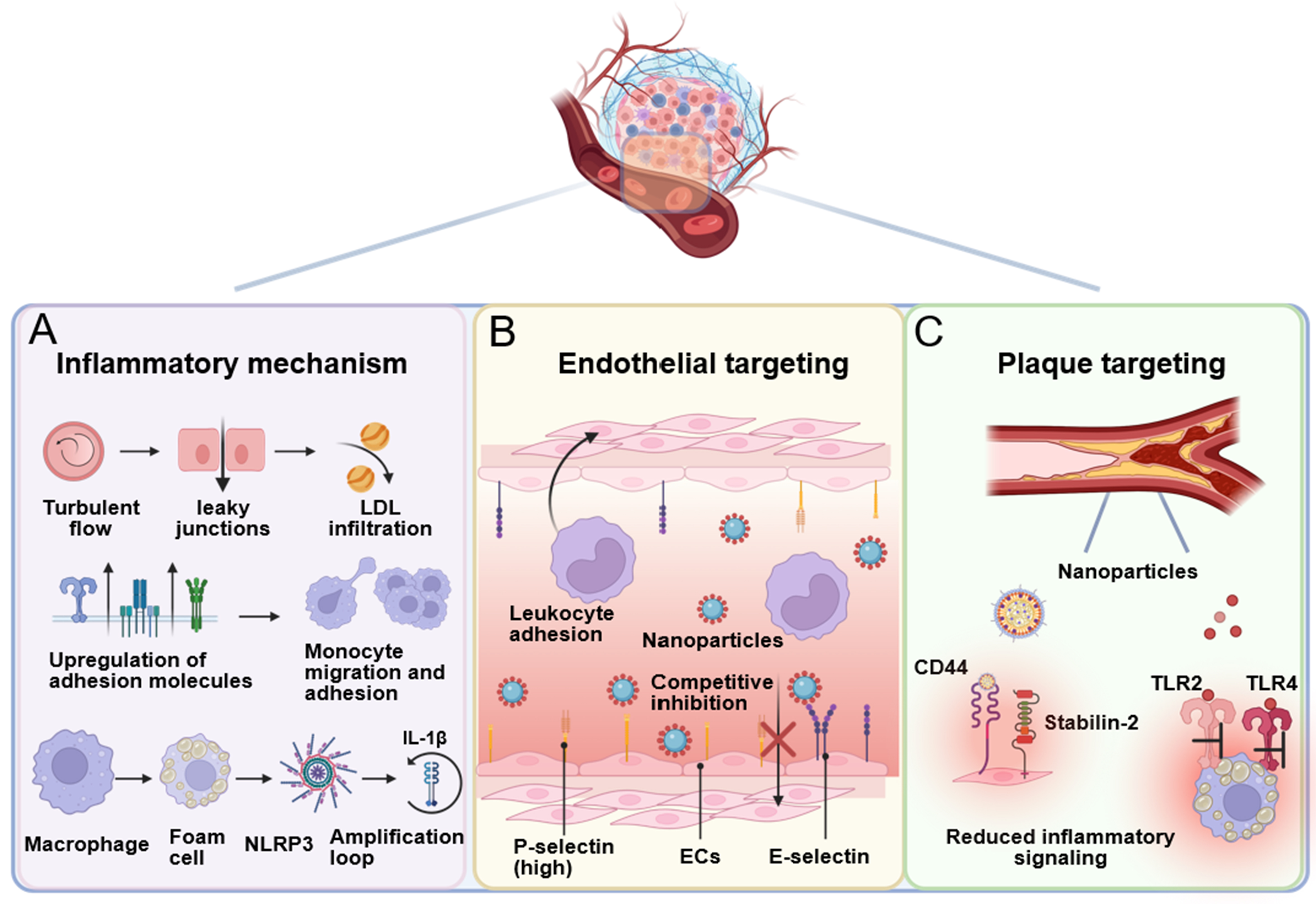
Figure 2. Illustration of inflammatory mechanisms and cellular targets in ASCVD: (A) the cascade from endothelial dysfunction to inflammatory amplification; (B) targeting adhesion molecules on endothelial cells to block leukocyte infiltration; and (C) plaque targeting and suppression of inflammatory signaling. Created with BioRender. Yang, G. (2026). https://BioRender.com/kp4oush. ASCVD: Atherosclerotic cardiovascular disease; LDL: low-density lipoprotein; NLRP3: NLR family pyrin domain containing 3; IL-1β: interleukin-1β; ECs: endothelial cells; CD44: cluster of differentiation 44; TLR2: Toll-like receptor 2; TLR4: Toll-like receptor 4.
Therapeutic targets for inflammatory intervention in ASCVD
The self-amplifying nature of ASCVD inflammation, together with the distinctive lesion microenvironment, makes inflammatory signaling pathways and their cellular mediators compelling therapeutic targets.
Key molecular signaling pathway targets: The NLRP3 inflammasome is a key link between lipid accumulation and innate immune activation. Its downstream effector, IL-1β, further drives the production of IL-6 and other inflammatory mediators, making the NLRP3–IL-1β–IL-6 axis an important target for anti-inflammatory therapy in ASCVD[22]. The CANTOS randomized controlled trial demonstrated that, in patients with a history of MI and persistent inflammatory risk, the IL-1β-neutralizing antibody canakinumab reduced recurrent cardiovascular events without lowering LDL-C levels, establishing that targeting residual inflammatory risk can provide cardiovascular benefits independent of lipid lowering[6]. Further mechanistic studies suggest that IL-1β inhibition not only reduces local inflammation but may also promote the accumulation of fibroblast-like cells within the fibrous cap and increase cap thickness, thereby favoring a more stable plaque phenotype[23]. However, this strategy primarily interrupts the downstream inflammatory amplification triggered by lipid accumulation and does not eliminate the upstream pathogenic process of ApoB-containing lipoprotein retention. It should therefore be considered a complement to, rather than a replacement for, lipid-lowering therapy.
Previous studies have primarily focused on three types of cells: endothelial cells, macrophages, and vascular smooth muscle cells (VSMCs). This review also discusses the distinct functions of immune-cell subgroups and their corresponding targeting requirements. Activated endothelial cells express high levels of P-selectin, VCAM-1, and integrin αvβ3 and are key targets for early anti-inflammatory intervention. Targeting these adhesion molecules may reduce leukocyte adhesion and infiltration into the vessel wall[24]. Nanoparticles that target endothelial adhesion molecules may interfere with leukocyte-endothelial interactions, thereby reducing leukocyte adhesion and subsequent inflammatory-cell recruitment [Figure 2B]. Plaque macrophage subgroups: although traditionally categorized as M1 (pro-inflammatory) and M2 (pro-resolving), recent single-cell RNA sequencing (scRNA-seq) studies have revealed a highly heterogeneous spectrum of macrophage phenotypes within plaques and infarcted myocardium, transcending this oversimplified binary model. Therefore, nanomedicine strategies should aim at modulating functional states based on specific transcriptomic signatures[25-27]. Nanomedicines should be used to selectively regulate macrophage polarization rather than suppressing their activity entirely. For instance, hyaluronic acid (HA) receptor-mediated plaque targeting at the atherosclerotic lesions is investigated by Lee et al. through its interactions with cluster of differentiation 44 (CD44) and stabilin-2. Following systemic administration in ApoE-/- mice, hyaluronic acid nanoparticles (HA-NPs) preferentially accumulated in atherosclerotic plaques and showed greater lesion accumulation than the passive-targeting nanoparticle control [Figure 2C][28]. In addition to CD44 or stabilin-2 targets, Toll-like receptor 2 (TLR2) and Toll-like receptor 4 (TLR4) that are overexpressed on macrophages are also attractive therapeutic targets[9,22]. Therefore, receptor-targeted nanoparticles may enhance drug accumulation within plaques and modulate inflammatory signaling, thereby reducing plaque inflammation and improving plaque stability. VSMCs: these cells undergo phenotypic switching from a contractile to a synthetic/proliferative state. While excessive VSMC proliferation contributes to restenosis, VSMC-derived cells also form the protective fibrous cap that stabilizes plaques. Nanomedicine strategies must therefore selectively inhibit pathological VSMC proliferation without impairing their protective functions. Adaptive immunity, clonal hematopoiesis, trained immunity, neutrophil extracellular traps, and defective efferocytosis also contribute to persistent inflammation and may generate targetable patient subgroups[29-31]. Details of nanodelivery systems targeting these cell types are provided in the section on targeting strategies below.
Inflammatory mechanisms underlying postprocedural ISR
PCI is the standard revascularization strategy for many patients with acute coronary syndromes. Drug-eluting stents (DES) substantially reduce restenosis and repeat revascularization compared with bare-metal stents, but ISR persists and varies with patient, lesion, procedural, and device factors[32,33]. ISR is not simply uncontrolled cell proliferation: mechanical underexpansion, inflammation, delayed healing, neointimal hyperplasia, and neoatherosclerosis can contribute, and intravascular imaging is often required to identify the dominant mechanism.
Stent deployment causes endothelial denudation and mechanical injury to the vessel wall. Platelets rapidly adhere to the exposed subendothelial matrix and, upon activation, release platelet-derived growth factor (PDGF), transforming growth factor-β (TGF-β), and other pro-inflammatory mediators that recruit circulating monocytes and neutrophils to the injury site, amplifying local inflammation[33,34]. Sustained inflammatory signaling then drives VSMCs to migrate from the media into the intima, where they proliferate and deposit extracellular matrix, ultimately resulting in neointimal hyperplasia and progressive luminal narrowing[33,34]. These mechanisms provide a pathological basis for the rational design of nanomedicines and nanostructured stent platforms for ISR intervention.
RATIONAL DESIGN OF NANOMEDICINES GUIDED BY ASCVD INFLAMMATORY MECHANISMS
The pathological cascade of ASCVD presents multiple inflammation-driven nodes amenable to targeting: endothelial activation triggers leukocyte recruitment; macrophage foam-cell transformation and oxidative stress mutually amplify one another; VSMC phenotypic switching drives plaque progression. Throughout this process, receptors overexpressed by inflammatory cells, reactive oxygen species (ROS) accumulation and local acidification in the lesion microenvironment, and homing signals on immune-cell membranes provide a rational basis for designing inflammation-targeted nanomedicines.
Targeting strategies: from ASCVD inflammatory mechanisms to nanomedicine design
Ligand-receptor targeting achieves active recognition of diseased cells through specific molecular interactions. Typical ligands include peptides [e.g., arginine-glycine-aspartic acid (RGD) motifs targeting integrin αvβ3 on activated endothelium], antibodies (e.g., anti-VCAM-1), polysaccharides (e.g., fucoidan targeting P-selectin), and HA (targeting CD44). RGD-conjugated nanoparticles markedly enhanced drug accumulation in atherosclerotic lesions and reduced plaque burden in ApoE-/- mice[35]. Liu et al. developed fucoidan–chitosan nanoparticles (CFNs) by exploiting the affinity of fucoidan for P-selectin. By recognizing P-selectin upregulated on activated endothelial cells, CFNs enhanced accumulation in atherosclerotic lesions, reduced local oxidative stress and inflammation, and attenuated plaque progression in ApoE-/- mice[36]. These designs exploit the aberrant expression of adhesion molecules at diseased sites to enable active recognition by nanocarriers, exemplifying ligand-mediated targeting of inflamed vasculature [Figure 3A].
Figure 3. Inflammation-guided targeting and responsive delivery strategies for atherosclerosis. (A) Ligand-mediated targeting: CFNs recognize P-selectin on activated endothelium. Adapted with permission from Ref.[36]. Licensed under CC BY 4.0; (B) pH-responsive theranostic delivery. Acid-triggered hydrolysis of benzoic-imine linkers induces disassembly of ST/NCP-PEG, releasing simvastatin and Gd complexes for anti-inflammatory therapy and enhanced T1-weighted MRI. Adapted with permission from Ref.[39]. Licensed under CC BY 4.0; (C) Biomimetic targeting: MM/RAPNPs use membrane-associated integrin α4β1 to interact with endothelial VCAM-1, promoting nanoparticle accumulation in atherosclerotic plaques. Adapted with permission from Ref.[40]. Licensed under CC BY 4.0. CFNs: Fucoidan–chitosan nanoparticles; ST/NCP-PEG: simvastatin-loaded, PEGylated lipid-coated nanoscale coordination polymer nanoparticles; MRI: magnetic resonance imaging; PLGA: poly(lactic-co-glycolic acid); MM/RAPNPs: macrophage membrane-coated, rapamycin-loaded poly(lactic-co-glycolic acid) (PLGA) nanoparticles; VCAM-1: vascular cell adhesion molecule-1; RAP: rapamycin.
Microenvironment-responsive strategies exploit the pathological ASCVD niche, which is characterized by elevated ROS, acidic pH, and overexpression of specific enzymes such as cathepsin K and hyaluronidase, to engineer nanocarriers that undergo trigger-dependent structural changes or drug release[24,37-39]. Xu et al. developed a dextran-based ROS-responsive nanoparticle (LFP/PCDPD) for targeted diagnosis of atherosclerosis and bifunctional therapy[37]. Dextran enables active lesion targeting through its high affinity for VCAM-1 and CD44, which are expressed on damaged endothelial cells. The nanoparticle incorporates cyclodextrin for lipid removal and poly(2-(methylthio)ethyl methacrylate) (PMEMA) blocks that undergo ROS-induced hydrophobic-to-hydrophilic conversion. Under elevated ROS conditions, the nanoparticles undergo structural swelling and release encapsulated cyclodextrin for lipid removal and prednisolone for anti-inflammatory action, thereby combining lipid-lowering and anti-inflammatory effects in a single strategy.
Beyond ROS responsiveness, acidic conditions and enzymatic activity offer additional triggers for controlled release. Lin et al. developed simvastatin-loaded, PEGylated lipid-coated nanoscale coordination polymer nanoparticles (ST/NCP-PEG) containing Gd3+ and pH-sensitive benzoic-imine linkers. Acid-triggered disassembly enabled drug release and magnetic resonance imaging (MRI) contrast generation, while the formulation attenuated atherosclerosis progression in ApoE-/- mice [Figure 3B][39]. Fang et al. combined c(RGDfC)-mediated endothelial targeting with cathepsin K-cleavable peptide linkers in rapamycin-loaded poly(lactic-co-glycolic acid) (PLGA) nanoparticles, coupling lesion recognition with enzyme-responsive release and reducing atherosclerotic lesion development in ApoE-/- mice[24].
Dual-responsive systems exploit complementary mechanisms within a single carrier. Lin et al. developed nebivolol-loaded silica nanoparticles coated with polydopamine and HA (NB/SPDA@HA). Hyaluronidase degraded the HA coating, whereas acidic conditions weakened drug–silica electrostatic interactions, accelerating release without pH-sensitive linkers. The formulation reduced oxidative stress, inflammation, and foam-cell formation in vitro[38].
The biomimetic strategy employs cell membrane coating to endow nanocarriers with both immune evasion and lesion-homing capabilities, enhancing their potential for clinical translation. Wang et al. developed macrophage membrane-coated, rapamycin-loaded poly(lactic-co-glycolic acid) (PLGA) nanoparticles (MM/RAPNPs), which enhanced lesion recognition through interactions between membrane-retained integrin α4β1 and VCAM-1 on activated endothelial cells. These nanoparticles increased plaque accumulation and attenuated atherosclerosis progression in ApoE-/- mice[40] [Figure 3C]. Li et al. developed nanoparticles coassembled from platelet and erythrocyte membranes to treat cardiac fibrosis. In this system, platelet membranes promoted targeting of cardiac myofibroblasts and collagen, whereas erythrocyte membranes prolonged nanoparticle circulation, supporting the use of hybrid-membrane carriers for targeted antifibrotic delivery in preclinical models of cardiac fibrosis[41]. Bu et al. reported an exosome-mediated delivery system carrying inflammation-responsive interleukin-10 (IL-10) mRNA for controlled treatment of atherosclerosis[42]. In this strategy, mRNA was designed to be translationally activated by inflammation-associated miR-155, thereby inducing IL-10 expression preferentially in inflamed plaque cells while limiting off-target expression in non-inflammatory tissues. The key features of these targeting strategies are summarized and compared in Table 1.
Comparison of inflammation-targeting strategies for ASCVD
| Strategy | Molecular basis | Representative evidence | Comparative value | Key limitation |
| Ligand-receptor | Specific binding to overexpressed receptors (integrin αvβ3, VCAM-1, CD44, P-selectin) | RGD-NPs[35]; fucoidan-NPs[36]; HA-NPs[28] | Can increase cell or lesion selectivity when the receptor is accessible | Receptor heterogeneity, corona masking, ligand immunogenicity, and shear-dependent adhesion |
| Microenvironment-responsive | ROS, low pH, enzymes (cathepsin K, hyaluronidase) | ROS-responsive dextran-PMEMA[37]; acid- and hyaluronidase-responsive silica nanoparticles[38]; pH-sensitive benzoic-imine linkers[39]; cathepsin K-cleavable NPs[24] | Links release to a disease-associated trigger and may reduce systemic exposure | Trigger variability, premature leakage, complex degradation, and limited human validation |
| Biomimetic | Cell membrane coating (macrophage) and exosome-mediated delivery | Macrophage membrane-RAP/PLGA NPs[40], MnO2[43]; inflammation-responsive exosomes[42] | Combines multiple native surface interactions and can prolong circulation | Source variability, challenges in potency testing, contamination risk, storage, and scale-up |
Interfacial engineering at the nano-bio interface
The success of inflammation-targeted nanomedicines depends strongly on interfacial events that occur upon contact with biological fluids and vascular surfaces. Adsorbed proteins create a dynamic corona that can shield ligands, change hydrodynamic size and charge, activate complement, and redirect uptake by the mononuclear phagocyte system (MPS). Protein-corona formation has reduced active targeting yield in experimental systems[44]. In atherosclerosis models, surface charge also changed corona composition and endothelial uptake under low-magnitude or oscillatory shear stress, demonstrating that hemodynamics and biological identity are coupled design variables[45]. Consequently, nanomedicine studies should assess particle-size distribution, zeta potential, ligand density and affinity, corona composition and exchange kinetics, complement activation, receptor expression, adhesion under physiological shear, endothelial transport, macrophage uptake, and off-target accumulation in the liver and spleen. Static-buffer binding assays alone cannot establish vascular targeting.
Nanocarrier platform selection and therapeutic cargo integration
Platform selection should be based on cargo, dosing route, degradation, lesion access, and manufacturing rather than novelty alone. Lipid and polymeric nanoparticles can encapsulate small molecules or nucleic acids; inorganic nanozymes may provide catalytic ROS scavenging; biomimetic vesicles may improve homing; and nanostructured stent coatings enable local delivery[46-53]. Table 2 compares representative evidence, translational maturity, and unresolved risks. Across platforms, efficacy studies should include free-drug and non-targeted carrier controls and assess pharmacokinetics, organ biodistribution, lesion exposure, and clinically relevant safety endpoints.
Nanocarrier types for ASCVD
| Platform and evidence | Delivery logic | Evidence level and reported findings | Principal unresolved issue |
| Liposomes or lipid nanoparticles[46,50] | Systemic delivery of small molecules or RNA; ligand modification optional | Established manufacturing precedents, but ASCVD therapeutic outcome evidence is mainly preclinical | Clearance by the MPS, leakage, infusion reactions, and uncertain lesion exposure |
| Polymeric nanoparticles[37,47] | Tunable degradation and ROS- or pH-responsive release | Flexible cargo loading and release control in rodent studies | Solvent residues, polymer heterogeneity, scale-up, and trigger variability |
| Biomimetic membranes or exosomes[40,42] | Natural homing and immune-evasion interfaces | Promising plaque targeting by macrophage membranes[40] and inflammation-responsive IL-10 mRNA delivery by exosomes[42] | Source control, batch variability, potency assays, storage, and biosafety |
| Inorganic nanoparticles or nanozymes[48,49] | Catalytic ROS scavenging, magnetic or imaging functions | Strong short-term efficacy signals in MIRI models | Persistence, metal release, long-term organ accumulation, and biodegradation |
| Nanostructured stent coatings[52] | Local reservoir or coating-controlled release | Direct access to the injured vessel and lower systemic exposure | Coating integrity, thrombogenicity, sterilization, release reproducibility, and combination-product regulation |
| Clinical benchmark: liposomal prednisolone[54] | Macrophage accumulation in human plaques | Plaque/macrophage accumulation without reduction in arterial-wall inflammation | Demonstrates that lesion accumulation does not necessarily translate into therapeutic benefit |
Synergistic therapy and theranostic integration
The multifactorial pathological network of ASCVD limits the efficacy of single-target interventions. Nanomedicines can combine multiple therapeutic agents or mechanisms within one platform to achieve synergistic effects. Several major synergistic strategies have been validated in preclinical models. The FeCur@TA nanozyme simultaneously scavenges free radicals via Fe3+/Fe2+ redox cycling and inhibits nuclear factor kappa B (NF-κB) signaling via curcumin release, interrupting the inflammation–free-radical cycle in MI hearts[55]. Co-delivery of cyclodextrin (for cholesterol efflux) and prednisolone (for cytokine suppression) from a single ROS-responsive nanoparticle reduced plaque area more effectively than either agent alone[37].
At the molecular level, synergistic anti-inflammatory effects often arise from pathway crosstalk rather than simple cargo co-loading. AMP-activated protein kinase (AMPK) activation simultaneously enhances nuclear factor erythroid 2-related factor 2 (Nrf2)-mediated antioxidant gene expression and suppresses NLRP3 inflammasome assembly[48,56]. Prussian blue nanozymes with enhanced superoxide dismutase-like activity activate AMPK signaling, achieving concurrent ROS scavenging and M2 macrophage polarization in MIRI models[48]. Selenium-based nanoparticles reduce cardiomyocyte death through dual inhibition of the signal transducer and activator of transcription 1 (STAT1) and ferroptosis pathways[49,56]. These examples illustrate that effective multi-mechanism designs require a deep understanding of disease pathway networks, not merely the co-formulation of therapeutic agents.
APPLICATION OF INFLAMMATION-TARGETED NANOMEDICINES ACROSS ASCVD PROGRESSION
The inflammatory state, accessible target, treatment window, and acceptable safety profile change across ASCVD progression. Chronic plaque disease favors repeated or sustained interventions that improve lipid handling and plaque stability. Acute MI and MIRI require rapid delivery strategies that preserve early debris clearance. ISR prevention favors short-range, device-associated delivery that suppresses pathological neointimal growth without delaying endothelial repair. The combination of nanocarriers and anti-inflammatory agents enables controlled dose delivery to targeted lesions, thereby offering therapeutic potential for full-spectrum ASCVD inflammatory management. Table 3 integrates these stage-specific requirements.
Stage-specific inflammatory conditions, targets, delivery requirements, and evidence endpoints across the ASCVD continuum
| Disease stage | Dominant inflammatory context | Priority target | Potential delivery strategy and treatment window | Evidence endpoint and maturity |
| Chronic plaque disease | Lipoprotein retention, endothelial activation, macrophage lipid loading, defective efferocytosis | Endothelium, defined macrophage states, cholesterol efflux, resolution pathways | Potential systemic, lesion-directed delivery; repeated or sustained dosing during chronic disease as an adjunct to LDL-C-lowering therapy | Plaque composition, cap thickness, necrotic-core fraction, imaging; mostly murine |
| Acute MI and early reperfusion | ROS burst, neutrophil recruitment, microvascular obstruction, essential debris clearance | ROS, microvascular endothelium, cardiomyocyte survival | Potential rapid delivery timed to reperfusion (minutes to hours); limit exposure to the early injury window while avoiding prolonged, non-selective immunosuppression | Infarct size, no-reflow, ventricular function, biodistribution; rodent and limited large-animal data |
| Repair and remodeling after MI | Inflammation resolution, angiogenesis, lymphangiogenesis, matrix deposition | Pro-resolving macrophage programs, lymphatic repair, fibrosis | Potential delayed or staged delivery during the repair phase (days to weeks), with controlled release matched to inflammation resolution and tissue repair | Edema, scar quality, remodeling, long-term function; preclinical |
| PCI and ISR | Device injury, platelet-leukocyte recruitment, VSMC switching, delayed endothelialization | Local inflammatory signaling, aberrant VSMC growth, endothelial recovery | Potential local device-based delivery at PCI, such as a drug-eluting or nanostructured stent; controlled early release to limit neointimal growth while permitting endothelial recovery | Neointimal area, endothelial coverage, thrombosis, coating stability; preclinical devices |
Atherosclerotic plaque stabilization and regression
The core therapeutic goals include suppressing foam cell formation, accelerating cholesterol efflux, reducing pro-inflammatory cytokine secretion, thickening fibrous caps and shrinking necrotic cores to lower the risk of plaque rupture [Figure 4A]. Assessment of therapeutic efficacy in ASCVD should consider both plaque stabilization and regression: plaque stabilization involves improvements in plaque structure and inflammatory status, whereas plaque regression requires evidence of a reduction in plaque burden relative to the pretreatment baseline. To evaluate the therapeutic efficacy of inflammation-targeted nanomedicines, measurements of plaque area or volume should be complemented by assessments of fibrous cap thickness, collagen content, and the proportion of plaque occupied by the necrotic core. Macrophage infiltration or matrix metalloproteinase (MMP) activity should also be assessed according to the mechanism of action to determine whether modulation of local inflammation is accompanied by structural improvements in the plaque[57-59]. In clinical practice, optical coherence tomography (OCT) can assess the fibrous cap, while intravascular ultrasound (IVUS) and near-infrared spectroscopy (NIRS) provide information on plaque burden and lipid content, respectively. Coronary computed tomography angiography (CCTA) can help identify high-risk plaque features, providing complementary information for assessment[59,60]. These measures can support the assessment of plaque stability but do not directly establish a reduction in cardiovascular events.
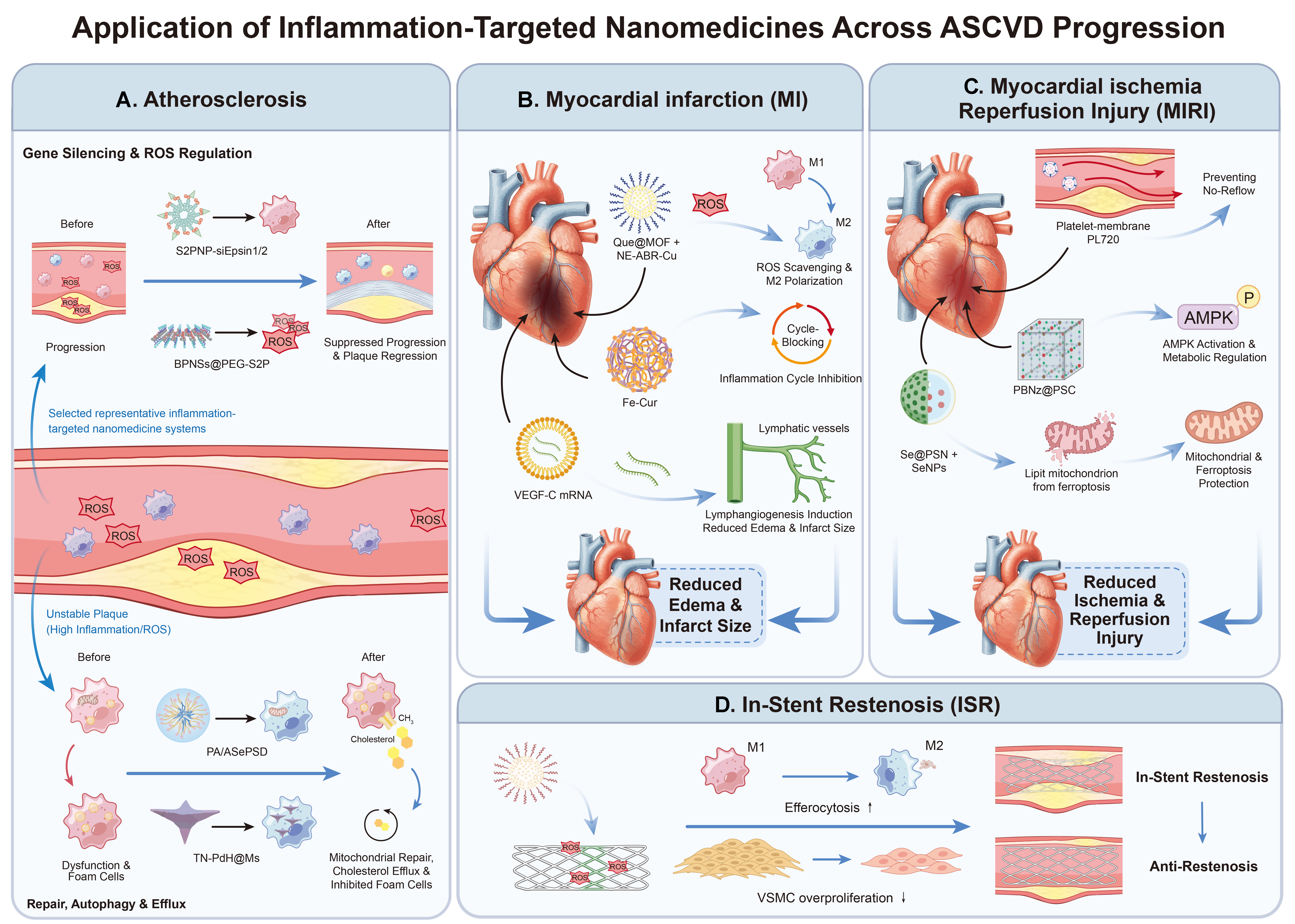
Figure 4. Applications of inflammation-targeted nanomedicines: (A) plaque stabilization and regression in atherosclerosis; (B) reduction of edema and infarct size after MI; (C) reduction of MIRI; and (D) inhibition of ISR. The schematics summarize preclinical mechanisms and therapeutic effects and do not establish clinical efficacy or patient-level therapeutic benefit. MI: Myocardial infarction; MIRI: myocardial ischemia-reperfusion injury; ISR: in-stent restenosis; ASCVD: atherosclerotic cardiovascular disease; ROS: reactive oxygen species; VEGF-C: vascular endothelial growth factor C; LNPs: lipid nanoparticles; AMPK: AMP-activated protein kinase; VSMC: vascular smooth muscle cell.
The macrophage-targeted gene-delivery system S2PNP-siEpsin1/2 silences Epsin expression to block lipid uptake in macrophages; its efficacy, however, has been verified only in ApoE-/- mice[61]. The dual inflammation-resolving nanocarrier BPNSs@PEG-S2P scavenges ROS and releases resolvin D1, promoting active resolution of inflammation while preserving the function of reparative M2 macrophages[62]. The mitochondria-protective multifunctional nanoplatform PA/ASePSD improves mitochondrial function and inhibits oxidized low-density lipoprotein (ox-LDL) internalization, thereby attenuating atherosclerotic plaque progression in preclinical models[63]. The macrophage-delivered TN-PdH nanozyme combines ROS scavenging, hydrogen-mediated anti-inflammatory effects, and autophagy activation to attenuate atherosclerosis[64]. Finally, the monocyte membrane-coated MoNP-VP system targets inflamed endothelium and suppresses Yes-associated protein (YAP)/transcriptional coactivator with PDZ-binding motif (TAZ) signaling, thereby reducing vascular inflammation and plaque development in mice[65]. Although these studies demonstrate substantial efficacy in rodent models, their long-term safety and therapeutic performance in clinically relevant large-animal models remain to be established.
Post-MI management: distal myocardial protection and local vascular repair
Post-MI myocardial management and reperfusion injury
Rupture of vulnerable plaques triggers coronary thrombosis and acute MI. Reperfusion after restoration of blood flow induces a massive burst of ROS and excessive activation of inflammatory cascades, ultimately leading to irreversible cardiomyocyte death. Nanomedicines intervene in multiple myocardial injury pathways [Figure 4B]. ROS-scavenging nanozymes, including Prussian blue[48] and selenium-based nanoparticles[49,56], have shown cardioprotective effects in MIRI by modulating oxidative stress, inflammatory signaling, and cell-death pathways [Figure 4C]. NE-ABR-CuxO attenuated myocardial injury by scavenging ROS and modulating NLRP3/IL-1β-mediated inflammation, thereby reducing infarct size in MI[66]. Vascular endothelial growth factor C (VEGF-C) mRNA-loaded lipid nanoparticles[50] stimulate functional cardiac lymphangiogenesis to alleviate myocardial edema and suppress interstitial inflammation; platelet membrane biomimetic nanocarriers accumulate in the microvascular no-reflow zone and restore microcirculatory perfusion at the early reperfusion stage[67]. Se@PSN alleviated MIRI by suppressing ferroptosis and mitochondrial dysfunction, thereby enhancing cardioprotection[49]. The post-MI inflammatory response is strictly time-dependent. Non-selective immune suppression during the early post-infarction phase can compromise debris clearance and repair; therefore, the timing of nanomedicine delivery is as critical as targeted localization. Current studies of MIRI have several limitations in experimental design. For example, many studies rely on a single short-term model involving 30 min of ischemia followed by 2 h of reperfusion, which fails to recapitulate chronic fibrosis and long-term low-grade inflammation after MI in humans. Long-term animal models with extended follow-up periods, such as 12 weeks, are therefore valuable for assessing sustained cardiac functional recovery, ventricular remodeling, scar evolution, and chronic post-infarction changes[68,69].
ISR
Current DES primarily inhibit VSMC proliferation by releasing paclitaxel or sirolimus-class drugs, substantially reducing the incidence of ISR[32,33]. However, these agents lack cell-type selectivity: while suppressing VSMC proliferation, they also delay re-endothelialization, increasing the risk of late thrombosis and necessitating long-term antiplatelet therapy. The next generation of anti-restenosis strategies must move beyond simple suppression of proliferation toward approaches that simultaneously reduce local inflammation, selectively inhibit aberrant VSMC growth, and lower thrombotic risk[33,34,52] [Figure 4D].
Combining biomimetic targeting with microenvironment-responsive release offers promise for ameliorating local inflammation at the implantation site and thereby lowering ISR risk. Zhao et al. constructed platelet membrane-coated ROS-responsive nanoclusters loaded with RVX-208 that, in a balloon injury model, demonstrated superior lesion targeting, tissue penetration, and anti-restenosis efficacy compared with conventional PLGA nanoparticles[51].
Beyond systemic administration, nanostructured stents and local drug-device combination approaches are also important strategies for ISR intervention. Nanoporous reservoirs, nanoscale coatings, and nanoparticle-eluting stents deliver therapeutic agents locally to the vessel wall, thereby reducing systemic exposure through sustained release. Yin et al. reviewed nanoparticle-based drug- and gene-eluting stents as platforms for localized, sustained delivery of therapeutic agents to the vessel wall to prevent and treat coronary restenosis[52]. Additionally, nanocellulose-gelatin-AS-IV[53] scaffolds can attenuate vein-graft restenosis by improving local mechanical support, inhibiting endothelial-to-mesenchymal transition (EndMT), and activating autophagy. This approach suggests a potential direction for scaffold materials that actively regulate vascular repair.
The role of nanomedicine in ISR is shifting from single-agent antiproliferative therapy toward integrated, locally active therapeutic platforms. However, key barriers to clinical application include long-term stent-coating stability, residual nanomaterials in the coatings, and complex regulatory requirements for these combination medical devices.
Despite robust preclinical efficacy in rodent models, the translational path for inflammation-targeted nanomedicines is fraught with hurdles. Clinical studies by van der Valk et al. showed that intravenously administered liposomal nanoparticles encapsulating prednisolone phosphate (LN-PLP) was detected in 75% of macrophages isolated from human iliofemoral atherosclerotic plaques[54]. However, in a separate randomized, placebo-controlled study, LN-PLP did not reduce arterial-wall permeability or inflammation, as assessed by multimodal imaging. These findings indicate that effective delivery to plaque macrophages does not necessarily translate into a measurable anti-inflammatory benefit.
Furthermore, long-term safety remains an important translational concern for inorganic nanomaterials because their persistence, degradation, and potential organ toxicity require systematic evaluation. Experimental studies of manganese oxide nanoparticles, for example, have identified dose- and material-dependent toxicological concerns[70]. Scalability and batch-to-batch reproducibility of cell-membrane biomimetic systems and exosomes pose major challenges to Good Manufacturing Practice (GMP)-compliant manufacturing. Additionally, the ApoE-/- mice fed a high-fat diet do not fully recapitulate human comorbidities (diabetes, hypertension, and polypharmacy) or the slow, spontaneous plaque progression seen in humans[71]. Therefore, validation in clinically relevant large-animal models should be considered an important step in assessing translational potential. Genetically modified porcine models, including ApoE/low-density lipoprotein receptor (LDLR)-deficient pigs, provide potential platforms for such studies[72].
CONCLUSION AND OUTLOOK
Inflammation-targeted nanomedicines have opened a new avenue for ASCVD treatment, with the potential to overcome the limitations of conventional drugs. Through ligand-mediated (e.g., peptides, antibodies, and aptamers), microenvironment-responsive (e.g., pH, ROS, and enzymes), and biomimetic (e.g., cell-membrane components) strategies, these nanomedicines achieve selective accumulation in inflamed ASCVD lesions, potentially reducing systemic toxicity. Future applications of nanomedicines should extend beyond isolated disease models and support integrated management across the full course of ASCVD.
Drug-device combinations warrant particular attention in future clinical translation. Compared with systemic administration, this approach confines nanomedicines to the lesion and enables sustained, controlled local release at low doses. Integrating stent-based local drug delivery with circulating cardioprotective nanomedicines may enable an integrated strategy for post-MI management.
Despite robust preclinical efficacy, several obstacles remain to be addressed before the clinical translation of inflammation-targeted nanomedicines. (a) Long-term safety requires systematic evaluation, particularly with respect to the biodistribution, degradation, and potential persistence of inorganic nanomaterials; (b) The scalability and reproducibility of biomimetic cell-membrane and exosome-based platforms remain major manufacturing challenges; (c) More clinically relevant animal models are needed, as ApoE-/- mice fed a high-fat diet do not fully recapitulate human ASCVD comorbidities, including diabetes, hypertension, and polypharmacy; (d) Combination nanomedicine formulations require rigorous quality control and regulatory assessment before clinical translation.
In addition, artificial intelligence (AI)-assisted digital cardiovascular twins and precision-medicine approaches may accelerate the development of inflammation-targeted nanomedicines, particularly by shortening the interval between carrier design and validation in cellular and animal models. Multi-omics data integration can identify inflammatory targets overexpressed in specific patient subgroups, while machine learning trained on high-throughput synthesis data can predict optimal nanoparticle composition, surface chemistry, and drug loading under varying blood-flow conditions, thereby reducing iterative trial-and-error cycles. On the patient side, combined analysis of imaging features (e.g., plaque MRI phenotypes) and circulating inflammatory markers could, in principle, identify subgroups most likely to respond to a given nanomedicine, enabling precision matching of targeting ligands to individual receptor expression profiles. Lesion imaging data acquired before and after nanomedicine administration can further be used to predict disease progression and guide regimen adjustment. Most of these applications remain at the proof-of-concept stage; substantial gaps remain before these approaches can meaningfully change clinical decision-making.
In summary, inflammation-targeted nanomedicines represent a promising therapeutic direction for ASCVD. Advances in precise inflammation targeting and stimuli-responsive drug delivery may facilitate the translation of these platforms from preclinical studies toward clinical investigation. Further improvements in safety, reproducibility, and clinically relevant validation will be essential for their future application in personalized ASCVD management.
DECLARATIONS
Authors’ contributions
Made substantial contributions to the conception: Guan, X.; Pan, M.; Chen, J.
Provided administrative and technical support: Li, Q.; Gu, N.
Wrote and revised the manuscript: Guan, X.; Luo, Y.; Yang, G.; Wang, F.
Availability of data and materials
Not applicable.
AI and AI-assisted tools statement
During the preparation of this manuscript, ChatGPT (OpenAI; GPT-6, released on 2026-10-07) was used solely for language editing. The tool did not influence the study design, data collection, analysis, interpretation, or the scientific content of the work. All authors take full responsibility for the accuracy, integrity, and final content of the manuscript.
Financial support and sponsorship
This work was supported by the National Natural Science Foundation of China (No. 32401084) and the Major Scientific and Technological Infrastructure Cultivation Projects of Jiangsu Province.
Conflicts of interest
All authors decleared that there are no conflicts of interest.
Ethical approval and consent to participate
Not applicable.
Consent for publication
Not applicable.
Copyright
© The Author(s) 2026.
REFERENCES
1. Vaduganathan, M.; Mensah, G. A.; Turco, J. V.; Fuster, V.; Roth, G. A. The global burden of cardiovascular diseases and risk. J. Am. Coll. Cardiol. 2022, 80, 2361-71.
2. Global Burden of Cardiovascular Diseases and Risks 2023 Collaborators. Global, regional, and national burden of cardiovascular diseases and risk factors in 204 countries and territories, 1990-2023. J. Am. Coll. Cardiol. 2025, 86, 2167-243.
4. Libby, P.; Ridker, P. M.; Hansson, G. K. Progress and challenges in translating the biology of atherosclerosis. Nature 2011, 473, 317-25.
5. Ference, B. A.; Ginsberg, H. N.; Graham, I.; et al. Low-density lipoproteins cause atherosclerotic cardiovascular disease. 1. Evidence from genetic, epidemiologic, and clinical studies. A consensus statement from the European Atherosclerosis Society Consensus Panel. Eur. Heart. J. 2017, 38, 2459-72.
6. Ridker, P. M.; Everett, B. M.; Thuren, T.; et al.; CANTOS Trial Group. Antiinflammatory therapy with canakinumab for atherosclerotic disease. N. Engl. J. Med. 2017, 377, 1119-31.
7. Libby, P. Interleukin-1 beta as a target for atherosclerosis therapy. J. Am. Coll. Cardiol. 2017, 70, 2278-89.
8. Smith, B. R.; Edelman, E. R. Nanomedicines for cardiovascular disease. Nat. Cardiovasc. Res. 2023, 2, 351-67.
9. Ajoolabady, A.; Pratico, D.; Lin, L.; et al. Inflammation in atherosclerosis: pathophysiology and mechanisms. Cell. Death. Dis. 2024, 15, 817.
10. Hu, B.; Boakye‐Yiadom, K. O.; Yu, W.; et al. Nanomedicine approaches for advanced diagnosis and treatment of atherosclerosis and related ischemic diseases. Adv. Healthc. Mater. 2020, 9, 2000336.
11. Zhang, J.; Tong, J.; Sun, Y.; et al. How advanced is nanomedicine to treat atherosclerosis? A comprehensive review of the literature. Int. J. Nanomedicine. 2026, 21, 563561.
12. Spadafora, L.; Quarta, R.; Martino, G.; et al. From mechanisms to management: tackling in-stent restenosis in the drug-eluting stent era. Curr. Cardiol. Rep. 2025, 27, 53.
13. Toldo, S.; Mezzaroma, E.; Buckley, L. F.; et al. Targeting the NLRP3 inflammasome in cardiovascular diseases. Pharmacol. Ther. 2022, 236, 108053.
14. Tardif, J.; Kouz, S.; Waters, D. D.; et al. Efficacy and safety of low-dose colchicine after myocardial infarction. N. Engl. J. Med. 2019, 381, 2497-505.
15. Nidorf, S. M.; Fiolet, A. T. L.; Mosterd, A.; et al. Colchicine in patients with chronic coronary disease. N. Engl. J. Med. 2020, 383, 1838-47.
16. Ridker, P. M.; Everett, B. M.; Pradhan, A.; et al. Low-dose methotrexate for the prevention of atherosclerotic events. N. Engl. J. Med. 2019, 380, 752-62.
17. Jolly, S. S.; d’Entremont, M. A.; Lee, S. F.; et al. Colchicine in acute myocardial infarction. N. Engl. J. Med. 2025, 392, 633-42.
18. Niu, Y.; Bai, N.; Ma, Y.; Zhong, P.; Shang, Y.; Wang, Z. Safety and efficacy of anti-inflammatory therapy in patients with coronary artery disease: a systematic review and meta-analysis. BMC. Cardiovasc. Disord. 2022, 22, 84.
19. Terkeltaub, R. A.; Furst, D. E.; Digiacinto, J. L.; Kook, K. A.; Davis, M. W. Novel evidence‐based colchicine dose‐reduction algorithm to predict and prevent colchicine toxicity in the presence of cytochrome P450 3A4/P‐glycoprotein inhibitors. Arthritis. Rheum. 2011, 63, 2226-37.
20. Ley, K.; Laudanna, C.; Cybulsky, M. I.; Nourshargh, S. Getting to the site of inflammation: the leukocyte adhesion cascade updated. Nat. Rev. Immunol. 2007, 7, 678-89.
21. Baumer, Y.; Irei, J.; Boisvert, W. A. Cholesterol crystals in the pathogenesis of atherosclerosis. Nat. Rev. Cardiol. 2025, 22, 315-32.
22. Tall, A. R.; Bornfeldt, K. E. Inflammasomes and atherosclerosis: a mixed picture. Circ. Res. 2023, 132, 1505-20.
23. Fidler, T. P.; Dunbar, A.; Kim, E.; et al. Suppression of IL-1β promotes beneficial accumulation of fibroblast-like cells in atherosclerotic plaques in clonal hematopoiesis. Nat. Cardiovasc. Res. 2024, 3, 60-75.
24. Fang, F.; Ni, Y.; Yu, H.; et al. Inflammatory endothelium-targeted and cathepsin responsive nanoparticles are effective against atherosclerosis. Theranostics 2022, 12, 4200-20.
25. Cochain, C.; Vafadarnejad, E.; Arampatzi, P.; et al. Single-cell RNA-seq reveals the transcriptional landscape and heterogeneity of aortic macrophages in murine atherosclerosis. Circ. Res. 2018, 122, 1661-74.
26. Winkels, H.; Ehinger, E.; Vassallo, M.; et al. Atlas of the immune cell repertoire in mouse atherosclerosis defined by single-cell RNA-sequencing and mass cytometry. Circ. Res. 2018, 122, 1675-88.
27. Fernandez, D. M.; Rahman, A. H.; Fernandez, N. F.; et al. Single-cell immune landscape of human atherosclerotic plaques. Nat. Med. 2019, 25, 1576-88.
28. Lee, G. Y.; Kim, J.; Choi, K. Y.; et al. Hyaluronic acid nanoparticles for active targeting atherosclerosis. Biomaterials 2015, 53, 341-8.
29. Kojima, Y.; Weissman, I. L.; Leeper, N. J. The role of efferocytosis in atherosclerosis. Circulation 2017, 135, 476-89.
30. Jaiswal, S.; Natarajan, P.; Silver, A. J.; et al. Clonal hematopoiesis and risk of atherosclerotic cardiovascular disease. N. Engl. J. Med. 2017, 377, 111-21.
31. Flores-Gomez, D.; Bekkering, S.; Netea, M. G.; Riksen, N. P. Trained immunity in atherosclerotic cardiovascular disease. Arterioscler. Thromb. Vasc. Biol. 2021, 41, 62-9.
32. Bønaa, K. H.; Mannsverk, J.; Wiseth, R.; et al.; NORSTENT Investigators. Drug-eluting or bare-metal stents for coronary artery disease. N. Engl. J. Med. 2016, 375, 1242-52.
33. Dangas, G. D.; Claessen, B. E.; Caixeta, A.; Sanidas, E. A.; Mintz, G. S.; Mehran, R. In-stent restenosis in the drug-eluting stent era. J. Am. Coll. Cardiol. 2010, 56, 1897-907.
34. Habib, A.; Finn, A. V. Endothelialization of drug eluting stents and its impact on dual anti-platelet therapy duration. Pharmacol. Res. 2015, 93, 22-7.
35. Li, Z.; Wang, Q.; Jing, H.; Luo, X.; Du, L.; Duan, Y. cRGD peptide-modified nanocarriers for targeted delivery of angiogenesis inhibitors to attenuate advanced atherosclerosis. ACS. Appl. Nano. Mater. 2021, 4, 11554-62.
36. Liu, M.; Zhang, Y.; Ma, X.; et al. Synthesis and characterization of fucoidan‐chitosan nanoparticles targeting P‐selectin for effective atherosclerosis therapy. Oxid. Med. Cell. Longev. 2022, 2022, 8006642.
37. Xu, H.; She, P.; Ma, B.; Zhao, Z.; Li, G.; Wang, Y. ROS responsive nanoparticles loaded with lipid-specific AIEgen for atherosclerosis-targeted diagnosis and bifunctional therapy. Biomaterials 2022, 288, 121734.
38. Lin, Y.; Huang, C.; Hou, X.; Kah, J. C. Y.; Wang, J. Polydopamine and hyaluronic acid-coated dual-responsive silica nanoparticles for targeted atherosclerosis imaging and therapy. Nanoscale. Adv. 2025, 7, 7134-44.
39. Lin, Y.; Liu, J.; Chong, S. Y.; et al. Dual‐function nanoscale coordination polymer nanoparticles for targeted diagnosis and therapeutic delivery in atherosclerosis. Small 2024, 20, 2401659.
40. Wang, Y.; Zhang, K.; Li, T.; et al. Macrophage membrane functionalized biomimetic nanoparticles for targeted anti-atherosclerosis applications. Theranostics 2021, 11, 164-80.
41. Li, Y.; Yu, J.; Cheng, C.; et al. Platelet and erythrocyte membranes coassembled biomimetic nanoparticles for heart failure treatment. ACS. Nano. 2024, 18, 26614-30.
42. Bu, T.; Li, Z.; Hou, Y.; et al. Exosome-mediated delivery of inflammation-responsive Il-10 mRNA for controlled atherosclerosis treatment. Theranostics 2021, 11, 9988-10000.
43. Chen, S.; Zhang, W.; Tang, C.; et al. Macrophage membrane-functionalized manganese dioxide nanomedicine for synergistic treatment of atherosclerosis by mitigating inflammatory storms and promoting cholesterol efflux. J. Nanobiotechnol. 2024, 22, 664.
44. Mirshafiee, V.; Mahmoudi, M.; Lou, K.; Cheng, J.; Kraft, M. L. Protein corona significantly reduces active targeting yield. Chem. Commun. 2013, 49, 2557.
45. Zhang, H.; Zhao, S.; Zhang, Q.; et al. Surface charge‐determined protein coronas of nanoparticles control endothelial cells uptake under low magnitude shear stress. Exploration 2026, 6, 20240248.
46. Allijn, I. E.; Czarny, B. M.; Wang, X.; et al. Liposome encapsulated berberine treatment attenuates cardiac dysfunction after myocardial infarction. J. Control. Release. 2017, 247, 127-33.
47. Yang, C.; Yang, S.; Fang, S.; et al. PLGA nanoparticles enhanced cardio-protection of scutellarin and paeoniflorin against isoproterenol-induced myocardial ischemia in rats. Int. J. Pharm. 2023, 648, 123567.
48. Long, M.; Wang, L.; Kang, L.; et al. Prussian blue nanozyme featuring enhanced superoxide dismutase-like activity for myocardial ischemia reperfusion injury treatment. ACS. Nano. 2025, 19, 4561-81.
49. Li, T.; Yang, B.; Liu, X.; et al. Silica nanoparticles loaded with selenium quantum dots reduce myocardial ischemia-reperfusion injury by alleviating ferroptosis and mitochondrial dysfunction. Int. J. Nanomedicine. 2025, 20, 1843-64.
50. Zhang, H.; Jiang, H.; Xie, W.; et al. LNPs-mediated VEGF-C mRNA delivery promotes heart repair and attenuates inflammation by stimulating lymphangiogenesis post-myocardial infarction. Biomaterials 2025, 322, 123410.
51. Zhao, Y.; Shirasu, T.; Yodsanit, N.; et al. Biomimetic, ROS-detonable nanoclusters - a multimodal nanoplatform for anti-restenotic therapy. J. Control. Release. 2021, 338, 295-306.
52. Yin, R.; Yang, D.; Wu, J. Nanoparticle drug- and gene-eluting stents for the prevention and treatment of coronary restenosis. Theranostics 2014, 4, 175-200.
53. Chu, T.; Li, Q.; Dai, C.; et al. A novel Nanocellulose-Gelatin-AS-IV external stent resists EndMT by activating autophagy to prevent restenosis of grafts. Bioact. Mater. 2023, 22, 466-81.
54. van der Valk, F. M.; van Wijk, D. F.; Lobatto, M. E.; et al. Prednisolone-containing liposomes accumulate in human atherosclerotic macrophages upon intravenous administration. Nanomedicine 2015, 11, 1039-46.
55. Liu, X.; Chen, B.; Chen, J.; et al. A cardiac‐targeted nanozyme interrupts the inflammation‐free radical cycle in myocardial infarction. Adv. Mater. 2024, 36, 2308477.
56. Chen, C.; Ma, J.; Duan, S.; et al. Mitigation of ischemia/reperfusion injury via selenium nanoparticles: suppression of STAT1 to inhibit cardiomyocyte oxidative stress and inflammation. Biomaterials 2025, 318, 123119.
57. Adkar, S. S.; Leeper, N. J. Efferocytosis in atherosclerosis. Nat. Rev. Cardiol. 2024, 21, 762-79.
58. Jansen, I.; Cahalane, R.; Hengst, R.; et al. The interplay of collagen, macrophages, and microcalcification in atherosclerotic plaque cap rupture mechanics. Basic. Res. Cardiol. 2024, 119, 193-213.
59. Vergallo, R.; Park, S. J.; Stone, G. W.; et al. Vulnerable or high-risk plaque: a JACC: Cardiovascular Imaging position statement. JACC. Cardiovasc. Imaging. 2025, 18, 709-40.
60. Mézquita, A. J. V.; Biavati, F.; Falk, V.; et al. Clinical quantitative coronary artery stenosis and coronary atherosclerosis imaging: a Consensus Statement from the Quantitative Cardiovascular Imaging Study Group. Nat. Rev. Cardiol. 2023, 20, 696-714.
61. Cui, K.; Gao, X.; Wang, B.; et al. Epsin nanotherapy regulates cholesterol transport to fortify atheroma regression. Circ. Res. 2023, 132, e22-42.
62. He, Z.; Chen, W.; Hu, K.; et al. Resolvin D1 delivery to lesional macrophages using antioxidative black phosphorus nanosheets for atherosclerosis treatment. Nat. Nanotechnol. 2024, 19, 1386-98.
63. Xu, H.; She, P.; Zhao, Z.; Ma, B.; Li, G.; Wang, Y. Duplex responsive nanoplatform with cascade targeting for atherosclerosis photoacoustic diagnosis and multichannel combination therapy. Adv. Mater. 2023, 35, 2300439.
64. Hu, R.; Dai, C.; Dong, C.; et al. Living macrophage-delivered tetrapod PdH nanoenzyme for targeted atherosclerosis management by ROS scavenging, hydrogen anti-inflammation, and autophagy activation. ACS. Nano. 2022, 16, 15959-76.
65. Huang, H.; Wang, T.; Rousseau, J.; et al. Biomimetic nanodrug targets inflammation and suppresses YAP/TAZ to ameliorate atherosclerosis. Biomaterials 2024, 306, 122505.
66. Su, F.; Ye, W.; Shen, Y.; et al. Immuno‐nanocomplexes target heterogenous network of inflammation and immunity in myocardial infarction. Adv. Sci. 2024, 11, 2402267.
67. Xu, X.; Li, M.; Yu, F.; et al. Platelet membrane nanocarriers cascade targeting delivery system to improve myocardial remodeling post myocardial ischemia–reperfusion injury. Adv. Sci. 2024, 11, 2308727.
68. Halkos, M. E.; Zhao, Z. Q.; Kerendi, F.; et al. Intravenous infusion of mesenchymal stem cells enhances regional perfusion and improves ventricular function in a porcine model of myocardial infarction. Basic. Res. Cardiol. 2008, 103, 525-36.
69. Schuleri, K. H.; Centola, M.; Evers, K. S.; et al. Cardiovascular magnetic resonance characterization of peri-infarct zone remodeling following myocardial infarction. J. Cardiovas. Magn. Reson. 2012, 14, 24.
70. Singh, S. P.; Kumari, M.; Kumari, S. I.; et al. Genotoxicity of nano- and micron-sized manganese oxide in rats after acute oral treatment. Mutat. Res. 2013, 754, 39-50.
71. Zhao, Y.; Qu, H.; Wang, Y.; Xiao, W.; Zhang, Y.; Shi, D. Small rodent models of atherosclerosis. Biomed. Pharmacother. 2020, 129, 110426.
Cite This Article
 Open Access
How to Cite
Guan, X.; Luo, Y.; Yang, G.; Chen, J.; Wang, F.; Li, Q.; Pan, M.; Gu, N. Inflammation-targeted nanomedicines for atherosclerotic cardiovascular disease: from atherosclerosis regression to post-myocardial infarction management. Soft Sci. 2026, 6, 104. https://dx.doi.org/10.20517/ss.2026.187
Download Citation
If you have the appropriate software installed, you can download article citation data to the citation manager of your choice. Simply select your manager software from the list below and click on download.
Export Citation File
Type of Import
Tips on Downloading Citation
Citation Manager File Format
Type of Import
Direct Import: When the Direct Import option is selected (the default state), a dialogue box will give you the option to Save or Open the downloaded citation data. Choosing Open will either launch your citation manager or give you a choice of applications with which to use the metadata. The Save option saves the file locally for later use.
Indirect Import: When the Indirect Import option is selected, the metadata is displayed and may be copied and pasted as needed.
About This Article
Special Topic
Copyright

Data & Comments
Data
0












Comments
Comments must be written in English. Spam, offensive content, impersonation, and private information will not be permitted. If any comment is reported and identified as inappropriate content by OAE staff, the comment will be removed without notice. If you have any queries or need any help, please contact us at [email protected].