

Ecology and mechanisms of the zebrafish gut microbiome
 0
Graphical Abstract
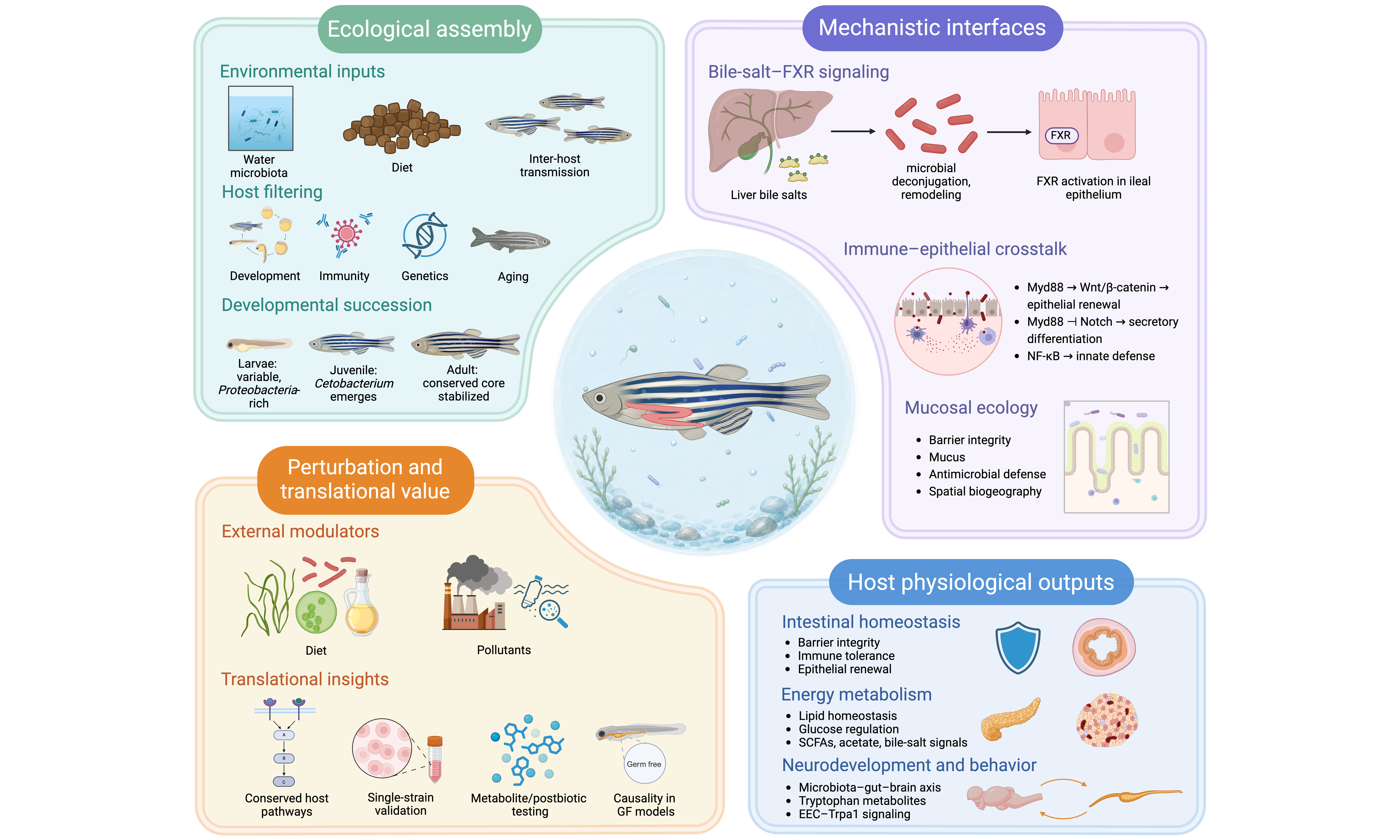
Abstract
The intestinal microbiota is increasingly viewed as an active organ that shapes diverse host physiological functions. Zebrafish have emerged as a unique vertebrate model for microbiome research, enabling manipulative, high-throughput in vivo tests that are challenging in mammals. Microbiota development in zebrafish is highly dynamic, driven by environmental inoculation, interhost transmission, and host filtering mechanisms - with critical developmental windows that set long-term community trajectories. This review elaborates on multiple mechanisms by which microbial activity affects the homeostasis of the intestinal epithelium, modulates lipid and glucose metabolism, and influences neurodevelopment and animal behavior via the microbiota-gut-brain axis, thereby highlighting the roles of microbes in metabolism, immunity, and neurological function. We also summarize the effects of various dietary components on the intestinal milieu and their roles in remodeling gut communities. In parallel, we catalog how diverse environmental toxicants elicit reproducible dysbiosis accompanied by inflammation, oxidative stress, and metabolic dysregulation. Future priorities include addressing species- and strain-level mechanisms by delineating molecular signaling cascades, expanding the focus beyond gut-brain axes to other organ systems, and developing precision microbiota-modulation strategies in zebrafish that can be translated to mammalian hosts. Together, these advances position zebrafish as a complementary vertebrate model for mechanistic microbiome biology and for preclinical screening of microbiota-directed interventions.
Keywords


 Download PDF
BACKGROUND
The gut microbiota is considered an extension of the host genome, playing crucial roles in host development, immune regulation, and metabolic balance, and complementing host functions in nutrient degradation and utilization[1-3]. Microbiota dysbiosis influences disease susceptibility and increases the incidence of various human diseases[4-8], and microbial signals engage gut-tissue pathways that modulate physiological responsiveness and behavior[9-11].
Zebrafish offer a unique model for studying how gut microbes act on host intestinal homeostasis and coordinate systemic physiology. Environmental seeding, dispersal among hosts, and host-driven selection jointly govern the composition of the zebrafish gut microbiota over time[12-14]. Across different facilities and genetic backgrounds, the zebrafish gut consistently contains recurring bacterial groups, often referred to as a “core” microbiome at the genus or phylum level, whereas variations in community structure reflect differences in rearing conditions. This pattern highlights the combined roles of environmental exposure and host-specific selection in shaping the microbiota[15,16]. Conversely, microbial cues regulate epithelial homeostasis, tune innate immune tone, reshape luminal chemistry, and link nutritional metabolism with neural outcomes in the host[17-19]. These interactions form reciprocal host-microbe feedback loops in which epithelial transport and barrier function constrain microbial growth and spatial organization, whereas microbial functions reprogram host transcriptional and signaling outputs[17,18]. Additionally, dietary composition and environmental contaminants represent two major extrinsic drivers that shape community structure and modulate host-microbe interactions[20-22]. Integrating these drivers with intrinsic mechanisms is essential for the coherent interpretation of findings from zebrafish microbiota studies.
This review focuses primarily on the bacterial gut microbiota of zebrafish, synthesizing ecological principles governing community assembly across development and in response to immune, dietary, and environmental influences. It consolidates pathway-resolved mechanisms linking microbial activities to epithelial homeostasis, energy metabolism, and neurobehavior, with an emphasis on mechanistic evidence and translational relevance. Viral and fungal components are briefly discussed to highlight gaps in our understanding of the wider intestinal ecosystem.
ZEBRAFISH INTESTINAL MICROBIAL COMPOSITION AND ECOLOGICAL ESTABLISHMENT
The zebrafish gut microbiome is shaped by host-mediated selective filtering that strengthens with age. Larval communities are variable and reflect environmental source pools, whereas juvenile and adult communities exhibit increasingly deterministic selection and converge toward a conserved core observed across laboratories and in wild populations[13,16].
Intrinsic filtering of zebrafish community composition
Community composition follows a reproducible stage-structured pattern across development. Larval communities are typically Proteobacteria-dominant, with nearly single-phylum profiles. Common early genera include Aeromonas, Plesiomonas, Vibrio, Pseudomonas, Shewanella, and Acinetobacter. Gut colonization begins soon after hatching, when the mouth and vent open at approximately 3-4 days post-fertilization (dpf), and the earliest communities are typically Proteobacteria-rich, with frequent detection of Aeromonas, Vibrio, Plesiomonas, Shewanella, and Pseudomonas[23-26]. During the juvenile stage, Cetobacterium emerges and increases in abundance, and signals from Fusobacteria and, in some datasets, Firmicutes (also referred to as Bacillota in updated nomenclature) or Actinobacteriota become more evident. In adults, the community stabilizes with Proteobacteria and Fusobacteria as the major phyla. Cetobacterium is frequently the most abundant genus, accompanied by Aeromonas, Plesiomonas, Acinetobacter, Vibrio, and the Firmicutes clade ZOR0006[27] [Figure 1]. In adult zebrafish, community profiles are reproducible across independent laboratory facilities and are congruent with those reported in wild zebrafish, indicating a conserved core of development-selected taxa[16,25,27].
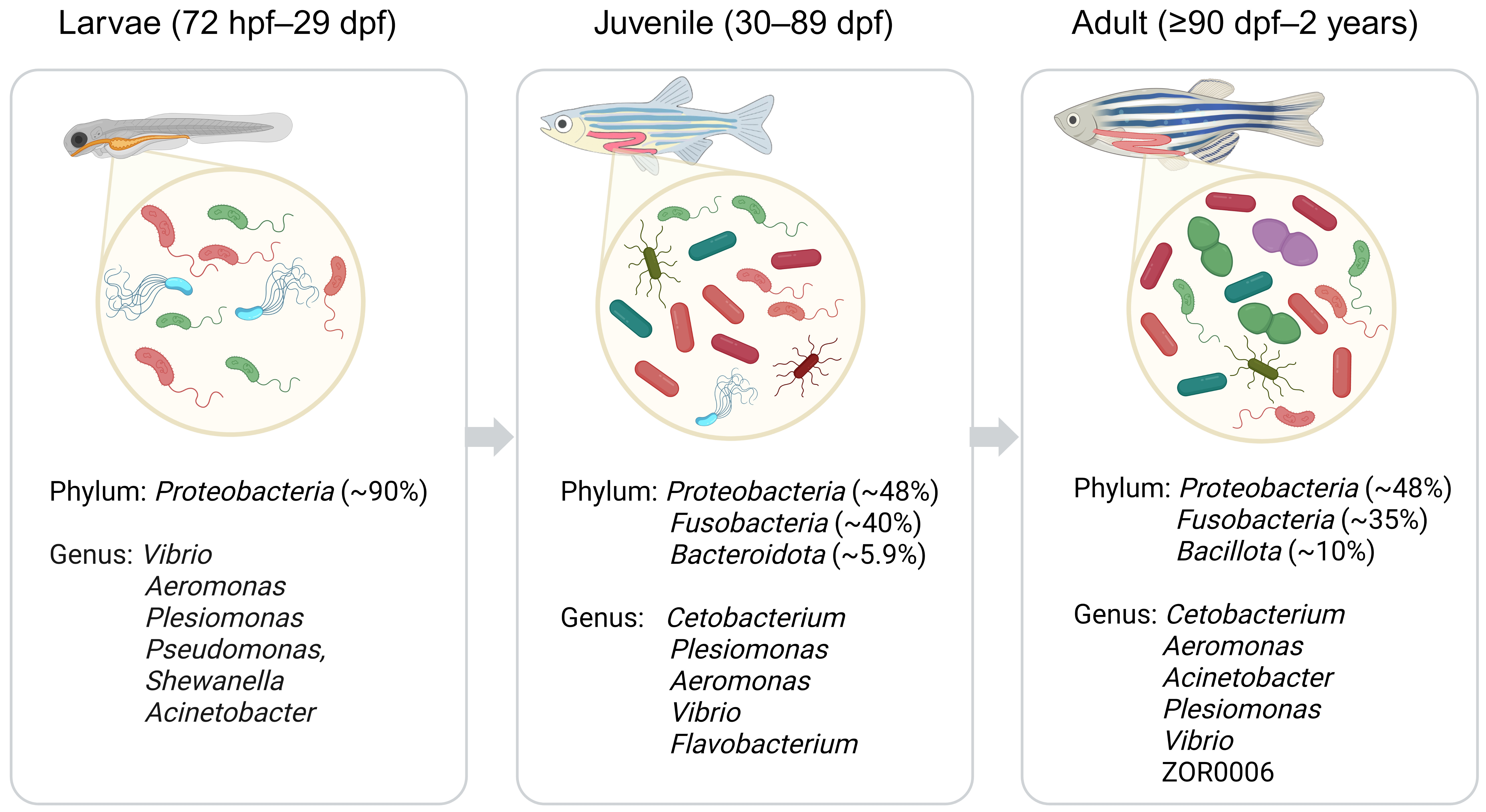
Figure 1. Dominant intestinal taxa across zebrafish development. Larvae (72 hpf-29 dpf) are Proteobacteria-dominant (~ 90%; representative genera: Vibrio, Aeromonas, Plesiomonas, Pseudomonas, Shewanella, Acinetobacter)[27]. Juvenile (30-89 dpf) profiles show Proteobacteria ~ 48%, Fusobacteria ~ 40%, and Bacteroidota ~ 5.9%, with increased relative abundance of Cetobacterium[27]. Adult (≥ 90 dpf-2 years) communities stabilize with Proteobacteria ~ 48%, Fusobacteria ~ 35%, and Bacillota (reported as Firmicutes in the original study) ~ 10% at the phylum level[27]; meta-analytic summaries identify Cetobacterium as the most abundant genus, together with Aeromonas, Acinetobacter, Plesiomonas, Vibrio, and ZOR0006[27]. hpf: Hours post fertilization; dpf: days post fertilization. Created in BioRender. Li, Y. (2026) https://BioRender.com/ik3knnb.
Beyond this conserved developmental program, aging introduces a distinct axis of gut microbiota remodeling, rather than simply extending adult stability. Between approximately 8 and over 33 months, alpha-diversity increases significantly by ~ 8 months, remains stable until ~ 32 months, and then decreases sharply after ~ 33 months. Community composition shifts significantly with age, including a marked expansion of Proteobacteria and coordinated changes in other dominant phyla. These phylum-level trajectories can be tracked by representative genera such as Aeromonas, Mycobacterium, and Cetobacterium, whose abundance peaks coincide with those of their corresponding phyla[28]. Mechanistically, an aging zebrafish model showed that telomerase deficiency was associated with reduced microbial diversity and dysbiosis, whereas delaying gut aging through gut-specific telomerase expression restored microbiota composition and ameliorated systemic aging phenotypes. These findings show that restoring intestinal telomerase can counteract age-associated changes in both the microbiota and host physiology[29].
Immune maturation imposes an additional intrinsic layer of selection that shapes both the composition and the functional output of the zebrafish gut microbiota. At the taxon level, a clear example is the adaptive control of Vibrio. As zebrafish mature, T lymphocytes restrict Vibrio expansion in the intestine, and this suppression is abolished in immunodeficient settings, indicating that immune competence can directly dictate the abundance of specific taxa[30]. At the community scale, adaptive immunity acts as an ecological filter that shifts assembly from neutral dynamics toward individualized adult states[12,31]. Immune-derived cytokine networks act in concert with this filtering to tune and stabilize microbiota homeostasis across life stages. Early in life, IL-26 from innate lymphoid cells shapes microbiota composition and supports epithelial homeostasis[32]. In larvae, enteroendocrine cell-derived IL-22 contributes to gut homeostasis through microbiota-linked control of intestinal physiology[33]. Collectively, these findings illustrate a multi-tiered immunological governance of the zebrafish microbial ecosystem.
Host species identity is another intrinsic determinant of zebrafish gut community assembly because the recipient gut habitat actively filters and reshapes incoming consortia. In germ-free (GF) reciprocal transplants between zebrafish and mice, donor lineages remained detectable, yet the resulting communities reproducibly shifted toward the recipient’s characteristic configuration, providing direct evidence for host selection over passive microbial takeover[34]. Consistently, attempts to introduce complex human communities into zebrafish larvae achieve only partial engraftment: a restricted subset of human phylotypes establishes in the fish gut, and colonization outcomes depend on the transplantation strategy and recipient background[35]. Even these limited microbial inputs can convey functional phenotypes. For example, transplantation of gut microbiota from high-fat diet-fed mice into microbiota-depleted zebrafish larvae increases susceptibility to diet-induced hyperlipidemia; the same study demonstrated that Enterococcus faecalis and the Gram-positive cell wall components peptidoglycan and lipoteichoic acid promote this phenotype through host MyD88 signaling[36]. Microbiota from cigarette smoke-exposed mice potentiates cigarette smoke extract-induced inflammation in zebrafish larvae[37]. Together, these studies indicate that species-specific ecological filters tightly govern stable community membership, whereas conserved host signaling pathways can translate admissible microbial signals into distinct physiological outcomes.
Spatial organization and microbial load across intestinal segments in zebrafish
Zebrafish gut colonization is spatially organized along the intestinal axis rather than being uniform, making segment-resolved analysis essential for mechanistic insight[38]. In gnotobiotic larvae, Vibrio populations were concentrated in the anterior bulb and Aeromonas was biased toward the mid-intestine, showing taxon-specific biogeography visible in vivo[38]. Spatial patterns also reflect biophysical traits: cohesive, aggregating bacteria localize more posteriorly than planktonic bacteria, linking spatial community organization to measurable population architecture alone[39]. In addition to imaging-based readouts, culture-dependent segment-level quantification has revealed consistently higher culturable bacterial load in posterior than in anterior intestinal segments in adult zebrafish across facilities. These findings suggest an anteroposterior gradient in intestinal carrying capacity, which may confound interpretation if segmental samples are pooled[40]. Even under controlled community conditions, inter-individual variation in colonization efficiency and spatial distribution remains significant, confirming that region-specific localization reflects biological signal rather than technical noise[41]. Taken together, these findings justify the use of segment-resolved sampling and absolute quantification when drawing functional inferences about zebrafish gut microbiota organization and host outcomes[38-40].
Ecological development of the zebrafish microbiome
Developmental stage is a dominant driver of gut microbiome succession in zebrafish[13,42]. In controlled environmental-transfer experiments, stage explains more community turnover than dispersal, and homogeneous selection increases from larva to adult. Larval assemblages mirror the surrounding water, whereas adult communities diverge from the environment and stabilize, reflecting progressively stronger host filtering with age[13,25].
Regarding diversity patterns, longitudinal data show a stage-linked increase in alpha diversity: taxonomic richness rises across developmental stages, and both phylogenetic diversity and Shannon Index are higher in late (56-98 days post-hatching) than in early (12-26 days post-hatching) periods[13,42]. Consistent with the stage-linked richness pattern, a cross-study meta-analysis reports no reproducible stage differences in Shannon Index or Simpson Index, yet detects lower richness in larvae than in juveniles and adults when richness is quantified as observed amplicon sequence variants (ASVs)[27]. Since differences in sampled populations and analytical methods may contribute to the divergent Shannon results, multiple metrics are needed to comprehensively assess changes in alpha diversity. For beta diversity, microbial communities differ significantly across developmental stages under both Bray-Curtis (abundance-weighted) and Jaccard (presence-absence) metrics. Further matrix correlation analysis showed that developmental stage has the strongest association with community dissimilarity even when environmental factors, transfer treatments, and diet are considered, supporting host development as the dominant driver of community turnover[13,42].
Technically, phylogenetic diversity metrics, such as the Faith index for alpha diversity and UniFrac for beta diversity, can add ecological and evolutionary resolution beyond taxonomic measures. Faith index has been used to assess how chronic microplastic exposure alters the phylogenetic breadth of zebrafish gut communities and their potential resilience under stress[43]. UniFrac, including weighted and unweighted variants, captures phylogenetically structured community differences and has been applied in cross-dataset analyses to define the zebrafish core gut microbiome[44]. Together, these studies support incorporating phylogenetic metrics in zebrafish microbiome research, particularly for developmental trajectories, environmental exposures, and cross-study comparisons.
A pronounced transition in mid-juvenile stages coincides with the emergence of adaptive immunity and the reshaping of nutrient niches, separating early variable communities from later stabilized assemblages. Network analyses further show age-dependent increases in interaction density and inferred stability, with rare keystone taxa exerting disproportionate effects in adults. The convergence of adult core membership across wild and laboratory populations underscores strong host habitat filtering and a conserved ecological endpoint[13,16,42].
MICROBIOTA-MEDIATED REGULATORY MECHANISMS IN THE ZEBRAFISH GUT
Importance of gut microbes in zebrafish intestinal development
Microbiota contributes to intestinal epithelial development and function in zebrafish[45-47]. Compared with conventionally raised (CV) fish, GF zebrafish larvae show reduced intestinal epithelial proliferation[45,46]. A previously published DNA microarray study identified 212 genes regulated by microbial colonization in larval digestive tracts, including genes involved in cell proliferation, nutrient metabolism, and innate immunity[45]. Reintroduction of a conventional microbiota restored epithelial proliferation in GF larvae, supporting a causal contribution of microbial colonization to this developmental response[45,46]. Microbial colonization also induces the brush-border enzyme intestinal alkaline phosphatase (IAP), demonstrating microbial regulation of epithelial function beyond proliferation[47]. Together, these findings support a role for microbial signals in intestinal epithelial development and functional maturation.
Microbial modulation in zebrafish intestinal mucosal homeostasis
Zebrafish intestinal mucosal homeostasis can be viewed as a dynamic steady state sustained by coordinated epithelial renewal and barrier integrity, a tolerogenic but controlled mucosal immune tone, a luminal chemical microenvironment shaped by host secretions and nutrient flow, and a stable microbial community with defined niche structure[27,48]. Microbial exposure remodels host growth, metabolic, and immune programs with strong cell-type specificity across epithelial and stromal compartments, providing an experimentally tractable in vivo readout of how homeostasis is established and maintained under controlled colonization[49]. Cross-vertebrate comparisons are supported by conserved gut architecture, epithelial regulatory programs, and the presence of mammalian-like immune cell types, including functional adaptive immunity in adult zebrafish[48,50].
Bile-salt metabolism and intestinal epithelial homeostasis
Among the host-derived factors contributing to the luminal chemical microenvironment, bile acids represent a key class of signaling molecules that integrate host metabolism with microbial activity. Zebrafish and other cypriniform fish utilize 5α-cyprinol 27-sulfate as the primary biliary bile salt, in contrast to the C24 bile acid-dominated pools typically found in mammals[18,51]. The most direct microbiota-linked evidence in zebrafish involves the farnesoid X receptor (FXR), for which bile-salt profiling combined with gnotobiotic manipulation allows quantitative mapping from microbial bile-salt modifications to epithelial transcriptional responses[18].
In zebrafish, epithelial bile salt-FXR signaling regulates ileal bile-acid uptake and trans-epithelial transport, suppresses lysosome-rich enterocyte (LRE) differentiation, and promotes epithelial secretion of fibroblast growth factor (fgf19) to the liver. Gut microbes act upstream by remodeling bile salts, for example, through deconjugation of taurocholic acid (TCA) to cholate (CA), thereby fine-tuning FXR activation and its downstream epithelial functions [Figure 2]. Single-cell RNA-seq further reveals that FXR upregulates bile-acid absorption genes (e.g., fabp6, slc10a2, slc51a), while suppressing lysosomal and LRE pathways in ileal epithelial clusters[18]. In larvae, the microbiome slows LRE-mediated protein uptake and lysosomal degradation[52], thereby refining regional epithelial specialization without altering ileal morphology. Taken together, these findings support a conserved paradigm in zebrafish in which microbial bile-salt remodeling modulates FXR signaling to orchestrate intestinal epithelial cell (IEC) function[18]. In an adult zebrafish model of short bowel syndrome, broad-spectrum antibiotic-driven microbiome perturbation was accompanied by increased intestinal fxr expression and altered FXR-axis transcriptional output, consistent with an effect of microbiota-linked bile-salt signaling on intestinal homeostasis in disease contexts[53]. Under high-lipid stress, intestinal homeostasis was impaired alongside reduced transcriptional readouts of the FXR pathway, and antibiotic treatment partially reversed these changes, supporting a microbiota contribution to bile-salt-linked FXR signaling in zebrafish[54].
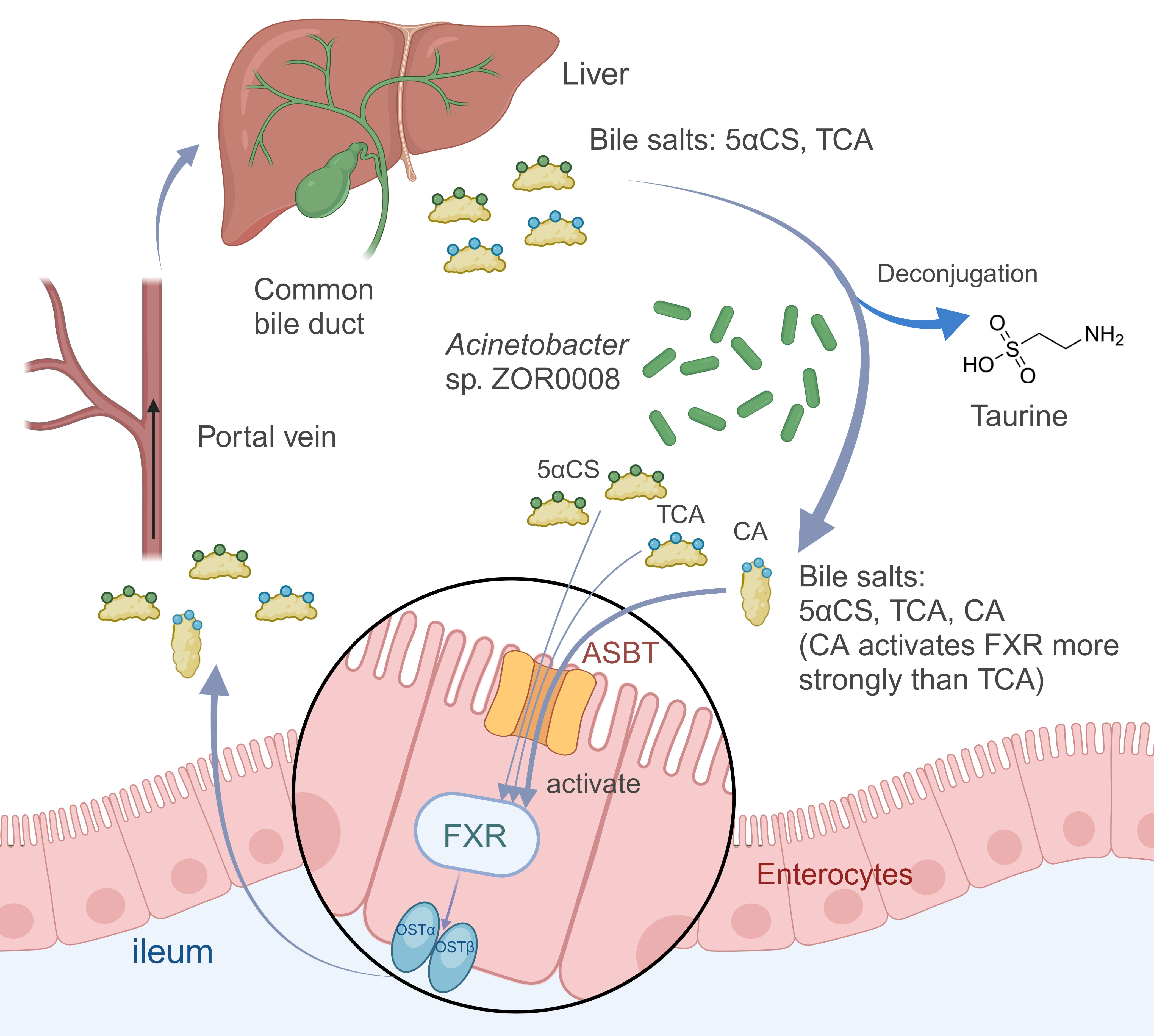
Figure 2. Microbial modulation of the enterohepatic circulation and ileal FXR signaling in zebrafish. Bile salts, dominated by 5αCS in zebrafish, are synthesized in the liver and secreted via the common bile duct into the intestinal lumen. In the gut, Acinetobacter sp. ZOR0008 deconjugates bile salts, liberating taurine and increasing the proportion of unconjugated cholate (CA), which is a stronger agonist of FXR in ileal enterocytes. Luminal bile salts are taken up across the apical membrane by ASBT, transported intracellularly, and exported basolaterally by the OSTα/OSTβ heterodimer to the portal vein, returning to the liver and completing the enterohepatic cycle. ASBT: Apical sodium-dependent bile acid transporter; FXR: Farnesoid X receptor; OSTα/OSTβ: organic solute transporter α/β; TCA: taurocholic acid; 5αCS: 5α-cyprinol sulfate. Created in BioRender. Li, Y. (2026) https://BioRender.com/8puqqah.
Immune-epithelial crosstalk in the maintenance of homeostasis
The maintenance of intestinal homeostasis relies on coordinated immune-epithelial circuits that convert microbial cues into regulated epithelial renewal, lineage allocation, and barrier defense while calibrating inflammatory tone to accommodate the microbiota.
During zebrafish larval development, gut microbes stimulate intestinal epithelial renewal by transmitting signals through the innate adaptor MyD88 to activate canonical Wnt/β-catenin signaling. This interaction stabilizes β-catenin, thereby activating cell-cycle gene expression and maintaining the pool of proliferative stem and progenitor cells in intestinal epithelia [Figure 3A]. Conversely, GF larvae exhibit fewer S-phase epithelial nuclei and a slower rate of epithelial turnover. Mono-association with Aeromonas veronii revealed that either the bacterium itself or its cell-free supernatant fraction is sufficient to boost epithelial proliferation, supporting a secreted effector activity that does not require direct bacterial contact. Furthermore, even a non-lethal dose of purified lipopolysaccharide (LPS) fails to raise proliferation, showing that LPS alone is not sufficient to recapitulate the microbiota-driven proliferative output in vivo and suggesting that commensal bacteria boost the endogenous Wnt/β-catenin pathway of the epithelium through a non-inflammatory mechanism[46]. At the level of signaling architecture, coupling MyD88-dependent microbial sensing with Wnt/β-catenin-mediated epithelial renewal reflects a conserved vertebrate logic, whereas the specific microbial effectors and their ecological availability are likely context-dependent.
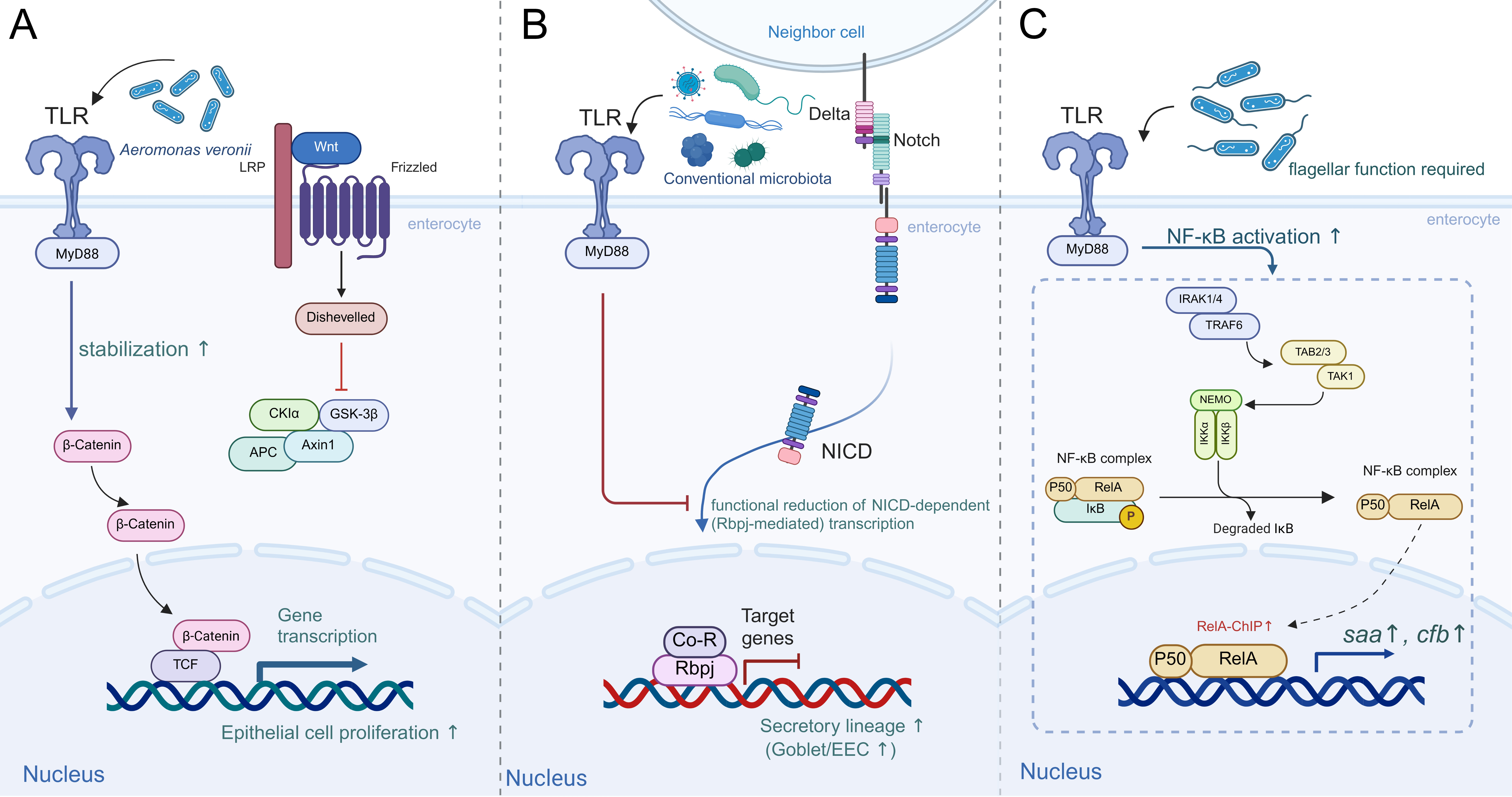
Figure 3. Microbiota-MyD88 regulatory signaling in the larval zebrafish intestine. (A) Microbial sensing via MyD88 stabilizes β-catenin to augment canonical Wnt output in the larval zebrafish intestine, which is required for epithelial proliferation in the intestine; (B) Microbiota-MyD88 signaling functionally reduces NICD-dependent Notch transcription to bias secretory fates in the larval zebrafish intestine, whereas NICD signaling via Rbpj normally limits secretory differentiation. This is reflected by changes in secretory cell markers; (C) Microbiota with flagellar function enhance the MyD88-dependent NF-κB cascade, increasing RelA signaling and promoter/enhancer occupancy at target genes such as saa and cfb and ultimately increasing expression of these genes. cfb: Complement factor B; IκB: inhibitor of κB; IKK: IκB kinase (IKK complex); IRAK: interleukin-1 receptor-associated kinase; MyD88: myeloid differentiation primary response protein 88; NF-κB: nuclear factor κB; NICD: Notch intracellular domain; p50: NF-κB1 p50 subunit; Rbpj: recombination signal-binding protein for immunoglobulin κJ region; RelA: NF-κB p65 subunit; saa: serum amyloid A; TAK1: TGF-β-activated kinase 1 (MAP3K7); TLR: toll-like receptor; TRAF6: TNF receptor-associated factor 6; Wnt: wingless/INT-1 signaling pathway; TCF: T-cell factor; APC: adenomatous polyposis coli; Axin1: axis inhibition protein 1; GSK-3β: glycogen synthase kinase 3 beta; Co-R: corepressor; NEMO: NF-κB essential modulator; TAB2/3: TAK1-binding protein 2/3; ChIP: chromatin immunoprecipitation. Created in BioRender. Li, Y. (2026) https://BioRender.com/lwq8avz.
Homeostasis also requires balanced epithelial specialization. Recent evidence in zebrafish showed that microbiota-induced, MyD88-dependent signaling inhibits epithelial Notch activity, which regulates the fate choice between absorptive and secretory epithelial cell lineages [Figure 3B]. Genetic perturbation studies confirmed that inhibition of Notch signaling promotes secretory cell differentiation, whereas Notch activation or myd88 deletion impairs it. GF larvae exhibit a marked reduction in secretory cells, including goblet and enteroendocrine cells (EECs), compared with CV larvae. This reduction is reversed upon microbial colonization, indicating a microbiota-dependent effect on lineage allocation[55]. This axis should be understood as a lineage-allocation mechanism rather than a narrow immune pathway. Given that secretory lineages are major contributors to mucus chemistry and hormone signaling, shifts in their fate choice plausibly reshape barrier function and the luminal habitat. Consistent with this broader functional role of secretory cells, EEC-derived IL-22 in larval zebrafish is promoted by microbial cues, helps sustain early-life gut motility, and shapes gut microbial composition, including enrichment of Lactobacillaceae, before lymphocytes become abundant in the gut[33]. In addition, IL-10 promotes epithelial Notch signaling to restrict goblet cell expansion during zebrafish intestinal development, extending immune regulation of secretory output beyond MyD88-dependent microbial sensing[56]. Furthermore, mammalian studies support microbiota-dependent regulation of epithelial differentiation through macrophage and mesenchymal niche signaling that converges on Wnt programs in early life, reinforcing conservation at the level of core epithelial fate pathways and cellular niches rather than taxonomic equivalence[57].
A complementary module of mucosal homeostasis involves an orchestrated inflammatory and antimicrobial tone, which restrains microbial expansion without compromising tissue function. In zebrafish, this balance is captured by the dynamic activity of a nuclear factor κB (NF-κB) reporter. NF-κB is a conserved transcriptional regulator that coordinates innate immune responses within the gut epithelium. The microbiota engages two complementary mucosal immune pathways: (1) an epithelial pathway that depends on NF-κB and, in part, on MyD88 and upregulates innate defense genes, including serum amyloid A (saa) and complement factor B (cfb) [Figure 3C]; and (2) an NF-κB- and MyD88-independent neutrophil pathway, reflected by induction of myeloperoxidase mRNA and consistent with microbiota effects on neutrophil development and/or survival. During intestinal development in zebrafish, NF-κB activity is detectable in both absorptive enterocytes and secretory cells. The resident microbiota increases epithelial NF-κB activity between 5 and 8 dpf. Colonization upregulates innate immune and metabolic transcripts in the digestive tract, including a subset homologous to mammalian NF-κB target genes. Functionally, NF-κB activation is sensitive to specific bacterial features, as mono-association with Pseudomonas aeruginosa is sufficient to induce reporter activity, and this induction requires a functional flagellar apparatus. Thus, canonical NF-κB signaling represents a conserved host module, whereas the upstream microbial determinants shaping its spatiotemporal activation are context-dependent in the zebrafish intestinal environment[17,58].
Immune homeostasis in zebrafish is supported by additional layers that directly interface with the microbiota. These include epithelial enzymatic editing, exemplified by intestinal alkaline phosphatase (IAP), which enforces epithelial tolerance by detoxifying microbial LPS. Deficiencies in this system predispose the host to inflammatory imbalance[47,59]. Another key layer involves localized redox control, in which dual oxidase mediates antibacterial activity at the mucosal surface, indicating a strategy of microbial containment through spatial control rather than systemic inflammatory escalation[60,61]. These mechanisms prompt a broader reconsideration of homeostasis as a dynamic equilibrium maintained through biochemical editing of the microenvironment and chemical restraint of microbial niches.
Microbial ecology at the mucosal interface
Intestinal homeostasis also depends on ecological properties of the gut community, including spatial biogeography along the intestinal axis, colonization stability, and context-dependent interactions that determine whether microbial cues are delivered as diffuse exposure or as structured, niche-localized signals[41]. From this perspective, these ecological features should be treated as quantitative variables, because even defined consortia can produce marked inter-individual differences in colonization efficiency and spatial organization, which correlate with host physiological processes such as mucus production[41]. Independent work further shows that generic physical and ecological processes, such as bacterial cohesion, are predictive of spatial distribution within the zebrafish gut, reinforcing that location and aggregation state are central determinants of host-microbe contact[39]. This ecological maturation continues over zebrafish development, with communities trending toward greater stability and structured interactions, reflecting increasing ecological constraints and niche partitioning during ontogeny[42].
In addition, robust ecological inference in aquatic models requires attention to the experimental environment. The aquatic housing system directly couples the gut to the water reservoir and can generate facility-dependent variation that extends beyond host genotype. Consequently, water source and husbandry should be viewed as defined experimental variables, because they systematically shape the exposure landscape and co-vary with intestinal profiles[62]. Accordingly, host-microbe interactions in aquatic models are best interpreted within a metacommunity framework, in which the host, gut microbiota, and environment form an integrated ecological unit.
Microbial modulation of zebrafish energy homeostasis
Intestinal microbiota plays an important role in energy homeostasis[63]. A previously published study shows that the intestinal microbiota broadly enhances neutral lipid deposition and lipid droplet formation in zebrafish intestinal epithelium. Specifically, GF larvae exhibit fewer epithelial lipid droplets than CV siblings; in antibiotic-depleted fish, intestinal Oil Red O signal is reduced, and expression of key lipid-handling genes is altered. These findings indicate that microbiota positively modulates intestinal lipid metabolism in the host[64].
In zebrafish, Firmicutes have been reported to exhibit a strong association with increased fatty-acid absorption and elevated lipid droplet numbers in enterocytes. Mono-association with a Firmicutes strain (Exiguobacterium sp.) is sufficient to enhance intestinal fatty acid uptake. Conditioned media from this strain also increase lipid droplet number without altering average size, suggesting the involvement of factors secreted by the bacterium and indicating that specific taxa differentially regulate droplet number and size[65,66].
Given the dynamic nature of microbial community composition, targeted modulation of the microbiota can reshape host lipid metabolism. Administration of the probiotic Lactobacillus rhamnosus increases the abundance of Firmicutes and reduces Actinobacteria, and concurrently downregulates genes involved in cholesterol and triglyceride metabolism and in acetate production[67,68]. These transcriptional changes correlate with phenotypic improvements, including reduced whole-body cholesterol and triglycerides, smaller lipid droplets, and improved intestinal epithelial ultrastructure reflected by elongated microvilli and enterocytes[67]. Mannose-enriched Cetobacterium somerae alleviated steatosis through microbiota-derived putrescine and host hepatic conversion to spermidine[69]. Together, these findings underscore the potential of microbiota reshaping to promote lipid homeostasis in zebrafish.
Glucose metabolism plays a central role in energy homeostasis, and emerging evidence underscores the influence of the gut microbiota on regulating glucose utilization and insulin signaling in zebrafish. Alterations in intestinal microbial composition can modulate glucose homeostasis, particularly through the production of short-chain fatty acids (SCFAs) and their effects on host neuroendocrine pathways.
Researchers found that omnivorous or herbivorous diets in zebrafish lowered postprandial glucose and increased insulin relative to a carnivorous diet, coinciding with enrichment of the acetate-producing bacterium Cetobacterium somerae. Colonization with Cetobacterium somerae in adult and GF zebrafish increased acetate, induced insulin gene expression, and improved glucose tolerance. Intracerebroventricular sodium acetate similarly reduced blood glucose and increased insulin, and both effects were abolished by atropine, indicating the involvement of parasympathetic signaling[70]. These findings are consistent with a previous study reporting that acetate enhances glucose-stimulated insulin secretion (GSIS) in mice by activating parasympathetic pathways[71]. Moreover, acetate has been identified as a ligand for the G-protein-coupled receptor FFAR2 (GPR43), which is expressed in pancreatic β-cells and contributes to the modulation of insulin secretion[72-74]. Beyond systemic neuroendocrine effects, microbiota-derived factors can also directly regulate pancreatic β-cell development and glucose homeostasis; in zebrafish, this has been mechanistically linked to the bacterial secreted β-cell expansion factor A (BefA)[75].
Taken together, current evidence indicates that the intestinal microbiota modulates lipid and glucose homeostasis mainly through taxon-dependent effects and metabolite-driven regulation, supporting microbiota-targeted strategies to improve metabolic imbalance.
MICROBIOTA REGULATION IN ZEBRAFISH BEHAVIOR AND NEURODEVELOPMENT
The gut microbiota has increasingly been recognized as a critical factor influencing neurodevelopment and behavior through the microbiota-gut-brain axis. The zebrafish is a suitable model for studying these interactions because neurobehavioral mechanisms are evolutionarily conserved between zebrafish and mammals[76-79].
Microbiota and behavioral regulation
Several studies have indicated that alterations in gut microbiota composition significantly affect zebrafish behaviors such as anxiety, social interaction, and locomotion[80,81]. For example, GF zebrafish larvae exhibit hyperactive behavior compared to CV controls, suggesting that microbial colonization during early development is critical for establishing normal locomotor and social behaviors[76,81].
Baseline behavioral phenotypes are independent of the gut microbiome’s overall community structure. This is evidenced by the absence of significant differences in gut-microbiome α- or β-diversity between adults selectively bred for bold versus shy phenotypes, suggesting a stable core microbiome despite behavioral divergence[82]. Nevertheless, targeted microbial interventions can modify behavior: administration of specific probiotics, such as Lactobacillus rhamnosus, significantly reduced anxiety-related behavior in adult zebrafish, as reflected by decreased geotaxis in a novel tank test[83]. Similarly, supplementation with Lactobacillus rhamnosus, alone or in combination with Bifidobacterium longum, alters shoaling behavior and exploration patterns in zebrafish, supporting a modulatory role of commensal bacteria[80,83]. Finally, in a pentylenetetrazol (PTZ)-induced sleep-disturbance model, fermented Gastrodia elata improved insomnia-like behaviors and restored circadian rhythms. Fecal microbiota transplantation (FMT) implicated the gut microbiota and amino acid metabolism in maintaining serotonin (5-HT) homeostasis as key mediators of these effects[84].
Microbiota-mediated neurodevelopmental mechanisms
In zebrafish, the presence of microbiota is important for the development and localization of forebrain microglia, and upregulates complement pathway transcripts (such as c1q) within microglia-enriched clusters. GF larvae or those with delayed microbial colonization fail to develop social orienting behavior at 14 dpf unless colonized during an earlier developmental window[76]. Mono-association with various zebrafish taxa partially rescues microglia and ventral telencephalon (vTel) structure, indicating that these effects are likely triggered by conserved microbial features[76]. Collectively, these findings support a model in which microbiota influences microglial function, which in turn regulates neural circuit development in the vTel and ultimately shapes social behavior [Figure 4A]. In mammals, the classical complement component C1q targets less-active synapses to trigger the complement cascade, thereby depositing C3b/iC3b on synaptic elements. Consequently, microglial complement receptor 3 (CR3; CD11b/CD18) recognizes iC3b to mediate engulfment during activity-dependent pruning[85,86]. Zebrafish data support the existence of a microbiota-microglia-complement axis that governs vTel circuit development and social behavior. However, the terminal step - microglial CR3 engulfment of iC3b-opsonized synapses - has been demonstrated only in mice and remains to be confirmed in zebrafish[76,85,86]. Recent work further suggests that microbiota-dependent neurodevelopmental regulation may also extend to early myelination, as GF zebrafish larvae show altered microglial distribution together with increased myelin-related gene expression and thicker myelin sheaths[87]. In contrast, EECs serve as rapid microbial sensors[88,89]. Within this population, bacterial tryptophan metabolites such as indole and indole-3-carboxaldehyde activate Trpa1b channels in tph1b-expressing EECs, eliciting intracellular Ca2+ signals and triggering serotonin (5-HT) release. This output engages local circuits by stimulating cholinergic enteric neurons and increases activity in vagal sensory ganglia [Figure 4B]. The same metabolites also activate TRPA1 in mammals, indicating functional conservation of this receptor across species[19].
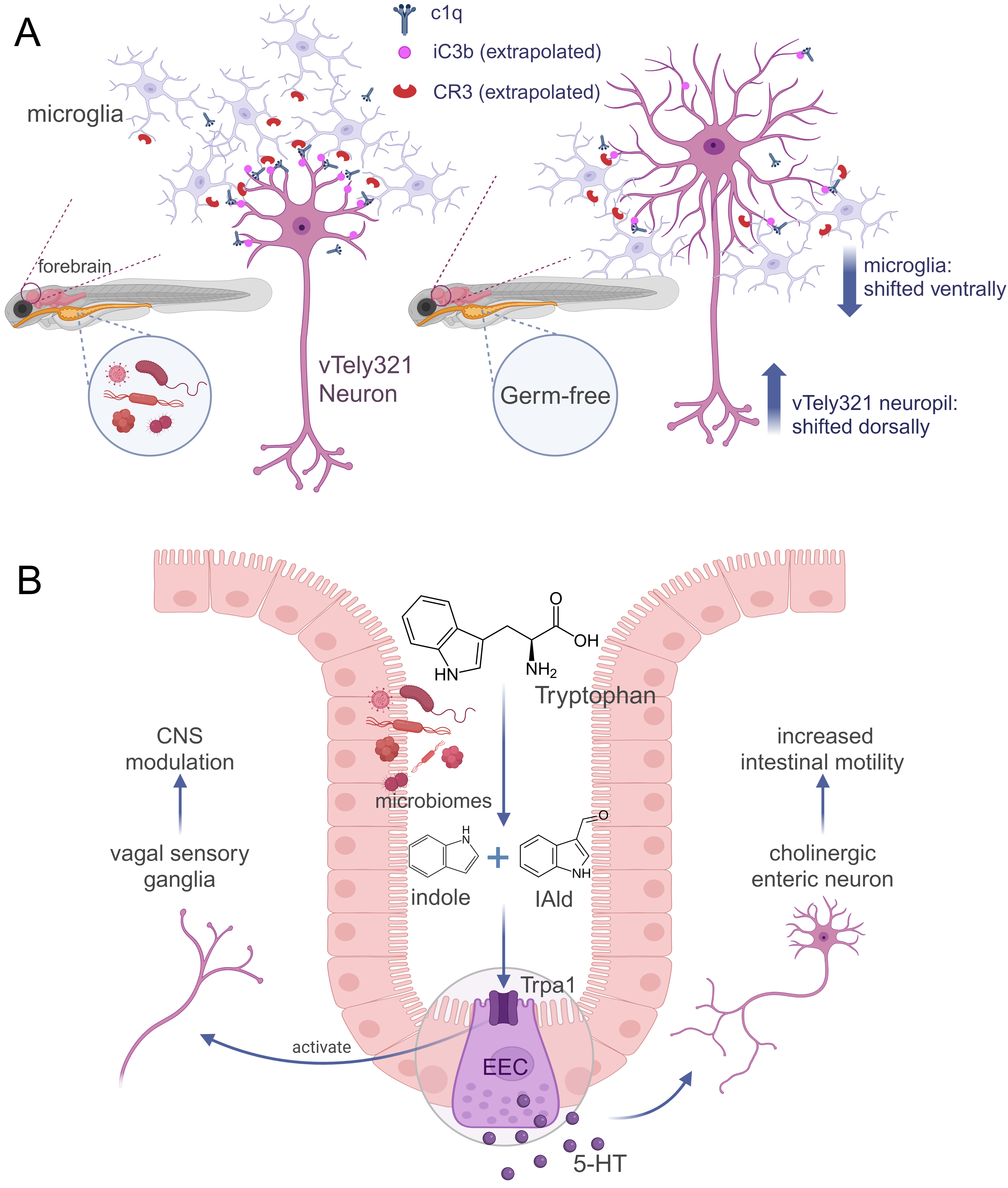
Figure 4. Microbiota regulates neural circuits. (A) Microbiota directs microglial remodeling of the vTely321 circuit through complement-associated mechanisms. In colonized larvae, forebrain microglia are more abundant and correctly positioned, and the microbiota promotes expression of complement pathway genes, including c1q. In zebrafish, c1q is required for normal vTely321 neurite density, and the complement pathway is implicated in microglial neurite remodeling. Downstream of c1q, the molecular recognition of iC3b-opsonized synapses by microglial CR3 has been demonstrated in mice but has not yet been directly validated in zebrafish and is therefore presented as an extrapolation. In GF larvae, microglia are reduced and shifted ventrally, the vTely321 neuropil is shifted dorsally, and arbors are exuberant (greater total length and branching depth with higher distal complexity); microglial c1q is decreased; (B) Microbial tryptophan catabolites engage the intestinal EEC-Trpa1 pathway to couple gut signaling to the enteric nervous system (ENS) and vagal pathways. Luminal tryptophan, from diet or microbial synthesis, is converted by gut bacteria into indole and indole-3-carboxaldehyde (IAld). Indole and IAld activate Trpa1 on intestinal enteroendocrine cells, triggering 5-HT release. The 5-HT signal drives the local enteric circuit, including cholinergic enteric neurons, and promotes intestinal motility. In parallel, bacterial tryptophan catabolites activate vagal sensory ganglia via EEC-Trpa1 signaling, linking epithelial chemosensation to a gut-brain afferent pathway. C1q: Complement component 1q; C3b: complement component 3 fragment b; CNS: central nervous system; CR3: complement receptor 3; iC3b: inactivated C3b; vTely321: y321-labeled ventral telencephalic neuron population; EEC: enteroendocrine cell; ENS: enteric nervous system; IAld: indole-3-carboxaldehyde; 5-HT: serotonin; Trpa1: transient receptor potential ankyrin 1. Created in BioRender. Li, Y. (2026) https://BioRender.com/8aylsv4.
In addition, microbes or their metabolites modify the expression of genes in GABAergic, serotonergic, dopaminergic, and neurotrophin-related pathways, often accompanied by behavioral changes. For example, expression of gad1, which encodes glutamic acid decarboxylase that converts glutamate to GABA, was increased in the adult brain after two weeks of exposure to Lactobacillus delbrueckii. These fish showed reduced anxiety-like behavior in the novel tank test, evidenced by increased time spent in and quicker entry into the upper section of the tank[90]. In zebrafish larvae, cell-free supernatants derived from Lactiplantibacillus plantarum APC2688 enhanced GABAergic signaling, as evidenced by upregulation of gad1 and gabra1 (GABA_A receptor)[91]. In another example, expression of brain-derived neurotrophic factor (bdnf), which supports neuronal growth and plasticity, was increased in the adult brain following feeding with Lactobacillus rhamnosus IMC501 for 28 days. Serotonergic gene expression was upregulated alongside altered shoaling behavior in this model[80]. Additionally, probiotic treatment has been reported to increase expression of genes regulating neurotransmitter systems, such as gad1b, tph1a, htr3a, th, and slc6a3, and improve behaviors[77]. However, further studies are needed to elucidate the underlying mechanisms.
EXTERNAL FACTORS MODULATE THE ZEBRAFISH GUT MICROBIOTA
Dietary effects on the gut microbiome composition
Distinct gut taxa select specific nutrients via substrate availability and host signaling, and certain taxonomic shifts feed back to modulate intestinal immunity through well-characterized microbial metabolites[92-94]. However, the persistence of these effects depends on several key factors, including the exposure window (differentiating larval from adult stages), the duration of intervention (typically weeks rather than months), and the biochemical nature of the dietary factor - whether it is a bulk nutrient, a metabolite, or a nutraceutical. In the following sections, we synthesize the available evidence to outline the most plausible mechanisms underpinning these changes.
Macronutrient availability shapes gut community composition in a manner modulated by host ontogeny. In larval zebrafish, a 25-day high-fat diet led to increased Bacteroidetes and upregulated inflammatory genes in the gut epithelium[20]. Microbiome composition shifts substantially across zebrafish development, underscoring the importance of life stage in mediating dietary effects and suggesting that outcomes observed at one age may not generalize to others without repeated interventions[25]. Extended dietary exposure can also elicit sustained community shifts in adult hosts: a gluten-based formulation administered over twelve weeks drove expansion of several Gram-negative bacteria, a decrease in Actinobacteria, and a contraction of Shewanella. This sustained, diet-induced shift reflects a chronic diet-mediated effect in mature hosts[95]. Consistent with these patterns, a controlled macronutrient-limitation study in adult zebrafish showed that reducing dietary protein or lipid altered body metrics and reconfigured gut-microbiome composition, alongside predicted functional shifts - protein restriction elevated steroid-hormone biosynthesis, while fat reduction increased secondary bile-acid and ketone-body metabolism and lowered steroid-hormone biosynthesis in females[96]. Taken together, these findings show that macronutrient composition, developmental stage, and exposure duration jointly shape the zebrafish gut microbiome and its metabolic output.
Micronutrients can also modulate host-microbe interactions in zebrafish. A four-week vitamin D3 intervention study in zebrafish reveals a direct circuit from a dietary micronutrient to a specific commensal bacterium and its metabolite, ultimately enhancing epithelial defense. Adequate vitamin D promotes the abundance of Cetobacterium (Fusobacteriota), reduces baseline infection susceptibility, and upregulates IL-22 and β-defensin expression. Antibiotic treatment abolishes this IL-22/antimicrobial peptide (AMP) response, confirming microbiota dependence. Acetate, a major metabolic end product of Cetobacterium somerae[97], increases intestinal il22 and β-defensin gene expression when administered intraperitoneally as sodium acetate to vitamin D-deficient cyp2r1-/- zebrafish. Sodium acetate also increases intestinal zfbd1 and zfbd2 expression in wild-type fish, whereas neither gene shows a significant increase in il22-/- fish[98]. Mono-association with Cetobacterium somerae following microbiota depletion elevates intestinal il22 expression and raises serum acetate levels, demonstrating that this commensal is sufficient to induce these effects. Furthermore, both vitamin D3 and 1,25(OH)2D3 directly stimulate C. somerae growth and acetate production in vitro, revealing a mechanism for vitamin D-mediated niche selection[98].
Dietary fiber and bioactive substances influence host metabolism and physiological functions through their ability to reshape the gut microbial community. In a low-fat overfeeding paradigm, supplementation with 5% agavins (a fructan dietary fiber) tempered hyperphagia and adiposity while producing modest, regimen-dependent shifts in gut taxa (e.g., Aeromonas, Paraburkholderia, Fusobacteriaceae), suggesting that specific fibers may link microbial remodeling to metabolic benefit[99]. Similarly, soybean hull polysaccharides improved lipid metabolic control in high-fat diet-fed zebrafish and alleviated gut dysbiosis, with enrichment of Cetobacterium and improved plasma lipid profiles, underscoring fiber-driven links between community restructuring and host metabolism[100]. As sources of biologically active compounds, microalgae serve as modifiable dietary interventions capable of inducing microbial and immune changes within weeks. In adult zebrafish fed a 15% microalgae diet (including Schizochytrium, Spirulina, Chlorella, Chromochloris, and Dunaliella) for four weeks, reduced intestinal pro-inflammatory cytokine levels (IL-1β, IL-6, IL-8) were reported alongside concurrent microbiota alterations - including increased Cetobacterium abundance with the Spirulina diet, elevated Stenotrophomonas with the Schizochytrium diet, and reduced Mycoplasma with most microalgae diets[101]. In zebrafish larvae, supplementation with specific microalgae ameliorated soybean diet-induced intestinal inflammation, as indicated by reduced neutrophil recruitment, and improved survival following bacterial infection[102]. Beyond microalgae, a bioactive extract from the seagrass Enhalus acoroides restored gut microbial balance and improved metabolic control in adult diabetic zebrafish. The improved metabolic control was evidenced by reduced body weight and fasting glucose, alongside altered expression of the metabolic regulators glucagon-like peptide-1 (GLP-1), peroxisome proliferator-activated receptor gamma (PPARγ), and sterol regulatory element-binding protein 1c (SREBP-1c), which are involved in glucose homeostasis, insulin sensitivity, and lipid metabolism. These findings provide parallel evidence that bioactives can reshape both the microbiota and host metabolism[103]. Exogenous organic acids, another class of bioactive compounds, can remodel the gut microbial community over multi-week periods, though effects vary across studies. In one-month-old zebrafish, dietary supplementation with 0.15% sodium acetate for four weeks reduced the relative abundance of Plesiomonas and significantly altered microbiome composition in multivariate analysis; this genus-level shift was attributed to the acetate intervention rather than stochastic background variation[104]. In a separate study, 0.15% succinate supplementation for four weeks improved growth and glucose tolerance but increased hepatic lipid accumulation, elevated the relative abundance of Proteobacteria, decreased that of Fusobacteria and Cetobacterium, and altered protein succinylation patterns in both intestine and liver[105]. Collectively, these data support the conclusion that dietary fibers and bioactive interventions remodel the zebrafish gut microbiota within weeks, with associated reductions in intestinal inflammation and improvements in metabolic control in both larval and adult zebrafish. However, the precise microbial molecular mechanisms underlying these host effects in zebrafish remain to be fully elucidated.
Impact of environmental pollutants on zebrafish gut microbiota
As an aquatic organism, zebrafish live in direct contact with their surrounding water, which often contains diverse contaminants capable of perturbing the intestinal milieu. This attribute has established zebrafish as a prominent model for assessing how environmental pollutants disrupt host-microbiota interactions[106]. Numerous studies have demonstrated that pollutants such as antibiotics, pesticides, heavy metals, micro- and nanoplastics, and endocrine-disrupting chemicals (EDCs) can induce intestinal dysbiosis in zebrafish, with potential consequences for metabolic, immune, and neurodevelopmental pathways[21,22,107].
Antibiotic residues, even at environmentally relevant concentrations, have been repeatedly shown to disrupt gut homeostasis and microbial communities in zebrafish. A six-week exposure to oxytetracycline (OTC) or sulfamethoxazole (SMX) significantly decreased goblet cell numbers, inhibited digestive and antioxidant enzyme activities, and was accompanied by intestinal inflammation[21]. Florfenicol, another widely used veterinary antibiotic, was linked to gut dysbiosis and depletion of SCFA-producing bacteria, and to impaired glucose homeostasis, possibly mediated through altered SCFA signaling[22].
Exposure to pesticides such as methomyl has been shown to compromise intestinal integrity, suppress nutrient and drug metabolism pathways, and increase inflammation and apoptosis. These changes in host physiology coincided with microbial community shifts, including reduced abundance of Shewanella and Pseudomonas, suggesting possible links between microbial disruption and host metabolic dysfunction[108]. Sublethal triflumezopyrim exposure (6.77 ppm; 21 days) altered adult gut communities, increasing Proteobacteria and Tenericutes and broadly shifting predicted functional capacities[109].
Emerging contaminants including microplastics and nanoplastics have drawn increasing concern owing to their environmental persistence and potential synergistic toxicity with co-occurring pollutants. Chronic co-exposure to triclosan (TCS) and polystyrene nanoplastics (PS-NPs) from embryogenesis to adulthood led to pronounced neurodevelopmental impairment and severe gut microbiota dysbiosis, with histological alterations including villus atrophy and intestinal wall thinning; 16S rRNA gene sequencing further indicated diminished microbial diversity and increased abundance of potentially pro-inflammatory taxa[107]. Chronic exposure to 50-nm carboxylated PS-NPs (0.1-10 mg/L for 14-28 days) increased Proteobacteria and decreased Firmicutes, depleted several beneficial genera, and weakened microbial co-occurrence networks in adult zebrafish[110]. Exposure to bisphenol A bis(diphenyl phosphate) for 21 days (10-100 μg/L) induced gut dysbiosis at the phylum and genus levels, accompanied by intestinal injury, impaired tight-junction and mucus integrity, and concurrent oxidative and pro-inflammatory responses in adult zebrafish[111]. Similarly, graphene oxide (GO) exposure induced sex-specific alterations in gut microbial diversity and community structure, accompanied by upregulated expression of pro-inflammatory genes such as tnf-α and il-1β[112].
Exposure to heavy metals such as antimony (Sb) has been shown to disrupt the gut microbiota by increasing the relative abundance of Fusobacteria and Actinobacteria while decreasing Proteobacteria and Firmicutes, with potential consequences for mucosal immune function and host metabolism. These community shifts were accompanied by redox imbalance and signs of potential neurotoxicity[113]. Moreover, chronic exposure to effluent from textile wastewater treatment plants led to growth retardation, oxidative damage, and decreased microbial diversity, with a reduction in probiotic genera (e.g., Lactobacillus, Akkermansia) and a concurrent increase in opportunistic pathogens (e.g., Mycoplasma, Vibrio), highlighting the potential ecological risks posed by industrial discharges[114].
Taken together, these studies indicate that environmental pollutants consistently disturb gut microbial composition, typically accompanied by host physiological or metabolic dysfunction. Although certain taxa - such as Cetobacterium, Lactobacillus, and SCFA-producing bacteria - have emerged as sensitive biomarkers of pollution, the mechanistic links between these microbial shifts and host phenotypes remain poorly understood and require further investigation. The consistent induction of inflammation, oxidative stress, and metabolic dysregulation across diverse pollutant classes suggests that gut dysbiosis may represent a central mechanism of chemical toxicity in zebrafish. Integrating GF models, microbiota transplantation, and multi-omics analyses will be essential to unraveling these mechanisms and evaluating whether microbiome-directed strategies could mitigate the adverse effects of environmental pollutants. These pollutant-induced inflammatory responses can be assessed not only by neutrophil infiltration and cytokine expression but also by upstream NF-κB and MAPK signaling, as demonstrated in toxicological studies of arsenic-exposed common carp[115]. Integrating these signaling readouts with microbial profiling would provide a more comprehensive view of host-microbe-toxicant interactions.
TRANSLATIONAL INSIGHTS
Gut microbial dysbiosis is closely associated with a variety of diseases. This section organizes studies of the zebrafish gut microbiota that elucidate the molecular mechanisms underlying related human diseases and discuss candidate interventions.
IBD-related epithelial and immune regulation
Zebrafish studies address epithelial maintenance and immune regulation relevant to inflammatory bowel disease (IBD). A conserved role for the microbiota in driving intestinal epithelial maturation is evident across vertebrates. In zebrafish, the intestinal microbiota promotes intestinal development by enhancing Wnt pathway activity and elevating epithelial cell proliferation rates[46]. Single-cell profiling further identifies microbiota-dependent transcriptional responses in intestinal and associated cell populations[60]. Mammalian evidence points in the same direction: in mice, Wnt proteins are essential for conferring basal competency to intestinal stem cells, thereby enabling their self-renewal[116], and microbial colonization supports Paneth cell differentiation through macrophage and mesenchymal niches during early postnatal development[57]. These findings identify processes relevant to epithelial maintenance, although their cellular organization differs between species.
Microbial sensing can support homeostasis or promote inflammation, depending on the cellular and regulatory context. In mice, Toll-like receptor (TLR)-MyD88 signaling supports epithelial homeostasis and protection from injury[117]. However, in interleukin-10-deficient mice, MyD88 signaling in colonic mononuclear phagocytes promotes inflammatory cytokine production and T-helper 17 responses[118]. Zebrafish provide complementary evidence for host mediators of microbial immune regulation. In gnotobiotic zebrafish, colonization strongly induces serum amyloid A (Saa) in digestive tissues[17]. Genetic studies show that Saa modulates neutrophil behavior in vivo, shaping their baseline activation and recruitment responses. These findings support investigating microbial regulation of inflammatory responses, without assuming that the same pathways have equivalent effects in every tissue or disease setting.
Zebrafish also retain interleukin-26 (IL-26), which is absent in rodents. Loss of il26 caused epithelial hyperproliferation and DNA damage in larvae; these abnormalities were absent under GF conditions and normalized after co-housing with CV wild-type fish[32]. This model links a cytokine associated with human inflammatory disease to microbiota-dependent epithelial homeostasis, providing a developmental window into the mechanisms relevant to chronic IBD.
Zebrafish provide a platform for evaluating microbial strains and their products as candidate interventions. For example, Lactobacillus-based interventions modified intestinal morphology, goblet-cell metrics, intraepithelial leukocyte proportions, and barrier-related gene expression, with effects depending on the strain and preparation[119]. Similarly, Bifidobacterium animalis subsp. lactis BL-99 improved intestinal histopathology and increased goblet cell numbers in diet-challenged adult zebrafish[120]. Combined SCFA administration before 2,4,6-trinitrobenzenesulfonic acid (TNBS) exposure improved larval survival and reduced intestinal neutrophil recruitment, without preventing epithelial damage or restoring goblet cell numbers[121]. A study combining human HT-29 cells and dextran sulfate sodium (DSS)-exposed zebrafish reported anti-inflammatory and barrier-related effects of indoleacrylic acid, together with reduced anxiety-like behavior in fish[122]. The Aeromonas-secreted protein AimA suppresses intestinal neutrophil accumulation and protects against septic shock in both bacterial and chemical models of inflammation[123]. AimA has lipocalin-like structural features, and mammalian lipocalin-2 has been evaluated as a fecal biomarker of intestinal inflammation in human IBD[123,124]. These zebrafish studies identify candidates for further testing of barrier and immune modulation, although their efficacy against IBD requires further validation.
Metabolic disorders
During early larval stages, the intestinal microbiota promotes pancreatic β-cell expansion, an effect attributable to specific bacteria that secrete BefA[75]. Subsequent work linked BefA activity to membrane permeabilization and demonstrated β-cell expansion in mice[125]. This cross-species evidence supports investigating microbial proteins that regulate β-cell mass, pointing to diabetes-relevant mechanisms whose efficacy remains to be evaluated in disease models and human islets.
In a high-cholesterol diet-induced hyperlipidemia model, Lactobacillus fermentum E15 reduced lipid accumulation in the liver and blood vessels and altered lipid metabolism-related gene expression[126]. Isovaleric acid supplementation also reduced lipid accumulation, and the effects of both interventions were sensitive to pertussis toxin. Together with receptor-expression measurements, these experiments suggested involvement of G protein-coupled receptor 43 (GPR43), but did not establish receptor-specific dependence or a requirement for bacterial SCFA synthesis. The study identifies L. fermentum E15 and isovaleric acid as candidates for further evaluation in hyperlipidemia.
Microbial proteins can also be evaluated for effects on hepatic lipid accumulation. Using a zebrafish model of high-fat diet-induced hepatic lipid accumulation, Ding et al. showed that the Akkermansia muciniphila outer membrane protein Amuc_1100 reduced hepatic lipid deposition[127]. Its protective effect was not detectable in GF larvae, whereas recipients of microbiota from Amuc_1100-fed donors showed less hepatic lipid accumulation than those receiving microbiota from high-fat-fed control donors. Protein-interaction assays and gene knockdown further implicated intestinal 14-3-3β/α-A in this response[127]. These experiments connect a defined microbial protein, microbiota-dependent effects, and a host mediator, providing a basis for investigating host-microbiota mechanisms relevant to fatty liver disease.
Neurodevelopmental disorders
Zebrafish studies allow microbial effects on neural development to be examined alongside behavioral outcomes. Early microbial exposure promotes microglial remodeling of forebrain neurons associated with later social behavior[76]. This provides a mechanistic basis for investigating how microbial exposure during sensitive developmental windows influences neural circuits.
Jia et al. used katnal2-deficient zebrafish, an autism spectrum disorder (ASD)-related genetic model, to assess microbial effects on behavioral phenotypes[128]. GF-derived mutant larvae receiving wild-type donor microbiota exhibited greater locomotor activity than CV mutants, although rearing conditions differed between groups. A. muciniphila supplementation partially improved locomotor activity in GF-derived katnal2 mutant larvae and altered serotonergic gene expression[128]. The neural mechanisms and genotype specificity of these responses remain to be established. Nevertheless, the study illustrates how a disease-related genetic model can be combined with microbial manipulation to evaluate candidate interventions for neurodevelopment-related phenotypes.
Infectious diseases
Microbiota-dependent host defense provides another setting for mechanistic and intervention studies. Cross-species functional genomics identified Elf3 as a conserved microbiota-responsive epithelial regulator. In zebrafish, elf3 is required for appropriate microbiota-dependent immune gene induction and protects against inflammation and infection of the swim bladder[129]. In addition to host regulators, specific microbial products can promote host defense. For example, a Cetobacterium somerae exopolysaccharide was shown to engage TLR2 and promote a type I interferon-dependent antiviral response against spring viremia of carp virus[130]. These studies identify host regulators and microbial products that can be tested for their contributions to infection resistance.
Single-strain interventions also provide candidate protective strategies. For example, in zebrafish infected with Aeromonas hydrophila, pre-supplementation with Lactobacillus plantarum SHY21-2 reduced mortality. This protection was accompanied by preserved intestinal barrier integrity and reduced inflammatory and oxidative stress responses[131]. Extending beyond barrier support, a translationally attractive strategy is anti-virulence intervention. The quorum-quenching bacterium Bacillus licheniformis T-1 protected zebrafish from Aeromonas hydrophila infection, supporting investigation of bacterial communication as an intervention target[132].
Finally, an infection ecology study demonstrated that Escherichia coli utilizes glucose in the zebrafish gut, producing acidic byproducts that lower intestinal pH, thereby inhibiting colonization of Vibrio cholerae under glucose-supplemented conditions[133]. This finding supports testing interventions that alter the local chemical environment to restrict pathogen colonization. Translation of these infection studies requires validation in relevant mammalian or human-derived systems, accounting for pathogen identity, microbial strain, and nutrient availability.
ZEBRAFISH ADVANTAGES FOR GUT MICROBIOME RESEARCH
In recent decades, zebrafish have emerged as a reliable and tractable vertebrate model for microbiome research, offering complementary approaches to addressing gaps in our understanding of the mechanistic links between the gut microbiota and human diseases. Several features make zebrafish well suited to these studies, as summarized in Figure 5.
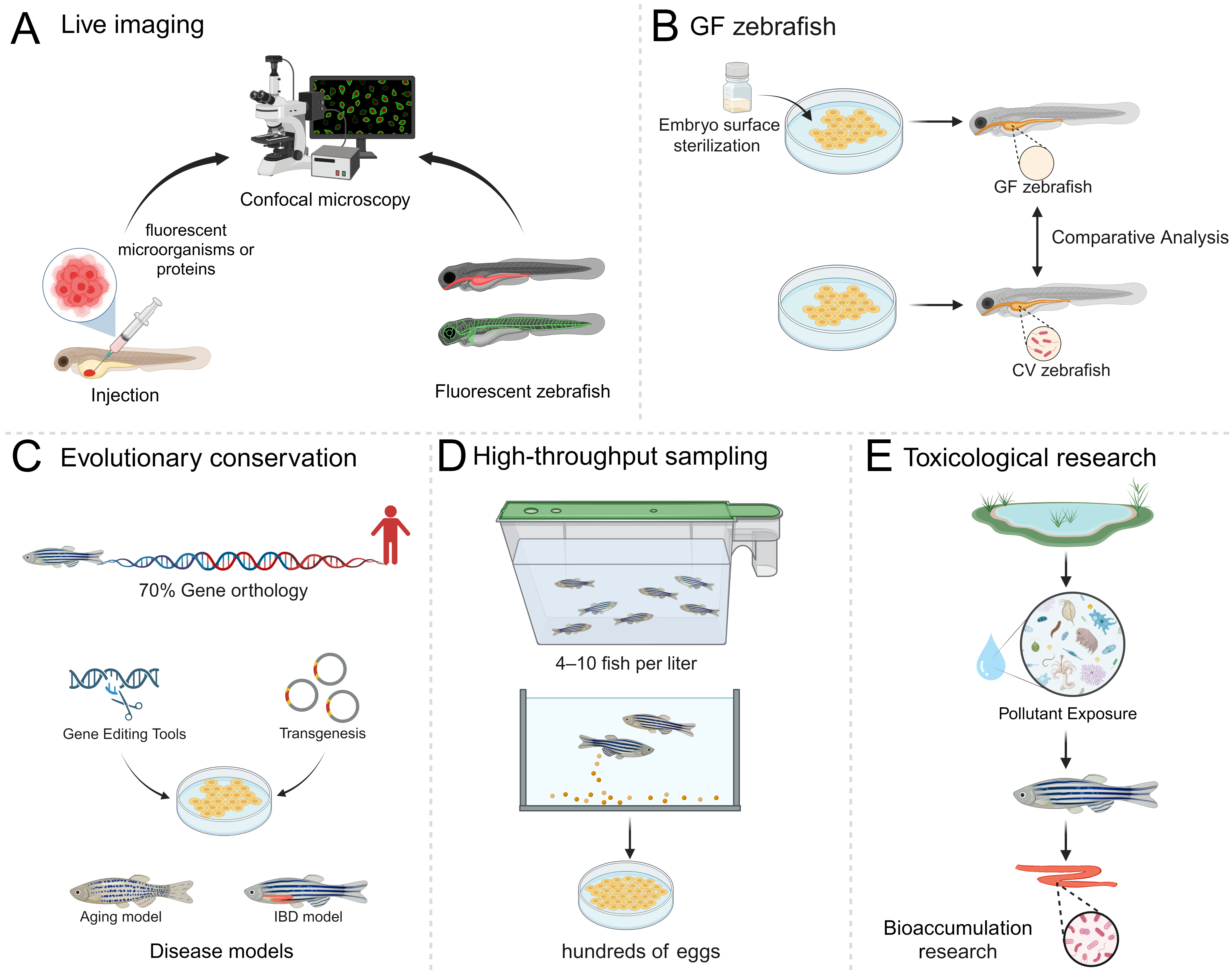
Figure 5. Advantages of zebrafish in gut microbiota research. (A) Fluorescence-based live imaging of microbial spatial organization and host cellular responses in zebrafish larvae; (B) GF and CV zebrafish for studying microbial contributions to intestinal development, metabolism, and neurobehavior; (C) Genetic conservation and genetic manipulation for disease modeling and translational research; (D) Zebrafish enable high-throughput gut microbiome research. Adult zebrafish are commonly maintained at 4-10 fish/L, and a single female can spawn hundreds of eggs per mating[134], supporting large-scale sampling; (E) Aquatic exposure models for studying toxicological mechanisms and pollutant effects on gut microbial communities and bioaccumulation. GF: Germ-free; CV: conventionally raised. Created in BioRender. Li, Y. (2026) https://BioRender.com/tqduw9z.
Particularly at the larval stage, zebrafish offer several distinctive advantages for studying gut microbiota. First, their optical transparency and external development permit live imaging of microbial dynamics with fluorescent labeling [Figure 5A]. For example, a live-imaging platform uses differential fluorescent labeling to distinguish phage generations, enabling in situ tracking of phage replication and inter-bacterial transmission at single-virion resolution[135]. Additionally, the availability of cell-specific fluorescent transgenic lines enables real-time imaging of host cellular responses to microbes[135,136]. For instance, studies have visualized how gut microbes influence host epithelial nutrient handling, including stimulation of fatty acid uptake and lipid droplet formation in both the intestine and extra-intestinal tissues[66]. These approaches allow microbial dynamics and host responses to be examined in intact larvae, with optical access that is more difficult to achieve in mice[137].
Second, GF larvae can be readily generated via surface sterilization of the chorion, allowing precise control of microbial exposure[45,138]. This enables functional interrogation of microbial effects by comparing GF and CV zebrafish [Figure 5B], or by introducing defined microbial stimuli into GF animals. For example, live imaging of fluorescent cargoes in GF and microbially colonized larvae showed that the microbiome slows soluble cargo uptake and lysosomal protein degradation in lysosome-rich enterocytes (LREs). These protein-absorption findings provide a basis for investigating microbial contributions to human protein malnutrition[52]. Single-cell analysis further uncovered a microbiome-dependent “Cloaca 3” population detected only in CV samples, enriched for bacterial sensing, immune signaling, and iron-transport programs[52]. These capabilities provide insights into host-microbiota interactions that are difficult to achieve with comparable throughput in mammalian models[137].
In addition to the larvae stage, zebrafish (both larvae and adults) offer several additional advantages. Third, zebrafish possess orthologues of approximately 70% of human genes and exhibit similar transcriptional regulation, making them suitable for investigating host genetic influences on the gut microbiota[139]. Modern genetic tools further enable targeted manipulation of host genes to study their impact on microbial composition and function [Figure 5C]. A specific advantage is the presence of IL-26, a cytokine associated with human intestinal inflammation that is absent in mice. In zebrafish larvae, il26 loss increased epithelial proliferation and DNA damage in the posterior gut. These increases were absent under GF conditions and were rescued by co-housing with CV wild-type larvae. Disruption of the receptor subunit il20ra did not reproduce these phenotypes, supporting a microbiota-dependent function independent of canonical IL-26 receptor signaling[32]. Zebrafish therefore enable investigation of an endogenous immune-microbiota interaction that conventional mouse models cannot directly reproduce. Because zebrafish serve as a better model than mice for studying telomere aging[140], one study used telomerase (tert)-deficient zebrafish together with gut-specific genetic rescue to show that restoring intestinal telomerase expression reversed gut microbiota dysbiosis and ameliorated systemic aging phenotypes[29].
Fourth, zebrafish are easy to breed in large numbers, facilitating high-throughput experimentation with extensive biological replication [Figure 5D]. This allows researchers to explore inter-individual variation and achieve remarkable statistical power in microbiota composition analyses. For instance, a study performed large-scale single-cell RNA sequencing on 160 zebrafish intestines, enabling statistically robust dissection of microbiome-host interactions at cellular resolution[60]. Another study analyzed gut microbiomes from more than 550 individual zebrafish spanning larval to adult stages, and achieved sufficient power to construct robust molecular ecological networks and to distinguish non-random microbial interactions from stochastic noise[42]. These studies illustrate how large-scale zebrafish sampling strengthens the statistical inference in gut microbiota research, whereas comparably large sample sizes are difficult to achieve in mouse models.
Fifth, as aquatic organisms, zebrafish are especially valuable for studying freshwater environmental microbiology and pollutant bioaccumulation[141] [Figure 5E], shedding light on toxicological risks to aquatic ecosystems and environmental health[43]. For example, Wray et al. combined waterborne azoxystrobin exposure with controlled microbial inoculation in zebrafish larvae[142]. Exposure induced transient hyperactivity after a light-to-dark transition in SIHUMIx-inoculated larvae but not in microbiota-depleted larvae[142]. This approach highlights the utility of zebrafish in aquatic environmental toxicology by enabling experimental assessment of microbial influences on host responses to waterborne pollutants.
Together, these features provide spatiotemporal resolution and experimental accessibility that complement rodent models, positioning zebrafish as a powerful system for gut microbiome research with translational potential in medicine and environmental ecology[143]. The key advantages of zebrafish for gut microbiome research, together with representative experiments and applicable research questions, are summarized in Table 1.
Advantages of zebrafish in gut microbiota research
| Advantage | Representative experiments | Applicable research questions |
| Live imaging | Fluorescently labeled microbes and host cells[58]; single-virion tracking | Microbial dynamics; host-microbe interactions[135] |
| GF zebrafish | Embryo surface sterilization and axenic rearing[138]; GF vs. CV comparison | Microbiota dependence of host responses[45] |
| Evolutionary conservation and genetic tractability | Human-zebrafish gene orthology[139]; gene editing and transgenesis | Host genetic influences on microbiota; microbiota-dependent epithelial homeostasis; functions of disease-associated host genes[29,32] |
| High-throughput sampling | Large-scale sampling; 16S rRNA gene sequencing | Microbial composition and inter-individual variation; developmental succession; inferred ecological networks[42] |
| Toxicological research | Chemical exposure; bioaccumulation studies[141] | Effects of environmental pollutants[141]; microbial contributions to neurobehavioral responses[142] |
LIMITATIONS IN ZEBRAFISH MICROBIOTA STUDIES
Although zebrafish provide a powerful platform for mechanistic microbiota research, their utility is tempered by specific challenges. (1) Over the past decade, microbiome-related research has advanced rapidly, with continual updates in sequencing technologies and computational methods. However, there remains substantial heterogeneity across studies in both the methods adopted and the way these methods are reported. This variability makes microbiome research difficult to reproduce, compare across projects, integrate, and share; these challenges are particularly pronounced in non-human host-associated microbiome research, including zebrafish[144]. Currently, there is no unified, zebrafish-specific technical platform for gut microbiome research. As a result, most studies rely on general-purpose tools designed for human or mouse microbiomes, which are not always applicable to zebrafish. This limitation has constrained the broader application of integrated multi-omics analyses in the field. (2) Compared with mammalian systems, zebrafish gut communities often exhibit lower taxonomic richness and reduced interaction complexity, which weakens claims that rely on higher-order ecological networks, rare-taxon effects, or human-specific niche specialists[16,145]. As with research using other model organisms, zebrafish microbiota studies can provide insights into evolutionarily conserved mechanisms; however, a considerable gap remains between basic discoveries and translational applications. (3) Although zebrafish exhibit some similarities to humans in intestinal function and cell composition[60,146,147], there are notable differences in overall gut morphology and physiology compared to mammals. For example, the zebrafish intestine lacks mammalian-type crypts and Paneth cells[147], limiting direct extrapolation of Paneth cell-mediated antimicrobial responses, such as angiogenin-4 secretion[148]. These distinctions modify the luminal physicochemical conditions, transit dynamics, and microbial niches, and therefore should be carefully considered and evaluated when interpreting results[145,149]. Candidate microbial factors identified in zebrafish therefore require functional validation in appropriate mammalian or human-derived systems, with attention to developmental stage, exposure, and disease context. (4) Similar to other model organisms, zebrafish are typically housed and maintained under strictly controlled experimental conditions that may not fully replicate the complexity of natural ecosystems, posing challenges for extrapolating findings to real-world ecological systems[43]. Therefore, claims based on the zebrafish model should be appropriately qualified, and extrapolation of gut microbiota findings to mammals requires consideration of these boundaries and limitations. (5) Variation in husbandry across laboratories presents an additional challenge to cross-study comparability. Comparative studies have identified facility-associated differences in zebrafish gut microbial communities and diet-dependent differences in gut community composition[150]. At the tank level, a comparison of wild-type and rag1-deficient zebrafish found that tank identity explained more variation in community composition than genotype[31]. Rearing-system design, feed, and water conditions therefore need to be considered when comparing findings across facilities. Sample processing can compound this variation: in larvae, sample collection and DNA extraction methods affected which taxa were associated with benzo[a]pyrene exposure, although its effect on overall community composition remained detectable across methods[151]. These findings warrant caution when interpreting inconsistent taxon-level results across studies. Reporting husbandry conditions and processing protocols, together with accounting for tank and processing-batch effects in study design and analysis, would improve comparability. (6) Viral and fungal contributions to the zebrafish gut ecosystem remain incompletely characterized. Viral metagenomics identified gut virome changes following polystyrene nanoplastic exposure, but correlations with host biochemical measures did not establish a causal role for these changes[152]. In larvae colonized with a defined two-member Escherichia coli community, antibiotic-induced phage lytic replication was accompanied by bacterial spatial reorganization[135]. Fungal internal transcribed spacer 2 (ITS2) profiling revealed differences in intestinal fungal composition between wild-caught and laboratory-reared zebrafish, while early exposure to fish-derived yeasts altered larval gut bacterial composition[153,154]. However, whether the bacterial changes following phage induction or yeast exposure affect host physiology was not tested. Experiments using defined bacterial communities with and without selected phages or yeasts could test their effects on bacterial colonization, intestinal barrier function, and inflammatory responses.
Conclusion and future directions
The zebrafish model provides a powerful system for investigating the association between microbial community shifts and physiological and pathological changes, as well as for elucidating the underlying mechanisms. Current evidence supports several conserved intestinal programs as recurring nodes of host-microbe interaction, including bile acid-dependent FXR signaling that shapes enterohepatic transport and hepatic transcriptional responses, MyD88-linked regulation of epithelial renewal and lineage allocation through Wnt and Notch programs, and epithelial NF-κB signaling that calibrates inflammatory set points and barrier function. Because intestinal perturbations propagate through endocrine and neural routes, microbiota-linked changes can scale to systemic phenotypes in lipid and glucose homeostasis and neurobehavioral outputs. These responses are further modulated by diet, probiotics, and environmental exposures, and depend on ligand concentration, microbial identity, host genetics, and developmental stage.
Despite rapid progress, a primary challenge is the incomplete elucidation of molecular mechanisms. For example, acute SCFA exposure produces bradycardia and selective vascular effects in larvae, yet the responsible host receptors and downstream signaling were not identified[155]. Vitamin D-related enrichment of Cetobacterium and acetate is associated with microbiota-dependent IL-22 and β-defensin programs, but the host mechanisms sensing acetate were not resolved[98]. Cetobacterium or acetate can also improve glucose homeostasis through atropine-sensitive parasympathetic activation, again without identifying the upstream acetate-sensing mechanism[70]. A specific priority is to test candidate host sensing pathways through genetic or pharmacological perturbation, using acetate-induced IL-22 expression or glucose responses as functional readouts. In parallel, future work should emphasize elucidating the intrinsic functional properties of gut microbes, including strain-specific traits, metabolic pathways, enzymatic activities, and secreted bioactive molecules that drive these host-relevant phenotypes. Finally, although mechanistic investigations have concentrated on the gut-brain and gut-liver axes, research connecting intestinal microbes to other tissues - such as skeletal muscle or the cardiovascular system - remains comparatively scarce, creating significant gaps in our understanding of systems-level physiology[156,157].
It is worth noting that, in host-microbe interaction research, moving from correlation to causation remains a central challenge. Across studies of metabolic phenotypes, dietary interventions, or pollutant exposure, shifts in microbial community composition - such as changes in the relative abundance of taxa - can be confounded by factors including dietary intake, growth and development, direct toxicant effects, and baseline environmental variation. As a result, compositional changes alone are insufficient to establish causality. The most compelling evidence must meet multiple criteria: it should directly link defined microbial factors (e.g., specific metabolites or functional modules) to conserved host cellular programs (e.g., neuroendocrine, metabolic, immune, or barrier pathways); these relationships should then be tested using GF animals, microbial transplantation, targeted depletion, or reconstitution designs. For a candidate microbial metabolite, disrupting its production and testing whether supplementation restores the host response would strengthen evidence for its causal contribution.
Taken together, current research on the zebrafish gut microbiota still leaves significant gaps in understanding the mechanisms and signaling pathways underlying host-microbe interactions. Despite these limitations, the zebrafish model demonstrates considerable value. The evolutionary conservation of key microbial associations and interaction mechanisms supports the utility of zebrafish as a model organism for investigating host-microbe dynamics. This model can provide insights into the mechanisms of human diseases linked to genetic disorders, metabolic imbalances, and environmental perturbations. In parallel, the distinct features of the zebrafish microbiome offer a unique opportunity to understand how lower vertebrates, particularly fish, have adapted to aquatic environments. These findings provide transferable biological insights relevant to aquaculture, environmental monitoring, and public health. Thus, with continued mechanistic progress, zebrafish will play an increasingly pivotal role in microbiome research across ecological, biomedical, and translational contexts.
DECLARATIONS
Acknowledgment
The graphical abstract was created with BioRender by Y. Li (2026): https://BioRender.com/ra7znbm
Authors’ contributions
Contributed to the design and drafted the main manuscript: Li Y (Yuchen Li)
Drew the figures: Li Y(Yuchen Li), Liu R
Assisted in writing: Liu R, Wang W
Provided professional guidance and funding support: Li Y (Yong Li)
Conceived the study, drafted the main manuscript, and provided professional guidance and funding support: Xia Z
Availability of data and materials
Not applicable.
AI and AI-assisted tools statement
During the revision of this manuscript, the AI tool Claude (Anthropic) (version Opus 4.8, released 2026-05-28) was used solely for language editing. The tool did not influence the study design, data collection, analysis, interpretation, or the scientific content of the work. All authors take full responsibility for the accuracy, integrity, and final content of the manuscript.
Financial support and sponsorship
This work was supported by grants from the National Natural Science Foundation of China (32371222 to Xia Z, 42477300 and 42330711 to Li Y).
Conflicts of interest
All authors declared that there are no conflicts of interest.
Ethics approval and consent to participate
Not applicable.
Consent for publication
Not applicable.
Copyright
© The Author(s) 2026.
REFERENCES
1. Muramatsu MK, Winter SE. Nutrient acquisition strategies by gut microbes. Cell Host Microbe. 2024;32:863-74.
2. O'Riordan KJ, Moloney GM, Keane L, Clarke G, Cryan JF. The gut microbiota-immune-brain axis: therapeutic implications. Cell Rep Med. 2025;6:101982.
3. Won TH, Arifuzzaman M, Parkhurst CN, et al.; JRI Live Cell Bank. Host metabolism balances microbial regulation of bile acid signalling. Nature. 2025;638:216-24.
4. Barrack KE, O'Toole GA. Cystic fibrosis and inflammatory bowel disease: parallels in gut physiology and microbiota. J Bacteriol. 2025;207:e0016725.
5. Farah A, Paul P, Khan AS, Sarkar A, Laws S, Chaari A. Targeting gut microbiota dysbiosis in inflammatory bowel disease: a systematic review of current evidence. Front Med (Lausanne). 2025;12:1435030.
6. Hou K, Wu ZX, Chen XY, et al. Microbiota in health and diseases. Signal Transduct Target Ther. 2022;7:135.
7. Ruiz-Malagón AJ, Rodríguez-Sojo MJ, Redondo E, Rodríguez-Cabezas ME, Gálvez J, Rodríguez-Nogales A. Systematic review: the gut microbiota as a link between colorectal cancer and obesity. Obes Rev. 2025;26:e13872.
8. Yu Y, Ding Y, Wang S, Jiang L. Gut microbiota dysbiosis and its impact on type 2 diabetes: from pathogenesis to therapeutic strategies. Metabolites. 2025;15:397.
9. Mayer EA, Tillisch K, Gupta A. Gut/brain axis and the microbiota. J Clin Invest. 2015;125:926-38.
10. Meerschaert KA, Chiu IM. The gut-brain axis and pain signalling mechanisms in the gastrointestinal tract. Nat Rev Gastroenterol Hepatol. 2025;22:206-21.
11. Tofani GSS, Leigh SJ, Gheorghe CE, et al. Gut microbiota regulates stress responsivity via the circadian system. Cell Metab. 2025;37:138-153.e5.
12. Burns AR, Miller E, Agarwal M, et al. Interhost dispersal alters microbiome assembly and can overwhelm host innate immunity in an experimental zebrafish model. Proc Natl Acad Sci U S A. 2017;114:11181-6.
13. Xiao F, Zhu W, Yu Y, et al. Host development overwhelms environmental dispersal in governing the ecological succession of zebrafish gut microbiota. NPJ Biofilms Microbiomes. 2021;7:5.
14. Evens KC, Bakke I, Bohannan BJM. Aquaculture facility-specific microbiota shape the zebrafish gut microbiome. Anim Microbiome. 2026;8.
15. Wu G, Xu T, Zhao N, et al. A core microbiome signature as an indicator of health. Cell. 2024;187:6550-6565.e11.
16. Roeselers G, Mittge EK, Stephens WZ, et al. Evidence for a core gut microbiota in the zebrafish. ISME J. 2011;5:1595-608.
17. Kanther M, Sun X, Mühlbauer M, et al. Microbial colonization induces dynamic temporal and spatial patterns of NF-κB activation in the zebrafish digestive tract. Gastroenterology. 2011;141:197-207.
18. Wen J, Mercado GP, Volland A, et al. FXR signaling and microbial metabolism of bile salts in the zebrafish intestine. Sci Adv. 2021;7:eabg1371.
19. Ye L, Bae M, Cassilly CD, et al. Enteroendocrine cells sense bacterial tryptophan catabolites to activate enteric and vagal neuronal pathways. Cell Host Microbe. 2021;29:179-196.e9.
20. Arias-Jayo N, Abecia L, Alonso-Sáez L, Ramirez-Garcia A, Rodriguez A, Pardo MA. High-fat diet consumption induces microbiota dysbiosis and intestinal inflammation in zebrafish. Microb Ecol. 2018;76:1089-101.
21. Zhou L, Limbu SM, Shen M, et al. Environmental concentrations of antibiotics impair zebrafish gut health. Environ Pollut. 2018;235:245-54.
22. Zhao F, Gong Z, Yang Y, et al. Effects of environmentally relevant concentrations of florfenicol on the glucose metabolism system, intestinal microbiome, and liver metabolome of zebrafish. Sci Total Environ. 2024;938:173417.
23. Bates JM, Mittge E, Kuhlman J, Baden KN, Cheesman SE, Guillemin K. Distinct signals from the microbiota promote different aspects of zebrafish gut differentiation. Dev Biol. 2006;297:374-86.
24. Murdoch CC, Rawls JF. Commensal microbiota regulate vertebrate innate immunity-insights from the zebrafish. Front Immunol. 2019;10:2100.
25. Stephens WZ, Burns AR, Stagaman K, et al. The composition of the zebrafish intestinal microbial community varies across development. ISME J. 2016;10:644-54.
26. Xia H, Chen H, Cheng X, et al. Zebrafish: an efficient vertebrate model for understanding role of gut microbiota. Mol Med. 2022;28:161.
27. Garibay-Valdez E, Olivas-Bernal CA, Vargas-Albores F, et al. Deciphering the gut microbiota of zebrafish, the most used fish as a biological model: a meta-analytic approach. Comp Biochem Physiol A Mol Integr Physiol. 2024;297:111713.
28. Xiao F, Zhu W, Liu H, et al. Effects of aging on gut microbiota nearly throughout zebrafish lifespan. Curr Zool. 2026;72:303-6.
29. El Maï M, Bird M, Allouche A, et al. Gut-specific telomerase expression counteracts systemic aging in telomerase-deficient zebrafish. Nat Aging. 2023;3:567-84.
30. Brugman S, Schneeberger K, Witte M, et al. T lymphocytes control microbial composition by regulating the abundance of Vibrio in the zebrafish gut. Gut Microbes. 2014;5:737-47.
31. Stagaman K, Burns AR, Guillemin K, Bohannan BJ. The role of adaptive immunity as an ecological filter on the gut microbiota in zebrafish. ISME J. 2017;11:1630-9.
32. Salloum Y, Gros G, Quintero-Castillo K, et al. IL-26 from innate lymphoid cells regulates early-life gut epithelial homeostasis by shaping microbiota composition. EMBO J. 2025;44:6832-56.
33. Rabahi S, Maurin L, Marachlian E, et al. IL-22 from enteroendocrine cells promotes early-life gut motility in zebrafish through the microbiota. Science. 2026;392:69-76.
34. Rawls JF, Mahowald MA, Ley RE, Gordon JI. Reciprocal gut microbiota transplants from zebrafish and mice to germ-free recipients reveal host habitat selection. Cell. 2006;127:423-33.
35. Valenzuela MJ, Caruffo M, Herrera Y, et al. Evaluating the capacity of human gut microorganisms to colonize the zebrafish larvae (Danio rerio). Front Microbiol. 2018;9:1032.
36. Manuneedhi Cholan P, Morris S, Luo K, et al. Transplantation of high fat fed mouse microbiota into zebrafish larvae identifies MyD88-dependent acceleration of hyperlipidaemia by Gram-positive cell wall components. Biofactors. 2022;48:329-41.
37. Morris S, Wright K, Malyla V, et al. Exposure to the gut microbiota from cigarette smoke-exposed mice exacerbates cigarette smoke extract-induced inflammation in zebrafish larvae. Curr Res Immunol. 2021;2:229-36.
38. Wiles TJ, Jemielita M, Baker RP, et al. Host gut motility promotes competitive exclusion within a model intestinal microbiota. PLoS Biol. 2016;14:e1002517.
39. Schlomann BH, Wiles TJ, Wall ES, Guillemin K, Parthasarathy R. Bacterial cohesion predicts spatial distribution in the larval zebrafish intestine. Biophys J. 2018;115:2271-7.
40. Cantas L, Sørby JR, Aleström P, Sørum H. Culturable gut microbiota diversity in zebrafish. Zebrafish. 2012;9:26-37.
41. Adade EE, Stevick RJ, Pérez-Pascual D, Ghigo JM, Valm AM. Gnotobiotic zebrafish microbiota display inter-individual variability affecting host physiology. bioRxiv. 2023:2023.02.01.526612.
42. Xiao F, Zhu W, Yu Y, et al. Interactions and stability of gut microbiota in zebrafish increase with host development. Microbiol Spectr. 2022;10:e0169621.
43. Medriano CA, Kim S, Kim LH, Bae S. Chronic exposure of adult zebrafish to polyethylene and polyester-based microplastics: metabolomic and gut microbiome alterations reflecting dysbiosis and resilience. J Hazard Mater. 2025;484:136691.
44. Sharpton TJ, Stagaman K, Sieler MJ Jr, Arnold HK, Davis EW 2nd. Phylogenetic integration reveals the zebrafish core microbiome and its sensitivity to environmental exposures. Toxics. 2021;9:10.
45. Rawls JF, Samuel BS, Gordon JI. Gnotobiotic zebrafish reveal evolutionarily conserved responses to the gut microbiota. Proc Natl Acad Sci U S A. 2004;101:4596-601.
46. Cheesman SE, Neal JT, Mittge E, Seredick BM, Guillemin K. Epithelial cell proliferation in the developing zebrafish intestine is regulated by the Wnt pathway and microbial signaling via Myd88. Proc Natl Acad Sci U S A. 2011;108 Suppl 1:4570-7.
47. Bates JM, Akerlund J, Mittge E, Guillemin K. Intestinal alkaline phosphatase detoxifies lipopolysaccharide and prevents inflammation in zebrafish in response to the gut microbiota. Cell Host Microbe. 2007;2:371-82.
48. Andersen-Civil AIS, Mantel M, Soh J, Mostowy S, Brugman S, Vanwalleghem GC. Novel and unscrutinized immune entities of the zebrafish gut. FEBS Lett. 2025;599:3444-55.
49. Massaquoi MS, Kong GL, Chilin-Fuentes D, et al. Cell-type-specific responses to the microbiota across all tissues of the larval zebrafish. Cell Rep. 2023;42:112095.
50. Jones LO, Willms RJ, Xu X, et al. Single-cell resolution of the adult zebrafish intestine under conventional conditions and in response to an acute Vibrio cholerae infection. Cell Rep. 2023;42:113407.
51. Kurogi K, Yoshihama M, Horton A, et al. Identification and characterization of 5α-cyprinol-sulfating cytosolic sulfotransferases (Sults) in the zebrafish (Danio rerio). J Steroid Biochem Mol Biol. 2017;174:120-7.
52. Childers L, Park J, Wang S, et al. Protein absorption in the zebrafish gut is regulated by interactions between lysosome rich enterocytes and the microbiome. Elife. 2025;13:RP100611.
53. Maselli KM, Gee K, Isani M, Fode A, Schall KA, Grikscheit TC. Broad-spectrum antibiotics alter the microbiome, increase intestinal fxr, and decrease hepatic steatosis in zebrafish short bowel syndrome. Am J Physiol Gastrointest Liver Physiol. 2020;319:G212-26.
54. Gao C, Wang H, Xue X, Qi L, Lin Y, Wang L. The potential role of intestinal microbiota on the intestine-protective and lipid-lowering effects of berberine in zebrafish (Danio rerio) under high-lipid stress. Metabolites. 2025;15:118.
55. Troll JV, Hamilton MK, Abel ML, et al. Microbiota promote secretory cell determination in the intestinal epithelium by modulating host Notch signaling. Development. 2018;145.
56. Morales RA, Rabahi S, Diaz OE, et al. Interleukin-10 regulates goblet cell numbers through Notch signaling in the developing zebrafish intestine. Mucosal Immunol. 2022;15:940-51.
57. Kim JE, Li B, Fei L, et al. Gut microbiota promotes stem cell differentiation through macrophage and mesenchymal niches in early postnatal development. Immunity. 2022;55:2300-2317.e6.
58. Murdoch CC, Espenschied ST, Matty MA, Mueller O, Tobin DM, Rawls JF. Intestinal serum amyloid A suppresses systemic neutrophil activation and bactericidal activity in response to microbiota colonization. PLoS Pathog. 2019;15:e1007381.
59. Yang Y, Millán JL, Mecsas J, Guillemin K. Intestinal alkaline phosphatase deficiency leads to lipopolysaccharide desensitization and faster weight gain. Infect Immun. 2015;83:247-58.
60. Willms RJ, Jones LO, Hocking JC, Foley E. A cell atlas of microbe-responsive processes in the zebrafish intestine. Cell Rep. 2022;38:110311.
61. Flores MV, Crawford KC, Pullin LM, Hall CJ, Crosier KE, Crosier PS. Dual oxidase in the intestinal epithelium of zebrafish larvae has anti-bacterial properties. Biochem Biophys Res Commun. 2010;400:164-8.
62. Breen P, Winters AD, Nag D, Ahmad MM, Theis KR, Withey JH. Internal versus external pressures: effect of housing systems on the zebrafish microbiome. Zebrafish. 2019;16:388-400.
63. Wu S, Pan M, Zan Z, et al. Regulation of lipid metabolism by gut microbiota in aquatic animals. Rev Aquacult. 2024;16:34-46.
64. Sheng Y, Ren H, Limbu SM, et al. The presence or absence of intestinal microbiota affects lipid deposition and related genes expression in zebrafish (Danio rerio). Front Microbiol. 2018;9:1124.
65. Quinlivan VH, Farber SA. Lipid uptake, metabolism, and transport in the larval zebrafish. Front Endocrinol (Lausanne). 2017;8:319.
66. Semova I, Carten JD, Stombaugh J, et al. Microbiota regulate intestinal absorption and metabolism of fatty acids in the zebrafish. Cell Host Microbe. 2012;12:277-88.
67. Falcinelli S, Picchietti S, Rodiles A, et al. Lactobacillus rhamnosus lowers zebrafish lipid content by changing gut microbiota and host transcription of genes involved in lipid metabolism. Sci Rep. 2015;5:9336.
68. Falcinelli S, Rodiles A, Hatef A, et al. Dietary lipid content reorganizes gut microbiota and probiotic L. rhamnosus attenuates obesity and enhances catabolic hormonal milieu in zebrafish. Sci Rep. 2017;7:5512.
69. Meng D, Zhang Z, Teame T, et al. Host-driven hepatic conversion of gut microbiota-derived putrescine to spermidine mediates mannose’s protective effects against hepatic steatosis in zebrafish. Imeta. 2026;5:e70101.
70. Wang A, Zhang Z, Ding Q, et al. Intestinal Cetobacterium and acetate modify glucose homeostasis via parasympathetic activation in zebrafish. Gut Microbes. 2021;13:1-15.
71. Perry RJ, Peng L, Barry NA, et al. Acetate mediates a microbiome-brain-β-cell axis to promote metabolic syndrome. Nature. 2016;534:213-7.
72. Brown AJ, Goldsworthy SM, Barnes AA, et al. The orphan G protein-coupled receptors GPR41 and GPR43 are activated by propionate and other short chain carboxylic acids. J Biol Chem. 2003;278:11312-9.
73. Priyadarshini M, Villa SR, Fuller M, et al. An acetate-specific GPCR, FFAR2, regulates insulin secretion. Mol Endocrinol. 2015;29:1055-66.
74. Xu K, Kumar P, Chang Stauffer SR, et al. Pancreatic β-cell FFA2 deficiency suppresses multiple low dose streptozotocin induced diabetes in male mice. Commun Biol. 2025;8:1389.
75. Hill JH, Franzosa EA, Huttenhower C, Guillemin K. A conserved bacterial protein induces pancreatic beta cell expansion during zebrafish development. Elife. 2016;5:e20145.
76. Bruckner JJ, Stednitz SJ, Grice MZ, et al. The microbiota promotes social behavior by modulating microglial remodeling of forebrain neurons. PLoS Biol. 2022;20:e3001838.
77. Bu LK, Jia PP, Huo WB, Pei DS. Assessment of probiotics’ impact on neurodevelopmental and behavioral responses in zebrafish models: implications for autism spectrum disorder therapy. Probiotics Antimicrob Proteins. 2025;17:3944-59.
78. James DM, Davidson EA, Yanes J, Moshiree B, Dallman JE. The gut-brain-microbiome axis and its link to autism: emerging insights and the potential of zebrafish models. Front Cell Dev Biol. 2021;9:662916.
79. Nagpal J, Cryan JF. Microbiota-brain interactions: moving toward mechanisms in model organisms. Neuron. 2021;109:3930-53.
80. Borrelli L, Aceto S, Agnisola C, et al. Probiotic modulation of the microbiota-gut-brain axis and behaviour in zebrafish. Sci Rep. 2016;6:30046.
81. Phelps D, Brinkman NE, Keely SP, et al. Microbial colonization is required for normal neurobehavioral development in zebrafish. Sci Rep. 2017;7:11244.
82. Ayayee PA, Wong RY. Zebrafish (Danio rerio) behavioral phenotypes are not underscored by different gut microbiomes. Ecol Evol. 2024;14:e70237.
83. Valcarce DG, Martínez-Vázquez JM, Riesco MF, Robles V. Probiotics reduce anxiety-related behavior in zebrafish. Heliyon. 2020;6:e03973.
84. Zhang C, Zhang X, Qiu H, et al. Fermented Gastrodia elata Bl. Intervenes gut microbiota and amino acid metabolism in zebrafish to promote 5-HT homeostasis against sleep disturbances. Food Res Int. 2025;217:116757.
85. Schafer DP, Lehrman EK, Kautzman AG, et al. Microglia sculpt postnatal neural circuits in an activity and complement-dependent manner. Neuron. 2012;74:691-705.
86. Stevens B, Allen NJ, Vazquez LE, et al. The classical complement cascade mediates CNS synapse elimination. Cell. 2007;131:1164-78.
87. Lynch CMK, Knox EG, Soong D, et al. The microbiota shapes central nervous system myelination in early life. Adv Sci (Weinh). 2026;13:e15671.
88. Bellampalli SS, Joshi V, Fonar G, Beyder A. Integration of signal transduction pathways in sensory epithelial enteroendocrine cells. Mol Biol Cell. 2025;36:re3.
89. Grundeken E, El Aidy S. Enteroendocrine cells: the gatekeepers of microbiome-gut-brain communication. NPJ Biofilms Microbiomes. 2025;11:179.
90. Olorocisimo JP, Diaz LA, Co DE, Carag HM, Ibana JA, Velarde MC. Lactobacillus delbrueckii reduces anxiety-like behavior in zebrafish through a gut microbiome - brain crosstalk. Neuropharmacology. 2023;225:109401.
91. Valderrama B, Daly I, Gunnigle E, et al. From in silico screening to in vivo validation in zebrafish - a framework for reeling in the right psychobiotics. Food Funct. 2025;16:2018-30.
92. Mann ER, Lam YK, Uhlig HH. Short-chain fatty acids: linking diet, the microbiome and immunity. Nat Rev Immunol. 2024;24:577-95.
93. Pickard JM, Maurice CF, Kinnebrew MA, et al. Rapid fucosylation of intestinal epithelium sustains host-commensal symbiosis in sickness. Nature. 2014;514:638-41.
94. Shao Y, Garcia-Mauriño C, Clare S, et al. Primary succession of bifidobacteria drives pathogen resistance in neonatal microbiota assembly. Nat Microbiol. 2024;9:2570-82.
95. Koo H, Hakim JA, Powell ML, et al. Metagenomics approach to the study of the gut microbiome structure and function in zebrafish Danio rerio fed with gluten formulated diet. J Microbiol Methods. 2017;135:69-76.
96. Green GBH, Williams MB, Chehade SB, et al. Body metrics and the gut microbiome in response to macronutrient limitation in the zebrafish Danio rerio. Curr Dev Nutr. 2023;7:100065.
97. Finegold SM, Vaisanen ML, Molitoris DR, et al. Cetobacterium somerae sp. nov. from human feces and emended description of the genus Cetobacterium. Syst Appl Microbiol. 2003;26:177-81.
98. Liao X, Lan Y, Wang W, et al. Vitamin D influences gut microbiota and acetate production in zebrafish (Danio rerio) to promote intestinal immunity against invading pathogens. Gut Microbes. 2023;15:2187575.
99. Navarro-Barrón E, Hernández-González C, Llera-Herrera R, García-Gasca A, López-Pérez M, Gómez-Gil B. Overfeeding, agavins, and dietary fat: factors that modulate the zebrafish gut microbiota. Zebrafish. 2025;22:141-51.
100. Chen C, Song H, Yang L, et al. Soybean hull polysaccharides ameliorate lipid metabolic disorders and gut microbiota dysbiosis in high-fat diet-fed zebrafish. Int J Biol Macromol. 2025;305:141123.
101. Ma K, Chen S, Wu Y, et al. Dietary supplementation with microalgae enhances the zebrafish growth performance by modulating immune status and gut microbiota. Appl Microbiol Biotechnol. 2022;106:773-88.
102. Bravo-Tello K, Ehrenfeld N, Solís CJ, et al. Effect of microalgae on intestinal inflammation triggered by soybean meal and bacterial infection in zebrafish. PLoS One. 2017;12:e0187696.
103. Kadharusman MM, Syahputra RA, Kurniawan R, et al. Seagrass Enhalus acoroides extract mitigates obesity and diabetes via GLP-1, PPARγ, SREBP-1c modulation and gut microbiome restoration in diabetic zebrafish. Diabetol Metab Syndr. 2025;17:235.
104. Zhang H, Ding Q, Wang A, et al. Effects of dietary sodium acetate on food intake, weight gain, intestinal digestive enzyme activities, energy metabolism and gut microbiota in cultured fish: Zebrafish as a model. Aquaculture. 2020;523:735188.
105. Ding Q, Lu C, Hao Q, et al. Dietary succinate impacts the nutritional metabolism, protein succinylation and gut microbiota of zebrafish. Front Nutr. 2022;9:894278.
106. Li P, Zhang J, Liu X, et al. The function and the affecting factors of the zebrafish gut microbiota. Front Microbiol. 2022;13:903471.
107. Wang D, Lan J, Meng W, et al. Co-exposure to triclosan and polystyrene nanoplastics on neurodevelopmental toxicity and gut microbiota dysbiosis in zebrafish (Danio rerio). Environ Pollut. 2025;381:126643.
108. Li M, Chen X, Song C, et al. Sub-chronically exposing zebrafish to environmental levels of methomyl induces dysbiosis and dysfunction of the gut microbiota. Environ Res. 2024;261:119674.
109. Iswari Das I, Sharma BM, Mahapatra M, et al. High-throughput sequencing of 16S rRNA gene analysis reveal insights into abundance, diversity, and function of the gut microbiota in zebrafish exposed to triflumezopyrim. Aquacult Rep 2025;40:102651.
110. Rehman A, Habumugisha T, Huang F, et al. Impacts of polystyrene nanoplastics on zebrafish gut microbiota and mechanistic insights. Ecotoxicol Environ Saf. 2025;299:118332.
111. Lyu Y, Wang X, Wang Y, Yang R, Zhong W, Zhu L. Bisphenol A bis(diphenyl phosphate) disrupts gut microbiota homeostasis and induces intestinal toxicity in zebrafish. Environ Pollut. 2025;386:127221.
112. Jia P, Sun T, Junaid M, et al. Chronic exposure to graphene oxide (GO) induced inflammation and differentially disturbed the intestinal microbiota in zebrafish. Environ Sci : Nano. 2019;6:2452-69.
113. Wang C, Yuan Z, Li J, Liu Y, Li R, Li S. Acute effects of antimony exposure on adult zebrafish (Danio rerio): from an oxidative stress and intestinal microbiota perspective. Fish Shellfish Immunol. 2022;123:1-9.
114. Wang C, Yuan Z, Sun Y, Yao X, Li R, Li S. Effect of chronic exposure to textile wastewater treatment plant effluents on growth performance, oxidative stress, and intestinal microbiota in adult zebrafish (Danio rerio). Front Microbiol. 2021;12:782611.
115. Wang Y, Zhao H, Liu Y, Nie X, Xing M. Zinc exerts its renal protection effect on arsenic-exposed common carp: a signaling network comprising Nrf2, NF-κB and MAPK pathways. Fish Shellfish Immunol. 2020;104:383-90.
116. Yan KS, Janda CY, Chang J, et al. Non-equivalence of Wnt and R-spondin ligands during Lgr5+ intestinal stem-cell self-renewal. Nature. 2017;545:238-42.
117. Rakoff-Nahoum S, Paglino J, Eslami-Varzaneh F, Edberg S, Medzhitov R. Recognition of commensal microflora by toll-like receptors is required for intestinal homeostasis. Cell. 2004;118:229-41.
118. Hoshi N, Schenten D, Nish SA, et al. MyD88 signalling in colonic mononuclear phagocytes drives colitis in IL-10-deficient mice. Nat Commun. 2012;3:1120.
119. Rawling M, Schiavone M, Mugnier A, et al. Modulation of zebrafish (Danio rerio) intestinal mucosal barrier function fed different postbiotics and a probiotic from Lactobacilli. Microorganisms. 2023;11:2900.
120. Chen M, Liu C, Dai M, Wang Q, Li C, Hung W. Bifidobacterium lactis BL-99 modulates intestinal inflammation and functions in zebrafish models. PLoS One. 2022;17:e0262942.
121. Morales Fénero C, Amaral MA, Xavier IK, et al. Short chain fatty acids (SCFAs) improves TNBS-induced colitis in zebrafish. Curr Res Immunol. 2021;2:142-54.
122. Amarasiri RPGSK, Hyun J, Lee SW, et al. Therapeutic potential of tryptophan metabolite indoleacrylic acid in inflammatory bowel disease: from cellular mechanisms to zebrafish stress-like behavior. Int Immunopharmacol. 2025;149:114207.
123. Rolig AS, Sweeney EG, Kaye LE, et al. A bacterial immunomodulatory protein with lipocalin-like domains facilitates host-bacteria mutualism in larval zebrafish. Elife. 2018;7:e37172.
124. Zollner A, Schmiderer A, Reider SJ, et al. Faecal biomarkers in inflammatory bowel diseases: calprotectin versus lipocalin-2-a comparative study. J Crohns Colitis. 2021;15:43-54.
125. Hill JH, Massaquoi MS, Sweeney EG, et al. BefA, a microbiota-secreted membrane disrupter, disseminates to the pancreas and increases β cell mass. Cell Metab. 2022;34:1779-1791.e9.
126. Chen Y, Zheng K, Leng Y, et al. Alleviating effect of Lactobacillus fermentum E15 on hyperlipidemia and hepatic lipid metabolism in zebrafish fed by a high-fat diet through the production of short-chain fatty acids. Front Nutr. 2025;12:1522982.
127. Ding Q, Lou R, Liang H, et al. Amuc_1100 alleviates HFD-induced hepatic lipid accumulation via gut microbiota in zebrafish: insights from the role of intestinal 14-3-3β/α-A. J Anim Sci Biotechnol. 2026;17:115.
128. Jia PP, Li Y, Yang HY, et al. Fecal microbiota transplantation and Akkermansia muciniphila restore neurodevelopment and behavior via the gut-brain axis in autism-like zebrafish. ISME J. 2026;20:wrag074.
129. Davis BR, Lickwar CR, Löhr CV, et al. Epithelial transcription factor Elf3 mediates host immune responses to microbiota and protects against aerocystitis in zebrafish. mBio. 2025;16:e0226725.
130. Liang H, Li M, Chen J, et al. The intestinal microbiome and Cetobacterium somerae inhibit viral infection through TLR2-type I IFN signaling axis in zebrafish. Microbiome. 2024;12:244.
131. Jiang Y, Liang M, Yang Y, Xue J, Suo H. Probiotic Lactobacillus plantarum SHY21-2 protected zebrafish against Aeromonas hydrophila infection by maintaining intestinal barrier integrity, inhibiting inflammatory and oxidative stress responses, and regulating intestinal microbiome. Aquaculture. 2024;582:740506.
132. Chen B, Peng M, Tong W, Zhang Q, Song Z. The quorum quenching bacterium Bacillus licheniformis t-1 protects zebrafish against Aeromonas hydrophila infection. Probiotics Antimicrob Proteins. 2020;12:160-71.
133. Nag D, Breen P, Raychaudhuri S, Withey JH. Glucose metabolism by Escherichia coli inhibits vibrio cholerae intestinal colonization of zebrafish. Infect Immun. 2018;86.
134. Aleström P, D’Angelo L, Midtlyng PJ, et al. Zebrafish: housing and husbandry recommendations. Lab Anim. 2020;54:213-24.
135. Ortiz de Ora L, Wiles ET, Zünd M, et al. Phollow reveals in situ phage transmission dynamics in the zebrafish gut microbiome at single-virion resolution. Nat Microbiol. 2025;10:1067-83.
136. Montanari A, Wittamer V. Live imaging and characterization of microglia dynamics in the zebrafish embryo. J Vis Exp. 2024:e66533.
137. Cornuault JK, Byatt G, Paquet ME, De Koninck P, Moineau S. Zebrafish: a big fish in the study of the gut microbiota. Curr Opin Biotechnol. 2022;73:308-13.
138. Melancon E, Canny SGDLT, Sichel S, et al. Best practices for germ-free derivation and gnotobiotic zebrafish husbandry. In: Methods in Cell Biology. Elsevier; 2017. pp. 61-100.
139. Howe K, Clark MD, Torroja CF, et al. The zebrafish reference genome sequence and its relationship to the human genome. Nature. 2013;496:498-503.
140. Martins RR, Besse S, Ellis PS, et al. Telomerase depletion accelerates ageing of the zebrafish brain. Aging Cell. 2025;24:e70280.
141. Sultan M, Cai ZX, Bao L, et al. Trophic transfer induced gut inflammation, dysbiosis, and inflammatory pathways in zebrafish via Artemia franciscana: a differential analysis of nanoplastic toxicity. J Hazard Mater. 2024;480:136030.
142. Wray C, Castañeda-Monsalve V, Engelmann B, et al. Integration of human microbiota (SIHUMIx) and zebrafish models reveals microbiome-mediated host responses to azoxystrobin. Toxicol Sci. 2026;209.
143. Abu-Siniyeh A, Khataibeh M, Al-Zyoud W, Al Holi M. Zebrafish as a model for human epithelial pathology. Lab Anim Res. 2025;41:6.
144. Kelliher JM, Mirzayi C, Bordenstein SR, et al.; STREAMS Consortium. STREAMS guidelines: standards for technical reporting in environmental and host-associated microbiome studies. Nat Microbiol. 2025;10:3059-68.
145. Lu H, Li P, Huang X, Wang CH, Li M, Xu ZZ. Zebrafish model for human gut microbiome-related studies: advantages and limitations. Med Microecol. 2021;8:100042.
146. Park J, Levic DS, Sumigray KD, et al. Lysosome-rich enterocytes mediate protein absorption in the vertebrate gut. Dev Cell. 2019;51:7-20.e6.
147. Wang Z, Du J, Lam SH, Mathavan S, Matsudaira P, Gong Z. Morphological and molecular evidence for functional organization along the rostrocaudal axis of the adult zebrafish intestine. BMC Genomics. 2010;11:392.
148. Hooper LV, Stappenbeck TS, Hong CV, Gordon JI. Angiogenins: a new class of microbicidal proteins involved in innate immunity. Nat Immunol. 2003;4:269-73.
149. Brugman S. The zebrafish as a model to study intestinal inflammation. Dev Comp Immunol. 2016;64:82-92.
150. Sieler MJ Jr, Al-Samarrie CE, Kasschau KD, Varga ZM, Kent ML, Sharpton TJ. Disentangling the link between zebrafish diet, gut microbiome succession, and Mycobacterium chelonae infection. Anim Microbiome. 2023;5:38.
151. Stagaman K, Kasschau KD, Tanguay RL, Sharpton TJ. Experimental methods modestly impact interpretation of the effect of environmental exposures on the larval zebrafish gut microbiome. Sci Rep. 2022;12:14538.
152. Teng M, Li Y, Zhao X, et al. Vitamin D modulation of brain-gut-virome disorder caused by polystyrene nanoplastics exposure in zebrafish (Danio rerio). Microbiome. 2023;11:266.
153. Siriyappagouder P, Galindo-Villegas J, Lokesh J, Mulero V, Fernandes JMO, Kiron V. Exposure to yeast shapes the intestinal bacterial community assembly in zebrafish larvae. Front Microbiol. 2018;9:1868.
154. Siriyappagouder P, Kiron V, Lokesh J, Rajeish M, Kopp M, Fernandes J. The intestinal mycobiota in wild zebrafish comprises mainly dothideomycetes while saccharomycetes predominate in their laboratory-reared counterparts. Front Microbiol. 2018;9:387.
155. Sree Kumar H, Wisner AS, Schiefer IT, Alviter Plata A, Zubcevic J. Chronotropic and vasoactive properties of the gut bacterial short-chain fatty acids in larval zebrafish. Physiol Genomics. 2024;56:426-35.
156. Chen J, Wu S, Wu R, et al. Essential oil from Artemisia argyi alleviated liver disease in zebrafish (Danio rerio) via the gut-liver axis. Fish Shellfish Immunol. 2023;140:108962.
Cite This Article
 Open Access
How to Cite
Li Y, Liu R, Wang W, Li Y, Xia Z. Ecology and mechanisms of the zebrafish gut microbiome. Microbiome Res Rep. 2026;5:29. https://dx.doi.org/10.20517/mrr.2026.73
Download Citation
If you have the appropriate software installed, you can download article citation data to the citation manager of your choice. Simply select your manager software from the list below and click on download.
Export Citation File
Type of Import
Tips on Downloading Citation
Citation Manager File Format
Type of Import
Direct Import: When the Direct Import option is selected (the default state), a dialogue box will give you the option to Save or Open the downloaded citation data. Choosing Open will either launch your citation manager or give you a choice of applications with which to use the metadata. The Save option saves the file locally for later use.
Indirect Import: When the Indirect Import option is selected, the metadata is displayed and may be copied and pasted as needed.
About This Article
Copyright

Data & Comments
Data
0









Comments
Comments must be written in English. Spam, offensive content, impersonation, and private information will not be permitted. If any comment is reported and identified as inappropriate content by OAE staff, the comment will be removed without notice. If you have any queries or need any help, please contact us at [email protected].