

fig2
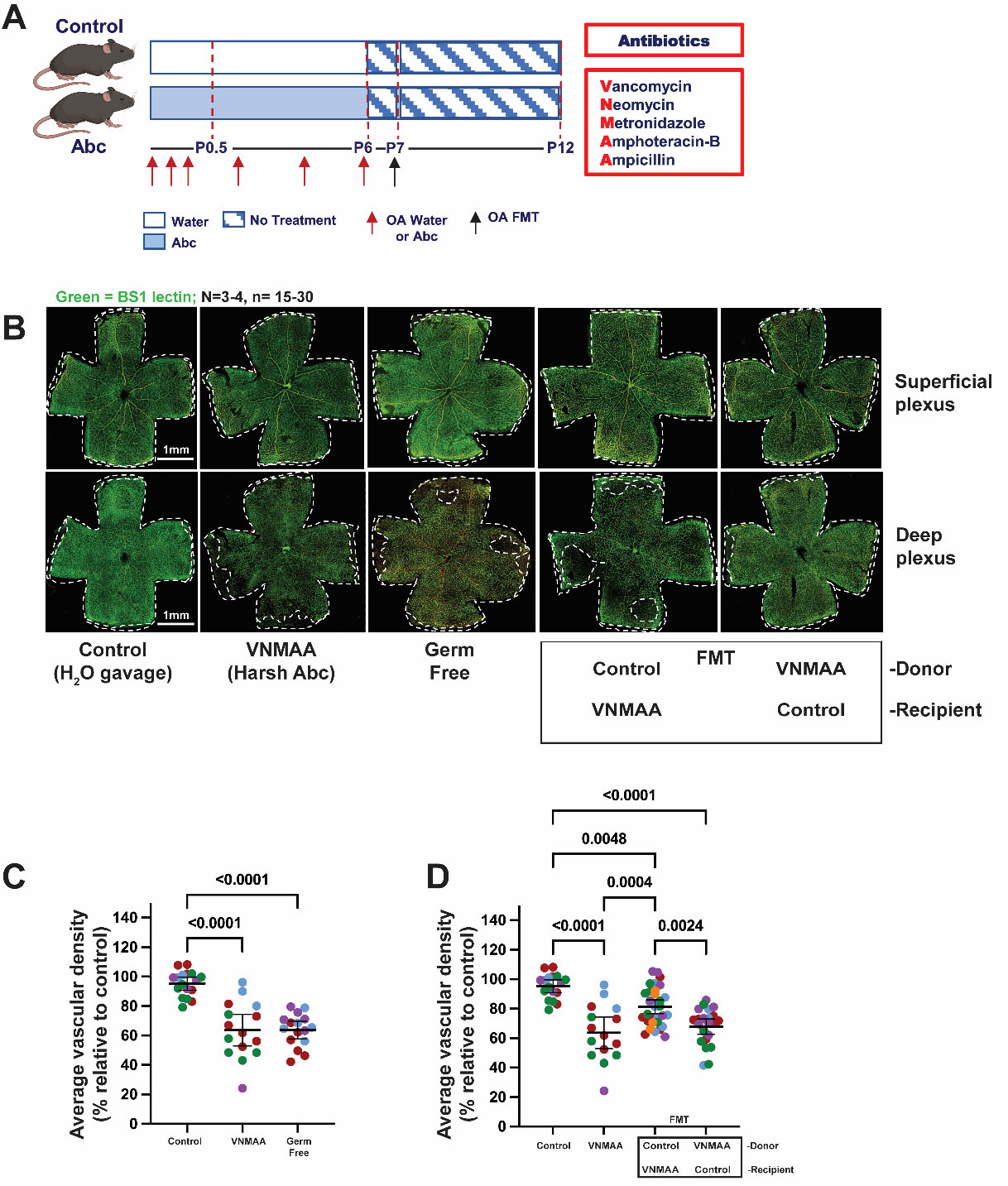
Figure 2. Reciprocal FMT modifies neonatal retinal angiogenesis. (A) Experimental design. Dams were orally gavaged with water or the VNMAA broad-spectrum antibiotic cocktail at the indicated time points during pregnancy, shown by red arrows. Following birth, dams and pups were orally administered water or VNMAA from P0.5 to P6 at the indicated time points, shown by red arrows. VNMAA consisted of vancomycin, neomycin, metronidazole, amphotericin B, and ampicillin. At P7, reciprocal FMT was performed, with VNMAA-treated dams receiving control microbiota and control dams receiving VNMAA-associated microbiota. No further treatments were administered after FMT. Retinas were collected at P12. The black arrow indicates oral FMT. Created in BioRender. Robinson, S. (2026) https://BioRender.com/isf8z0b; (B) Representative BS-1-labelled P12 retinal whole-mount images from control, VNMAA-treated, germ-free, VNMAA-treated (donor) to control (recipient) FMT, and control (donor) to VNMAA (recipient) FMT groups. Retinal vasculature is shown in green. Images show superficial and deep vascular plexus preparations; white outlines indicate the retinal edge. Scale bar, 1.0 mm; (C) Quantification of average deep plexus vascular density at P12, expressed as a percentage relative to control. VNMAA-treated and germ-free mice showed reduced vascular density compared with controls. Each point represents an individual retina and each colour represents an independent biological repeat/litter; black horizontal lines indicate mean ± 95% CI; n = 15-17 retinas from N = 3-4 independent litters per group (litters are colour-coded); (D) Quantification of average deep plexus vascular density following reciprocal FMTs, expressed as a percentage relative to control. Transfer of control microbiota into VNMAA-treated dams partially restored offspring vascular density, whereas transfer of VNMAA-associated microbiota into control dams shifted vascular density towards the VNMAA-associated phenotype. Each point represents an individual retina; black horizontal lines indicate mean ± 95% CI; n = 15-31 retinas from N = 4-5 independent litters per group (litters are colour-coded). Statistical significance is indicated on the graphs. These experiments demonstrate that altering the maternal microbial ecosystem is associated with changes in offspring retinal vascular development but do not establish the specific microbial taxa or products responsible. Statistically significant P values are indicated on the graphs. Comparisons for which no P value is shown were not statistically significant. FMT: Faecal microbiota transplantation; CI: confidence interval.