
fig1
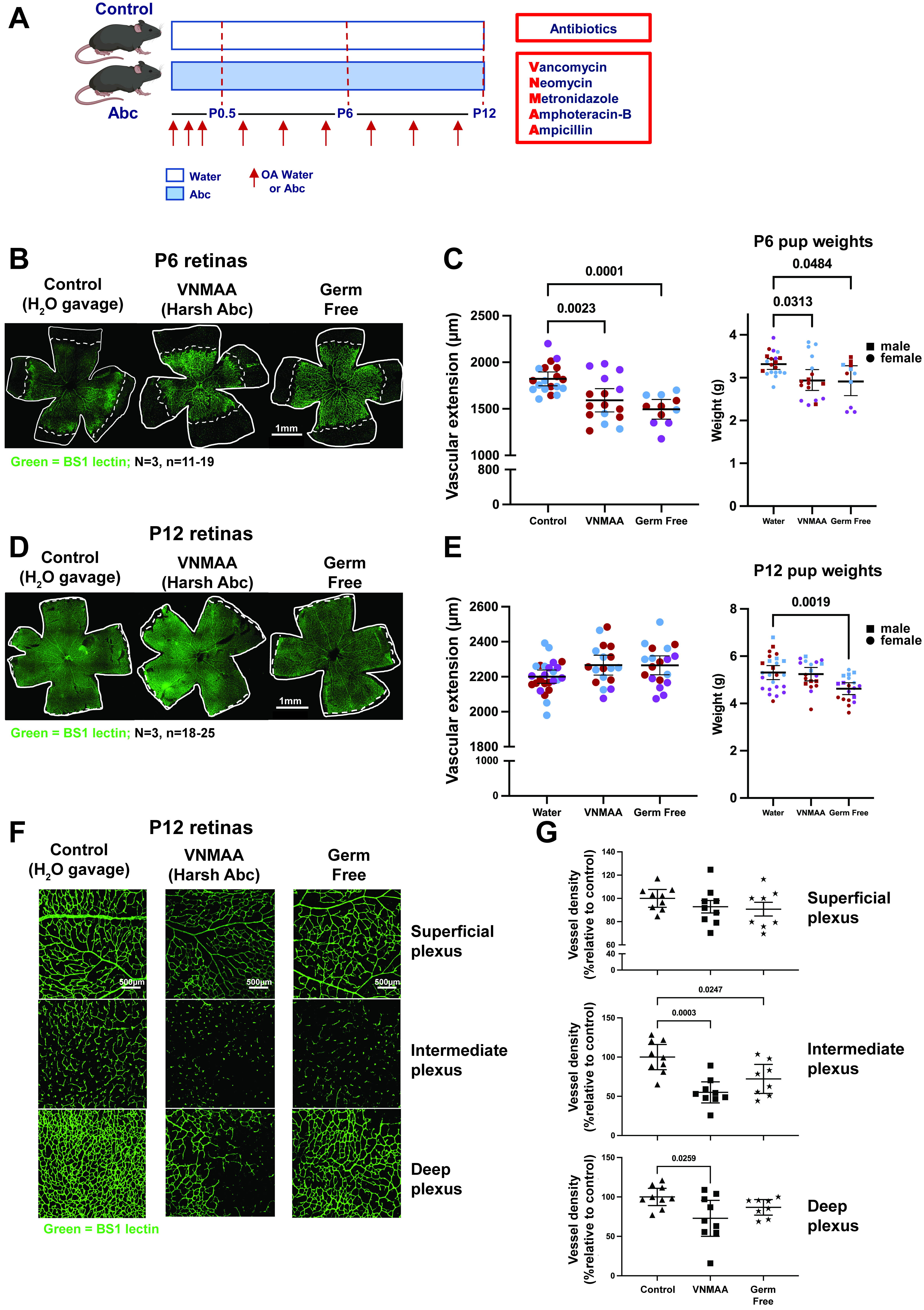
Figure 1. Perinatal microbiome perturbation delays retinal vascular extension and alters retinal vascular plexus development. (A) Experimental design. Mid-gestation dams were orally gavaged with water or the VNMAA broad-spectrum antimicrobial cocktail at the indicated time points during pregnancy, shown by red arrows. Following birth, dams and pups were orally administered water or VNMAA until the indicated experimental endpoint. VNMAA comprised vancomycin, neomycin, metronidazole, amphotericin B and ampicillin. In the schematic, “OA” denotes oral administration and “Abc” denotes the VNMAA cocktail; “OA Water or Abc” therefore indicates oral administration of water or VNMAA. The label “Harsh Abc” in the image panels refers to this same broad-spectrum cocktail and does not indicate a separate treatment. Retinas were collected at postnatal day (P)6 or P12. Germ-free mice were analysed in parallel as an independently maintained microbiota-deficient comparator. Created in BioRender. Robinson, S. (2026) https://BioRender.com/p6zsidr; (B) Representative Bandeiraea simplicifolia lectin (BS-1)-labelled whole-mount P6 retinas from water-treated control, VNMAA-treated and germ-free mice. “BS1” in the figure labels denotes the same lectin referred to as BS-1 throughout the text and legend. Retinal vasculature is shown in green; white outlines indicate the retinal edge and dashed white lines indicate the vascular front used to measure radial vascular extension. Scale bar, 1.0 mm; (C) Quantification of P6 vascular extension, measured as the radial distance from the optic nerve head to the vascular front (left), and pup body weight (right). VNMAA-treated and germ-free mice showed reduced vascular extension compared with water-treated controls, accompanied by a modest reduction in body weight. Each point represents an individual retina or pup, as appropriate; black horizontal lines indicate mean ± 95% confidence interval (CI). n = 11–19 retinas for vascular extension and n = 11–20 pups for body weight, from N = 3 independent litters per group; litters are colour-coded.; (D) Representative BS-1-labelled whole-mount P12 retinas from water-treated control, VNMAA-treated and germ-free mice. Retinal vasculature is shown in green; white outlines indicate the retinal edge and dashed white lines indicate the vascular front. Scale bar, 1.0 mm; (E) Quantification of P12 vascular extension, measured as the radial distance from the optic nerve head to the vascular front (left), and pup body weight (right). Radial vascular extension did not differ significantly among groups at P12. Body weight did not differ significantly between water-treated controls and VNMAA-treated pups, whereas germ-free pups had significantly lower body weight than water-treated controls (P = 0.0019). Each point represents an individual retina or pup, as appropriate; black horizontal lines indicate mean ± 95% CI. n = 18–25 retinas for vascular extension and n = 18–25 pups for body weight, from N = 3 independent litters per group; litters are colour-coded; (F) Representative confocal z-stack images of BS-1-labelled P12 retinal vasculature showing the superficial, intermediate/descending branch and deep vascular layers. Scale bar, 500 μm; (G) Quantification of P12 retinal vascular architecture. Vessel density was quantified in the superficial, intermediate and deep plexuses; values are expressed as percentages relative to water-treated controls. Each point represents an individual analysed retina; black horizontal lines indicate mean ± 95% CI. n = 3 mice per group from N = 3 independent litters per group. Statistically significant P values are indicated on the graphs. Comparisons for which no P value is shown were not statistically significant. CI: Confidence interval; Abc: VNMAA antimicrobial cocktail; BS-1: Bandeiraea simplicifolia lectin; OA: oral administration; P: postnatal day; VNMAA: vancomycin, neomycin, metronidazole, amphotericin B and ampicillin. “Harsh Abc” is an alternative figure label for VNMAA.