

fig4
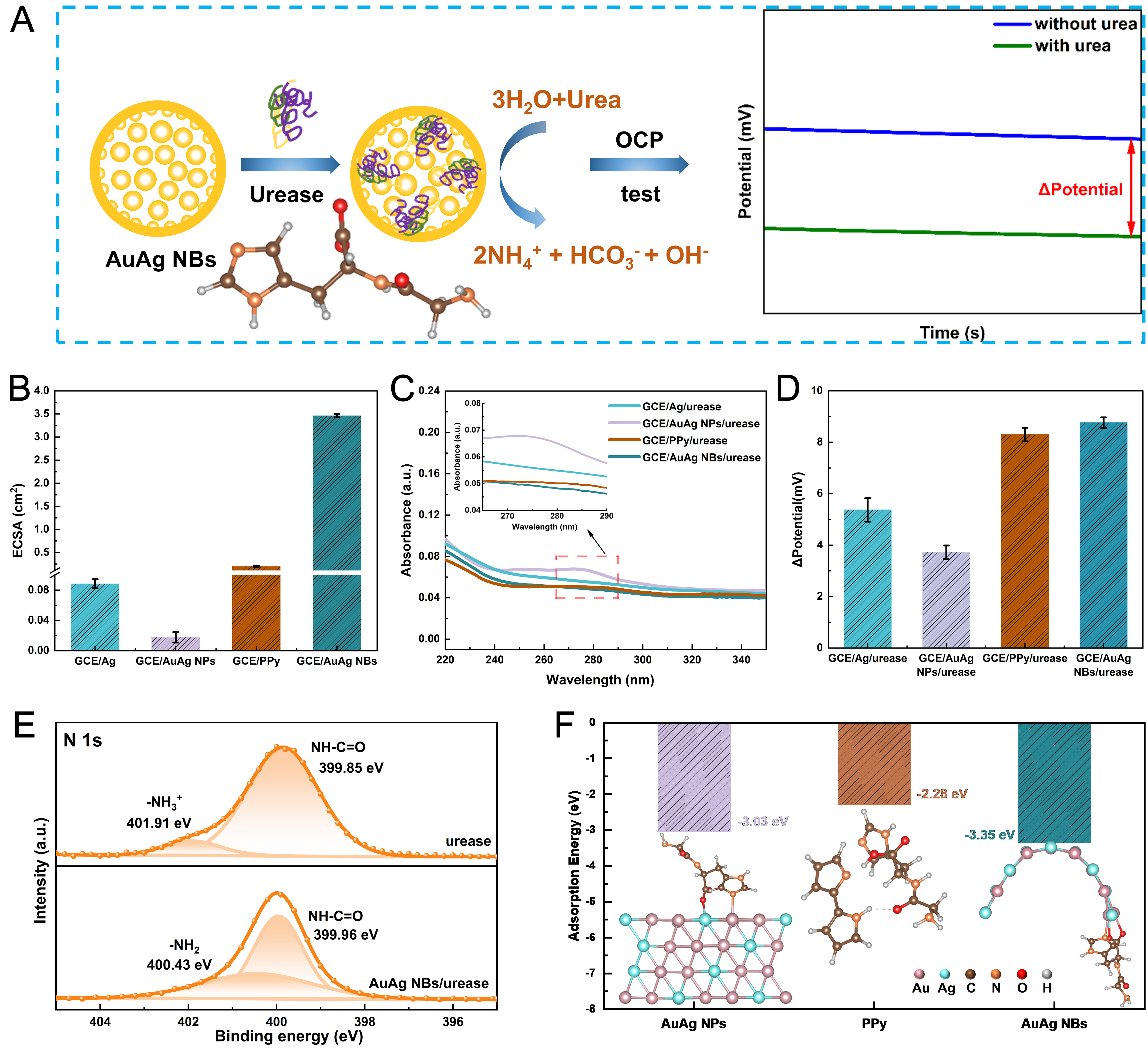
Figure 4. (A) Schematic illustration of the urea detection mechanism. The inset in panel (A) illustrates the structure of the Gly-His dipeptide; (B) ECSA values of different electrodes (SD of n = 3); (C) UV-vis spectra of eluents collected by rinsing enzyme-modified electrodes with 5 mL of PBS solution; (D) Potentiometric responses of different electrodes to 800 μM urea (SD of n = 3). (E) High-resolution N 1s spectra of urease and AuAg NBs/urease; (F) Comparison of adsorption energies of the Gly-His model on AuAg NPs, PPy, and AuAg NBs. AuAg NPs: AuAg alloy nanoparticles; PPy: polypyrrole; AuAg NBs: AuAg alloy nanobowls; UV-vis: ultraviolet-visible; ECSA: electrochemically active surface area; PBS: phosphate buffer solution; GCE: glassy carbon electrodes; OCP: open-circuit potential; SD: standard deviation.