

fig3
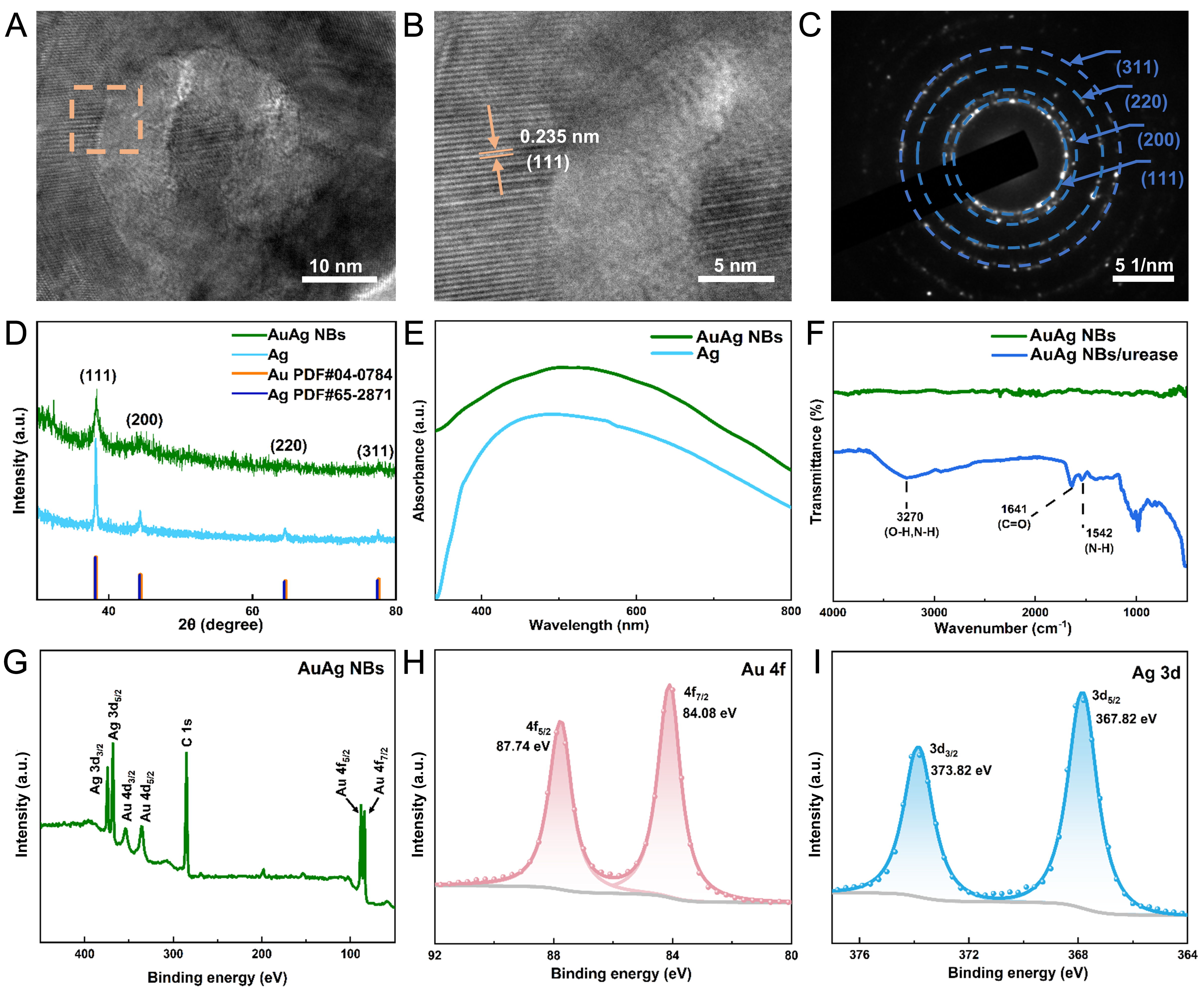
Figure 3. (A) TEM image of a small AuAg nanobowl, (B) crystal lattice spacing within the dashed box of panel (A), (C) SAED pattern; (D) XRD patterns, (E) UV-vis spectra of AuAg NBs and Ag; (F) FTIR spectra of AuAg NBs and AuAg NBs/urease; (G-I) XPS spectra of AuAg NBs. TEM: Transmission electron microscopy; SAED: selected area electron diffraction; XRD: X-ray diffraction; FTIR: Fourier-transform infrared; UV-vis: ultraviolet-visible; AuAg NBs: AuAg alloy nanobowls; XPS: X-ray photoelectron spectroscopy.