


Tunable self-recoverable near-infrared mechanoluminescence from platelike strontium-aluminate: SrAl12O19:Cr3+
 0
0 
Abstract
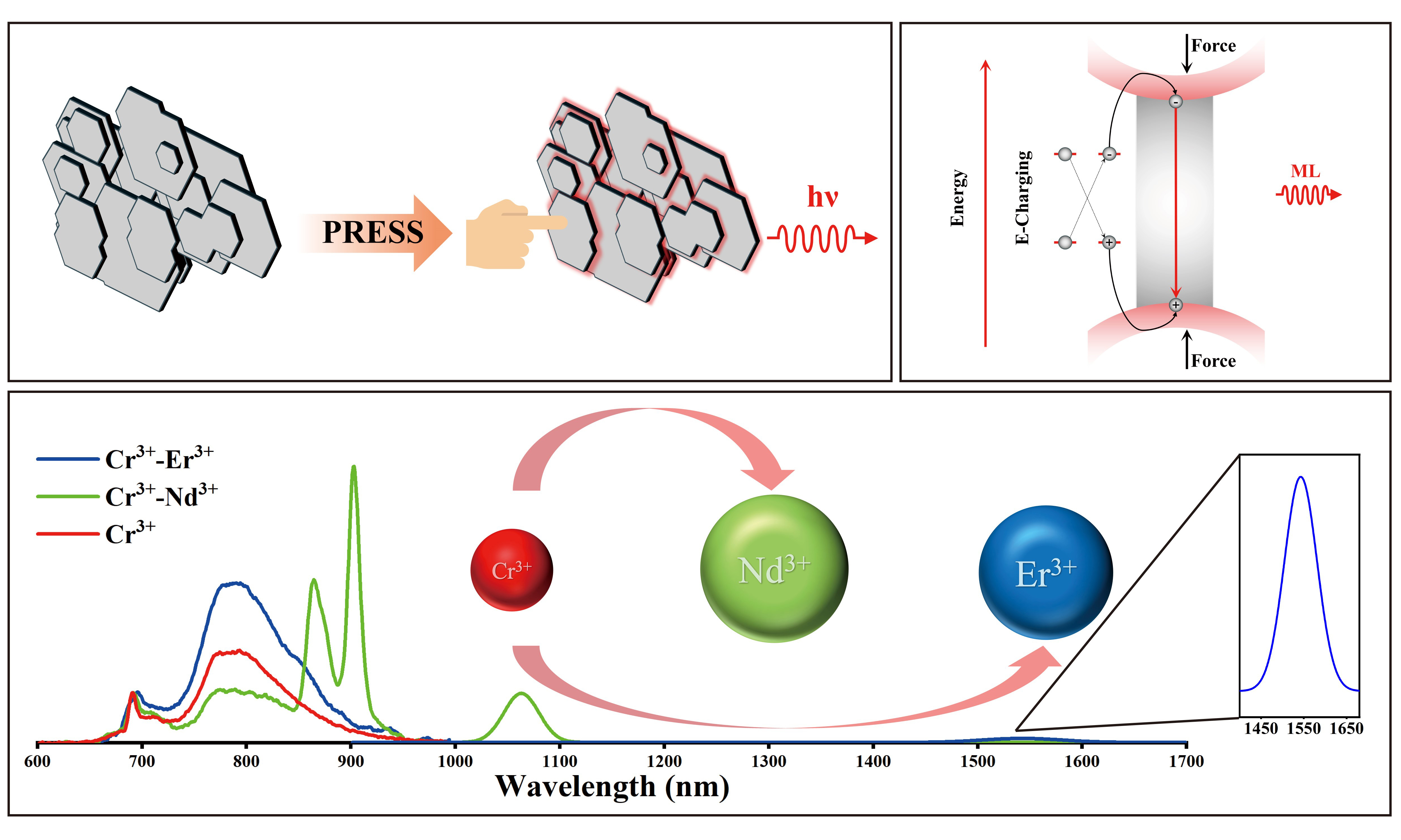
The development of high-performance self-recoverable near-infrared (NIR) mechanoluminescent materials is crucial for advancing applications. In this work, we presented a self-recoverable NIR mechanoluminescent material, platelike SrAl12O19, through singly doped with Cr3+ and co-doped with lanthanide ions (Nd3+, Yb3+, Er3+) in one step. By modulating the Cr3+ doping concentration, we achieved precise control over the mechanoluminescence (ML) intensity as well as the spectral tunability between characteristic R-line emission (~ 690 nm) and the broadband emission (750-950 nm). Moreover, energy transfer from Cr3+ to lanthanide ions enables multispectral ML emission extending into the NIR-II window (1,000-1,700 nm). The resultant material exhibits excellent ML self-recoverability and high chemical stability. The co-doped system was demonstrated with great potential in dynamic stress visualization, naked-eye-invisible information encryption and special identification under challenging conditions (e.g., underwater). We further demonstrated practical applications by fabricating dual-mode flexible NIR mechanoluminescent paper sheets and sprayable coatings. This work contributes to the advancement of new NIR mechanoluminescent materials with unique morphological features for various scenarios, including the advancement of intelligent sensing and multi-level anti-counterfeiting technologies.
Keywords
INTRODUCTION
Mechanoluminescence (ML) describes the form of light emission that directly responds to dynamic mechanical stimuli[1]. Distinct from traditional forms of light emission - such as photoluminescence (PL), electroluminescence (EL), or reaction-driven processes like chemiluminescence (CL) and bioluminescence (BL)[2], ML can directly convert mechanical energy (e.g., compression, friction, or twisting[3]) into light energy in one step, making it an efficient and clean energy conversion technology[4]. The energy sources for ML behavior are diverse, ranging from kinetic energy generated by human sports activities to natural mechanical energy. Consequently, ML materials hold significant promise for a wide range of applications such as structural health monitoring[5], smart sensors[6] and human-machine interactive interfaces[7]. Specifically, the NIR spectrum presents minimal interference from ambient light, enabling more precise measurements in bright environments. Besides, the NIR spectrum exhibits superior tissue penetration, favorable for in vivo imaging applications[8,9]. These characteristics have recently spurred considerable research interest in developing high-performance NIR ML materials. The NIR ML materials can emit beyond the visible spectrum, present promising new solutions to overcome the current ML applications’ limitations. Currently, NIR ML materials, such as CaZnOS:Nd3+[10], LiNbO3:Nd3+[11], LiGa5O8:Pr3+[12] and SrZn2S2O:Yb3+[13] rely exclusively on rare-earth dopants which manifest high cost and limited spectral tunability of rare-earth elements. Consequently, extensive research has been conducted on Cr3+-activated phosphors, such as Y3Al5O12:Cr3+[14], CaAl12O19:Cr3+[15,16], Ga2O3:Cr3+[17,18], LaAlO3:Cr3+[19] and Lu3Ga5O12: Cr3+[20]. The luminescence of Cr3+ is uniquely governed by crystal field strength, enabling a broad tunable emission from approximately 700 to 1,200 nm. This attribute facilitates the development of high-performance NIR ML materials via adjusting the coordination environment of Cr3+ ions.
Cr3+-activated NIR ML materials are broadly classified into two categories: trap-controlled and self-recoverable types. Trap-controlled NIR ML materials require pre-irradiation with ultraviolet (UV) light to populate charge carriers in traps prior to ML. After multiple stress cycles, these traps are largely de-trapped, preventing sustained emission. In contrast, self-recoverable NIR ML materials exhibit stable and continuous luminescence upon repeated mechanical stimulation, holding unique potentials for in vivo biological imaging[21], rapid stress detection in large-scale structures, and sports biomechanics analysis. Although significant progress has been made in achieving self-recoverable Cr3+-activated ML, research reports on the controlled synthesis with well-defined morphologies are scarce[22-24]. The physical properties of Cr3+-activated ML materials are governed by crystal anisotropy, which also influences the processability and long-term stability in practical applications. Platelike structure possesses highly uniform morphology and excellent substrate compatibility, allowing conformal adhesion to both flat and irregular surfaces. By capturing its NIR ML images, stress distribution across complex components can be obtained intuitively. However, current platelike NIR ML materials are relatively scarce. The predominant synthesis strategies involve secondary processing after a high-temperature solid-state or molten salt shielding synthesis[25], which requires a large amount of solvent to improve morphology with additional washing steps. A preliminary synthesis of Cr3+-doped matrix with varying Sr/Al ratios was conducted to compare their ML intensity and morphological characteristics. SrAl12O19 was selected as the ideal matrix, exhibiting superior ML intensity and unique platelike morphology [Supplementary Figures 1-3]. SrAl12O19, due to its magnetite structure, exhibits uniform and extensive platelike structures via a single-step high-temperature solid-state reaction[26]. Unlike other irregular particle morphologies that form gaps during accumulation, platelike SrAl12O19 can align parallel to the substrate surface, forming dense and continuous layers. These characteristics highlight the potential of platelike SrAl12O19 in advanced NIR ML applications.
In this work, we synthesized a series of platelike SrAl12O19:Cr3+ self-recoverable NIR ML samples via a one-step high-temperature solid-state method. First, adjusted the ML intensity by modulating the Cr3+ doping concentration, which enables variation between the R-line narrow emissions and broadband wide emissions. Then systematically investigated the ML properties under different synthetic conditions. Furthermore, co-doped with Nd3+, Yb3+ and Er3+ ions to achieve multi-spectral ML. Based on Cr3+ and Nd3+ co-doped samples, we designed NIR ML paper sheet and spray coating - for anti-counterfeiting, thereby proposing a novel strategy for the application of NIR ML.
MATERIALS AND METHODS
Preparation of phosphor
A series of SrAl12O19:x mol% Cr3+ (x = 0.1, 0.5, 1, 2, 4) phosphors were synthesized via a one-step high-temperature solid-state method. The high-purity raw materials include SrCO3 (99%, Sinopharm Chemical Reagent), Al2O3 (99.99%, Sinopharm Chemical Reagent), Cr2O3 (99.95%, Aladdin), Nd2O3 (99.99%, Aladdin), Yb2O3 (99.99%, Macklin), and Er2O3 (99.99%, Macklin).
These reagents were weighed according to the stoichiometric ratio and transferred to an agate mortar. An appropriate amount of ethanol was added as a mixing medium. The mixture was thoroughly ground and homogenized until the ethanol largely evaporated. After that, the precursors were subsequently transferred to the forced-air drying oven and set to 80 °C to ensure complete ethanol removal. The dried precursor powder was then loaded into an alumina crucible and sintered in a high-temperature box furnace at 1,250-1,650 °C for 4 h under an air atmosphere. All samples were heated from room temperature to 1,000 °C at a rate of 10 °C/min, and then heated to 1,250-1,650 °C at a rate of 5 °C/min. After holding at 1,250, 1,350, 1,450, 1,550, 1,650 °C for 4 h, the samples were subsequently cooled down naturally within the furnace chamber. Finally, the as-synthesized block samples were ground into fine powder using an agate mortar and passed through a 150-mesh stainless steel sieve for subsequent characterizations and application designs.
Characterization
The phase purity of the synthesized SrAl12O19:x%Cr3+ compounds was analyzed by X-ray diffraction (XRD) using an Ultima IV high-resolution diffractometer (Rigaku Corporation, Japan), with data collected in the range of 10-120°. The Rietveld refinement was performed using the GSAS-II software. The platelike morphology of the samples was captured by a field emission scanning electron microscope (FESEM) on a Gemini SEM 560 instrument (ZEISS, Germany). Elemental analysis was conducted using energy-dispersive X-ray spectroscopy (EDS) with an Oxford cryogenically cooled spectrometer ULTIM MAX (Oxford, UK). Microstructural characterization was performed by aberration-corrected transmission electron microscopy (TEM) on a Titan Cubed Themis G2 300 microscope (Thermo Fisher Scientific, The Netherlands). PL spectra and fluorescence decay curves were measured with an Edinburgh FLS1000 spectrometer (Edinburgh Instruments, UK). Quantum efficiency (QE) was measured with Quantaurus-QY Plus C13534-12 (Hamamatsu Photonics, Japan). The ML experiments were conducted with a custom-built setup including a digital push-pull gauge, a metal slider driven by a linear motor, and an optical fiber connected to the spectrometer. The ML spectra were acquired using an QE65 PRO high-sensitivity spectrometer (Ocean Optics, USA) for the visible range and an NIR QUEST 512 spectrometer (Ocean Optics, USA) for the NIR region. For ML testing, the films were prepared using a PTFE mold (5 × 5 × 0.03 cm) featuring five parallel 10 × 2 mm rectangular sections. The material block was first crushed and sieved through a 150-mesh stainless steel screen to obtain a uniform powder. This powder was then evenly distributed into the mold’s rectangular sections. After removing any excess, the shaped powder strips were thermally sealed with PET laminating film to create the final test samples. The ML signal was subsequently obtained by scraping the film surface with the metal slider at a distance of 20 mm and a speed of 16 mm/s. The PDMS used in the application section was prepared by mixing powder and PDMS in a 1:1 mass ratio. After being placed in an oven for 6 h, it was cured for subsequent testing. All the NIR images and videos were captured using an ONV3+ night vision device (ORPHA, Germany) and a smartphone. The infrared thermal video was captured by a TI25 instrument (Fluke, USA).
RESULTS AND DISCUSSION
Structure characterization of SrAl12O19:x%Cr3+
The magnetite-type SrAl12O19 matrix belongs to the hexagonal crystal system with a space group of P63/mmc. Rietveld refinement of the XRD pattern demonstrates an excellent agreement between the calculated (red line) and observed (black circles) data for SrAl12O19:2%Cr3+ sample [Figure 1A]. The refinement parameters were Rp = 5.70%, Rwp = 7.85%, and GOF = 2.25. After introducing Cr3+ into the host lattice, the unit cell parameters of the SrAl12O19:2%Cr3+ sample increased (a = b = 5.5685 Å, c = 22.0114 Å) compared with the undoped SrAl12O19 matrix (a = b = 5.5666 Å, c = 22.0018 Å). This lattice expansion can be attributed to the replacement of Al3+ ions (0.535 Å, CN = 6) by larger Cr3+ ions (0.615 Å, CN = 6)[27]. These results provide clear evidence that Cr3+ ions successfully replaced Al3+ sites in the host structure. Supplementary Figure 4 shows the XRD patterns of SrAl12O19:x%Cr3+ synthesized with varying Cr3+ doping concentrations (x = 0, 0.1, 0.5, 1, 2, 4) at 1,650 °C for 4 h. All diffraction patterns remain nearly identical and closely match the standard card of SrAl12O19 (PDF#80-1195), confirming the doping of Cr3+ does not induce any secondary phases or alter the host crystal structure. Similarly, Supplementary Figure 5 shows the XRD patterns of samples SrAl12O19:2%Cr3+ synthesized at different sintering temperatures (1,250, 1,350, 1,450, 1,550 and 1,650 °C) for 4 h. At 1,250 °C, several peaks from Al2O3 impurity (PDF#88-0826) are evident. However, these peaks are largely eliminated when the temperature reaches 1,350 °C, and a pure SrAl12O19 phase is gradually obtained (PDF#80-1195), indicating that a synthesis temperature of 1,350 °C or higher is required to achieve phase-pure SrAl12O19 material.
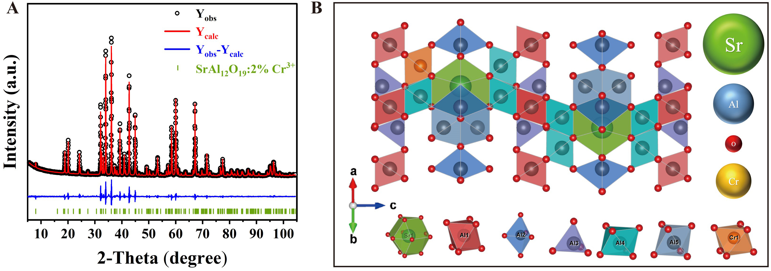
Figure 1. (A) Rietveld refinement of SrAl12O19:2%Cr3+ synthesized at 1,650 °C; (B) Crystal structure of SrAl12O19:Cr3+.
As illustrated in Figure 1B, the crystal structure of SrAl12O19:Cr3+ comprises one [AlO4] tetrahedron, one [AlO5] trigonal bipyramid, and three [AlO6] octahedra, while the Sr2+ occupies the 12-oxygen-coordinated site[28]. The rich crystal structure diversity provides an extremely high compatibility host lattice for doping with various transition metals and rare-earth ions. Furthermore, SrAl12O19:Cr3+ is composed of alternately stacked mirror-image dense spinel structural units and relatively loose mirror layers. The oxygen ions are densely packed in the spinel structural units but sparsely distributed in the mirror layers[29]. This structural anisotropy promotes preferential crystal growth along the a-b plane, while the large c/a axial ratio (3.95) suppresses growth along the c-axis, ultimately resulting in a stable hexagonal layered structure for SrAl12O19:Cr3+[30].
Due to the hydrolysis of the high-performance ML material SrAl2O4:Eu2+, Dy3+, we specifically conducted comparative experiments and evaluated the aqueous stability of SrAl12O19:2%Cr3+. The sample synthesized at 1,650 °C was immersed in deionized water for a month. Afterward, the powder was dried and characterized as shown in Supplementary Figure 6, the XRD pattern of the water-immersed SrAl12O19:2%Cr3+ sample remains nearly identical to the non-water-immersed sample and the standard card SrAl12O19 (PDF#80-1195), with no detectable phase transitions or structural changes. The result indicates the outstanding chemical stability of the SrAl12O19:2%Cr3+ in aqueous environments, supporting its potential for applications in underwater NIR ML devices. The excellent waterproof performance is intrinsically related to its unique crystal structure.
Morphology of SrAl12O19:x%Cr3+
To identify the anisotropic growth, Figure 2A presents the SEM image of the SrAl12O19:2%Cr3+ sample. The result reveals a stacked platelike structure, each comprising multiple single-crystal layers. These platelike morphologies are attributed to the preferential crystal growth along the (001) plane. Notably, the platelike structure of SrAl12O19 is achieved through one-step high-temperature solid-state synthesis, eliminating the need for molten salt or secondary processing, thereby offering considerable advantages for NIR ML coating applications.
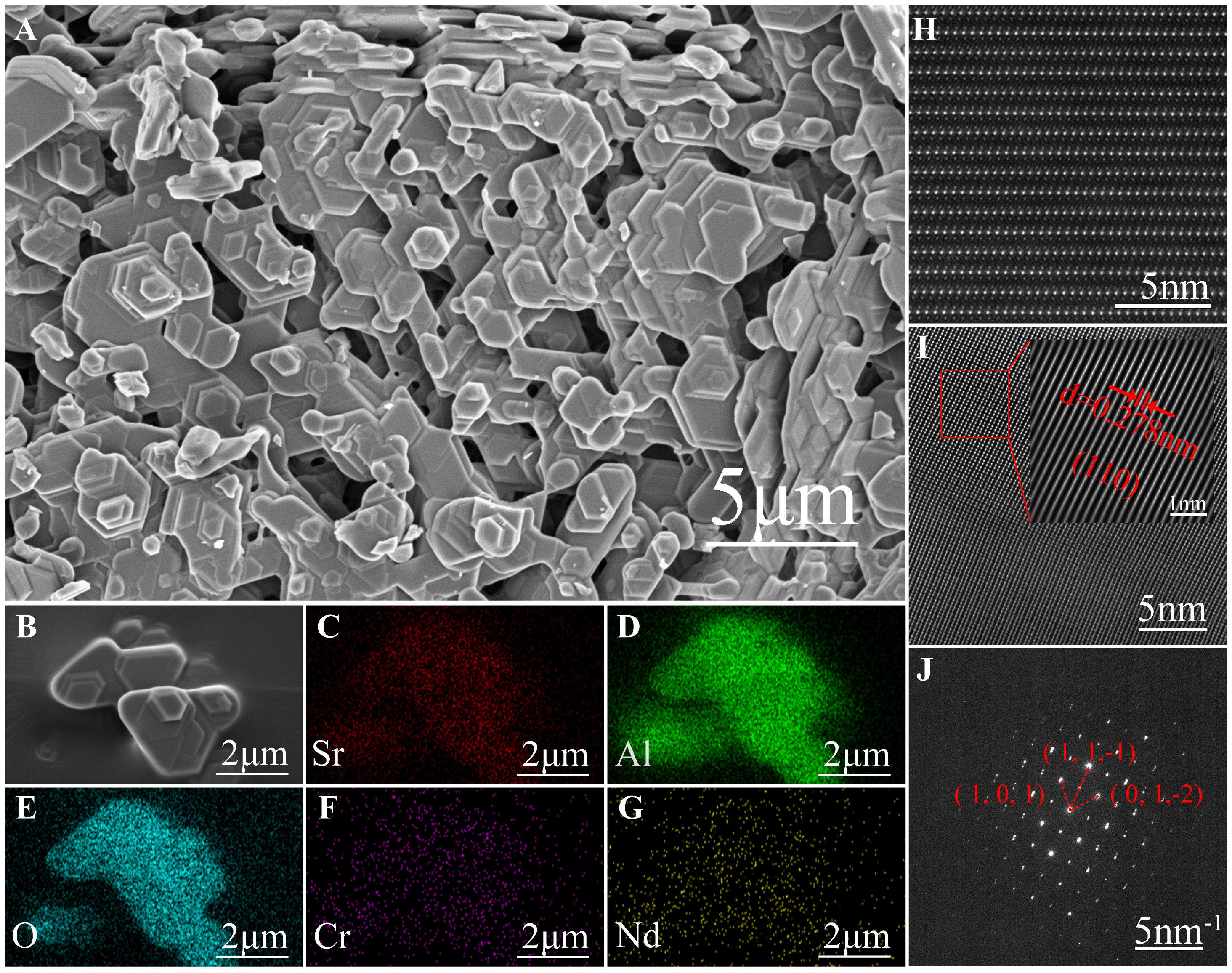
Figure 2. (A) SEM image of SrAl12O19:2%Cr3+ sample; (B-G) SEM image and element mappings of SrAl12O19:2%Cr3+, 1%Nd3+ sample; (H) High-resolution TEM image of SrAl12O19:2%Cr3+; (I) Fourier transform image of the selected region for SrAl12O19:2%Cr3+; (J) Selected area electron diffraction image of SrAl12O19:2%Cr3+. SEM: Scanning electron microscope; TEM: transmission electron microscopy.
Elemental mappings via EDS analysis [Figures 2B-G] confirm the homogeneous distribution of Sr, Al, and O, indicating the successful co-doping of both Cr3+ and Nd3+ into the SrAl12O19 matrix. Figure 2H presents a high-resolution TEM image. Specifically, since Sr atoms present a significantly higher atomic mass than Al atoms, the heavier Sr atoms in the SrAl12O19 matrix appear as brighter white spots due to pronounced scattering effects than Al atoms under dark-field conditions. The elemental mappings of the enlarged area in Supplementary Figure 7 further show that Sr, Al, O atoms exhibit a uniform layered distribution, and the Cr atoms are successfully doped into the Al layer. Figure 2I reveals the Fourier transform image, with the corresponding (110) lattice spacing of 0.278 nm. Figure 2J displays the selected area electron diffraction (SAED) pattern of the SrAl12O19:2%Cr3+ sample. The periodic diffraction spots further indicate a high crystallinity.
PL and ML properties of SrAl12O19:x%Cr3+
The PL properties of SrAl12O19:Cr3+ were systematically investigated. Figure 3A presents the photoluminescence excitation (PLE) and PL spectra of SrAl12O19:2%Cr3+. The PLE spectrum, monitored at 688 nm emission, exhibits two intense excitation bands centered at 422 and 584 nm, corresponding to the spin-allowed [4A2 → 4T1] and [4A2 → 4T2] transitions of Cr3+. Under excitation at 422 nm, the emission spectrum is characterized by both the R-line and broadband emission. Specifically, the sharp peaks at 688 and 694 nm arise from the spin-forbidden [2E → 4A2] transition, and the broadband emission from 750 to 950 nm is ascribed to the spin-allowed [4T2 → 4A2] transition[31]. The co-existence of sharp R-line and the broadband emissions indicates that the Cr3+ ions occupy the distorted octahedral sites in the SrAl12O19 matrix, with an intermediate crystal field strength[32].
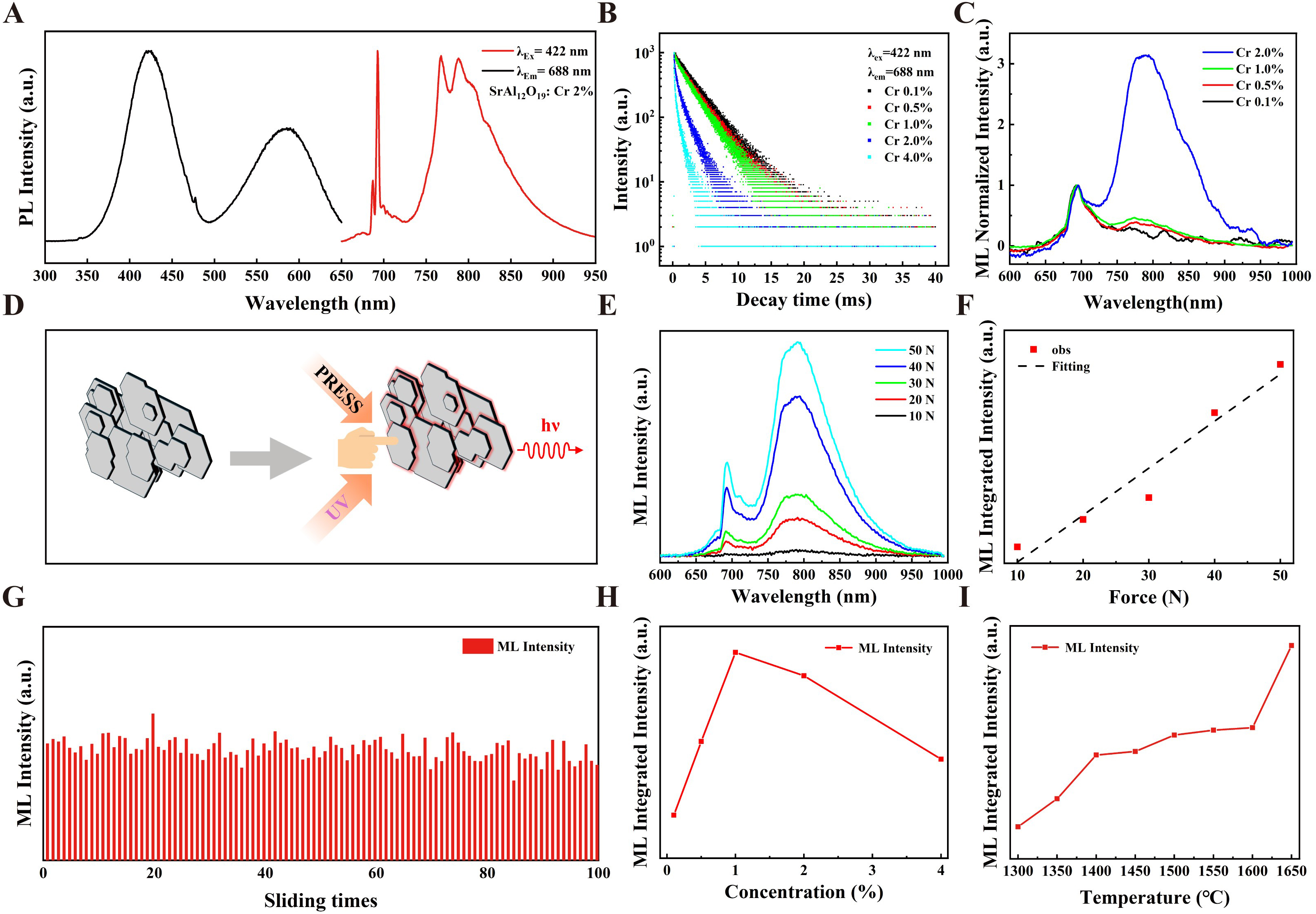
Figure 3. (A) PLE and PL spectra of SrAl12O19:2%Cr3+; (B) PL decay curve of SrAl12O19:x%Cr3+; (C) Normalized ML spectra of SrAl12O19:2%Cr3+; (D) Schematic diagram of platelike SrAl12O19:x%Cr3+ NIR luminescence under different stimuli; (E) ML spectra of SrAl12O19:2%Cr3+ recorded under the applied force from 10 to 50 N; (F) Integrated ML intensity as a function of applied load and corresponding linear fitting; (G) ML Repeatability for SrAl12O19:2%Cr3+ under periodic load; (H) Integrated ML intensity of SrAl12O19:x%Cr3+ under an applied load of 30 N; (I) Integrated ML intensity of SrAl12O19:2%Cr3+ synthesis at different temperature. PL: Photoluminescence; ML: mechanoluminescence; PLE: photoluminescence excitation; NIR: near-infrared.
The PLE and PL spectra of SrAl12O19:x%Cr3+ are presented in Supplementary Figure 8. The PL spectra are dominated by sharp R-line emissions at 688 and 694 nm with Cr3+ concentration below 1%. As the Cr3+ concentration increases, the PL intensity of broadband emission gradually increases and finally surpasses that of sharp emissions. Specifically, the PL intensity reaches a maximum at 1% Cr3+ concentration, followed by a concentration quenching with increasing Cr3+ concentration. Supplementary Figure 9 presents the PLE and PL spectra of SrAl12O19:2%Cr3+ synthesized at different temperatures. The PL intensities show monotonic increase as the synthesis temperature is raised. The PL decay curve of SrAl12O19: x%Cr3+samples are presented in Figure 3B and Supplementary Figure 10 as a function of Cr3+ concentration. The corresponding lifetimes were calculated using Equation 1[33].
where A1 and A2 are the amplitudes, τ1 and τ2 are the decay lifetimes of the individual components, respectively. As a result, the lifetime decreases from 3.22 ms to 0.57 ms as the Cr3+ concentration increases. The reduction is attributed to an enhanced probability of energy migration among adjacent Cr3+ ions and finally to quenching sites, which collectively promote non-radiative decay pathways. Table 1 summarizes the quantum efficiency measured within 650-950 nm for SrAl12O19:x%Cr3+. As Cr3+ concentration increases, the internal quantum efficiency (IQE) presents a continuous decrease, whereas the external quantum efficiency (EQE) initially increases and then decreases. Notably, the SrAl12O19:2%Cr3+ sample demonstrates the highest EQE value of 29%.
Quantum efficiency of SrAl12O19:x%Cr3+ samples under 404 and 422 nm excitation
| Concentration | IQE (404 nm) | IQE (422 nm) | EQE (404 nm) | EQE (422 nm) |
| 0.1% | 85% | 90% | 11% | 12% |
| 0.5% | 85% | 85% | 19% | 19% |
| 1.0% | 83% | 84% | 23% | 24% |
| 2.0% | 80% | 77% | 29% | 27% |
| 4.0% | 53% | 52% | 24% | 24% |
Consistent with the PL spectra, the ML spectra of SrAl12O19:x%Cr3+ are composed of a sharp R-line emission at 690 nm and a broadband emission spanning 750-950 nm [Supplementary Figure 11]. The sharp R-line corresponds to the spin-forbidden [2E → 4A2] transition of Cr3+, whereas the broadband emission arises from the spin-allowed [4T2 → 4A2] transition of Cr3+. As shown in Figure 3C and Supplementary Figure 11, the ML intensity of SrAl12O19:x%Cr3+ increases with the Cr3+ doping concentration increases from 0.1% to 1.0%, reaching maximum at 1.0%. The enhancement is attributed to the increasing quantity of available Cr3+ luminescent centers. At higher Cr3+ doping concentrations from 1.0% to 4.0%, the ML intensity decreases due to concentration quenching. The reduced interionic distance between Cr3+ ions facilitate enhanced energy migration, which promotes non-radiative energy transfer and consequently diminishes the luminescence efficiency. Furthermore, the spectrum exhibits a concentration-dependent shift: the ML is dominated by the R-line emission at Cr3+ concentrations below 1.0%, while the broadband emission becomes predominant at Cr3+ concentrations exceeding 1.0%. This unique spectral shift properties arouse our research interest. The SrAl12O19:2%Cr3+ sample, in which the sharp peak and broadband intensities are comparable, was selected as the primary subject for further investigation. The dual-mode NIR luminescence of platelike material SrAl12O19:x%Cr3+ were further explored, as schematically shown in Figure 3D. To explore the relationship between applied force and ML intensity, Figure 3E displays the ML spectra of the SrAl12O19:2%Cr3+ sample under linearly increasing applied loads (F = 10-50 N). The corresponding fitting curve demonstrates a nearly linear correlation between the ML intensity and the applied force (R2 = 0.95), favorable for stress feedback applications [Figure 3F]. Besides, the self-recoverable property of SrAl12O19:2%Cr3+ was also verified by subjecting the prepared PET film to 100 sliding cycles under 5 N load, as shown in Figure 3G. The recorded ML signals remained stable throughout the test, demonstrating robust ML cyclic stability, the ML intensity remains at 86% of its average value even after 100 cycles. To eliminate the influence of thermal effects on ML, we recorded the SrAl12O19:2%Cr3+ PET scraping process using an infrared thermal imaging camera. The results showed no significant heating phenomenon [Supplementary Video 1]. These results indicate that SrAl12O19:Cr3+ belongs to the category of elastico ML materials. Moreover, the material exhibits exceptional chemical stability, as evidenced by the negligible change in the ML before and after water-immersed [Supplementary Figure 12].
Temperature-dependent ML measurements were also performed, and the result shows that the ML intensity progressively diminishes as temperature increases [Supplementary Figure 13]. This phenomenon is attributed to the enhanced lattice vibrations, which generally promote non-radiative relaxation of excited electrons. The ML integrated intensity as shown in Figure 3H for different Cr3+ doping concentrations indicate that excessively high doping concentrations weaken the ML intensity, which is attributed to the concentration quenching effect. Additionally, the effect of different sintering temperatures (1,250-1,650 °C) on the ML intensity for the SrAl12O19:2%Cr3+ sample is also shown in Figure 3I and Supplementary Figure 14. The result indicates that no ML signal can be detected below 1,250 °C, whereas a continuous enhancement is observed with further increasing temperature. The peak position of the ML emission remains unchanged across temperatures. Furthermore, the holding time during reaction was found to have a negligible impact on the ML intensity and spectral curve [Supplementary Figure 15]. Besides, the SrAl12O19:2%Cr3+ sample was also sintered in a reducing N2/H2 atmosphere and exhibits a slight enhancement in ML intensity [Supplementary Figure 16]. This observation is primarily ascribed to limited oxygen incorporation during synthesis, thereby stabilizing oxygen vacancies, which is favorable for promoting local lattice distortion and constructing piezoelectric potential, leading to enhanced ML performance.
Multispectral ML and energy transfer of SrAl12O19:x%Cr3+
Figure 4A illustrates a proposed mechanism for the ML process. Although the host lattice of SrAl12O19 is centrosymmetric, the doping Cr3+ ions and oxygen vacancies result in local structural distortion. Under external mechanical force, an internal piezoelectric field is generated, leading to the separation of electron-hole pairs. These electrons subsequently relax to the valence band maximum and recombine with holes. The released energy in the form of photons excites the Cr3+ ions to produce the NIR ML. Furthermore, the incorporation of lanthanide ions (Nd3+, Yb3+, Er3+) enables distinct NIR-Ⅱ ML via efficient energy transfer from the excited Cr3+.
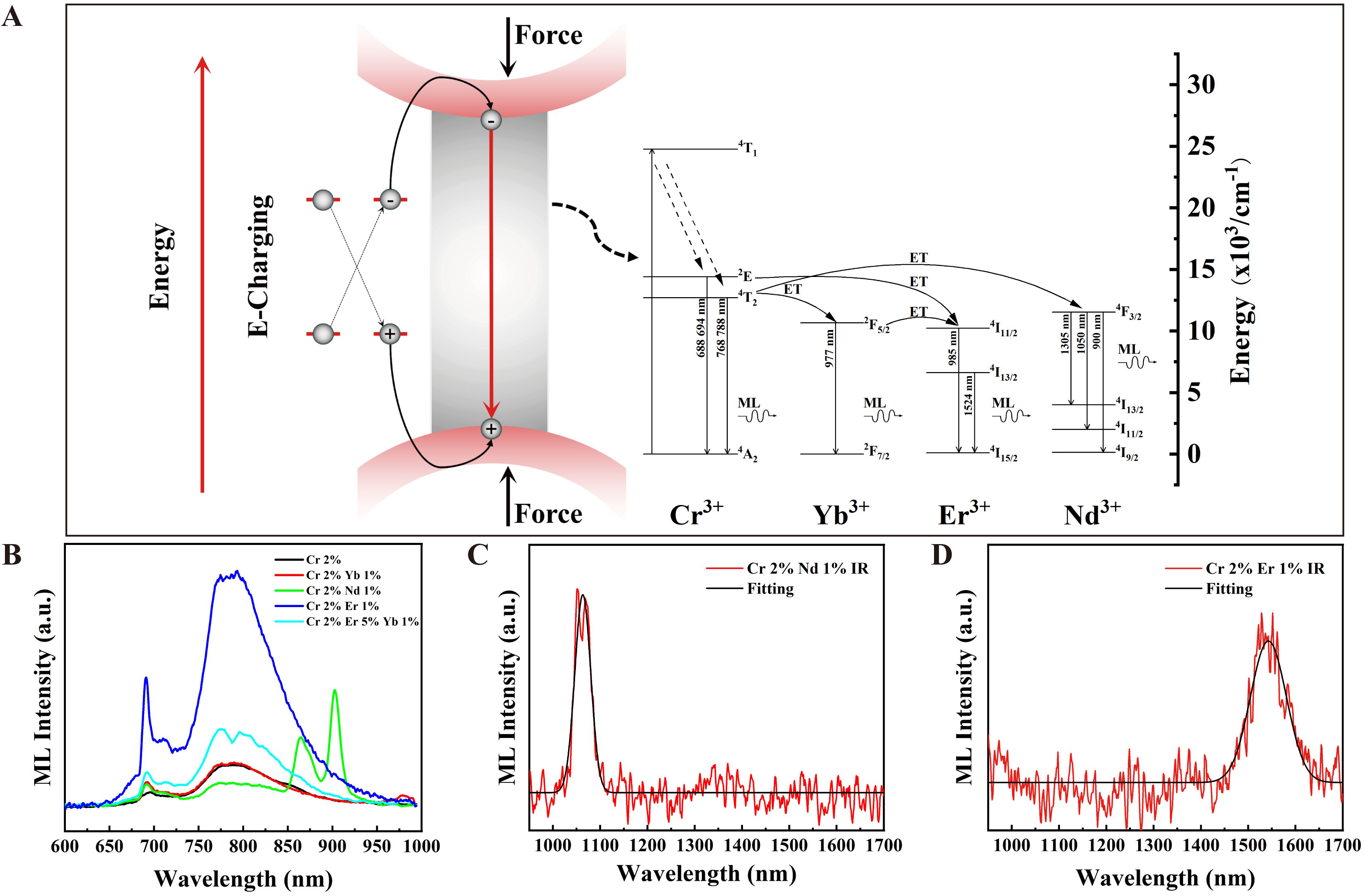
Figure 4. (A) Schematic diagram of the ML mechanism and energy transfer for SrAl12O19: Cr3+, Nd3+ (or Yb3+ or Er3+ or Yb3+, Er3+); (B) ML spectra of SrAl12O19:2%Cr3+, 1%Nd3+ (or 1%Yb3+ or 1%Er3+ or 1%Yb3+, 5%Er3+) samples employed a visible-NIR detector (600-1,000 nm); (C) ML spectra of SrAl12O19:2%Cr3+, 1%Nd3+ and (D) ML spectra of SrAl12O19:2%Cr3+, 1%Er3+ using a NIR detector (900-1,700 nm). ML: mechanoluminescence; NIR: near-infrared.
Additionally, for SrAl12O19, the Sr2+ site is favorable for doping lanthanide ions to construct energy transfer pathway and realize NIR-Ⅱ ML. Therefore, the co-doped and tri-doped SrAl12O19:2%Cr3+, 1%Nd3+ (or 1%Yb3+ or 1%Er3+ or 1%Yb3+, 5%Er3+) samples were synthesized to modulate the emission spectra[34,35]. Compared with Cr3+ singly doped sample, the co-doped samples present distinct emission at 900/1,050, 977, and 1,524 nm under 422 nm excitation, ascribed to the intra-configurational 4F3/2→4I9/2/4I11/2, 2F5/2→2F7/2, and 4I13/2→4I15/2 transitions of Nd3+[36,37], Yb3+[38], and Er3+, respectively [Supplementary Figure 17]. Besides, the introduction of lanthanide ions leads to reduced luminescence lifetimes [Supplementary Figures 18-21], which provide evidence for energy transfer from Cr3+ to lanthanides. Specifically, the lifetime of Cr3+, Nd3+ co-doped sample decreases substantially, indicating a significantly higher energy transfer efficiency. The corresponding ML spectra are presented in Figure 4B-D. In contrast to SrAl12O19:2%Cr3+ sample, the co-doped samples exhibit additional ML emissions within the NIR-II region, which is consistent with the PL spectra. For example, the SrAl12O19:2%Cr3+,1%Nd3+ sample presents pronounced ML emissions at 864, 903 and 1,064 nm, while weak emission ranging 700-900 nm, which can be ascribed to the efficient energy transfer from Cr3+ to Nd3+ ions. Specifically, the emission at 903 nm, originating from the 4F3/2 → 4I9/2 transition, exhibits a doublet structure attributed to the Stark effect. The SrAl12O19:2%Cr3+, 1%Yb3+ sample presents a significantly weak ML at 980 nm of Yb3+ ions, indicating a much weaker energy transfer efficiency from Cr3+ to Yb3+. The SrAl12O19:2%Cr3+, 1%Er3+ sample presents a substantially red-shifted ML at 1,543 nm of Er3+ ions. These results collectively demonstrate that the ML spectrum can be effectively tuned via doping lanthanide ions and controlled energy transfer pathways.
Applications of SrAl12O19:2%Cr3+, 1%Nd3+
Due to the excellent mechanical-to-photon conversion, ML materials have already been developed for information encryption[39,40], self-powered illumination[41,42], and so on. Leveraging the NIR ML properties of SrAl12O19:2%Cr3+,1%Nd3+, a dual-mode flexible NIR ML paper was fabricated. The fine powder was homogeneously blended with dehydrated paper pulp block in water at a powder/block mass ratio of 5:1. The mixture was subsequently transferred using a paper-making screen, followed by drying to obtain the final product. Figure 5A and B respectively compare the difference of this paper sheet under ambient light and UV illumination. Under ambient light, the result shows that the resultant paper exhibits off-white bodycolor under ambient light, while dark red PL under UV excitation. Besides, the uniform PL across the paper sheet indicates the homogeneous distribution of the ML powder. A piece of paper sheet was cut and encapsulated in a PET for the ML test. As shown in Figure 5C, the ML spectrum confirms that the paper sheet composite retains the characteristic emission from the pristine SrAl12O19:2%Cr3+,1%Nd3+ powder.
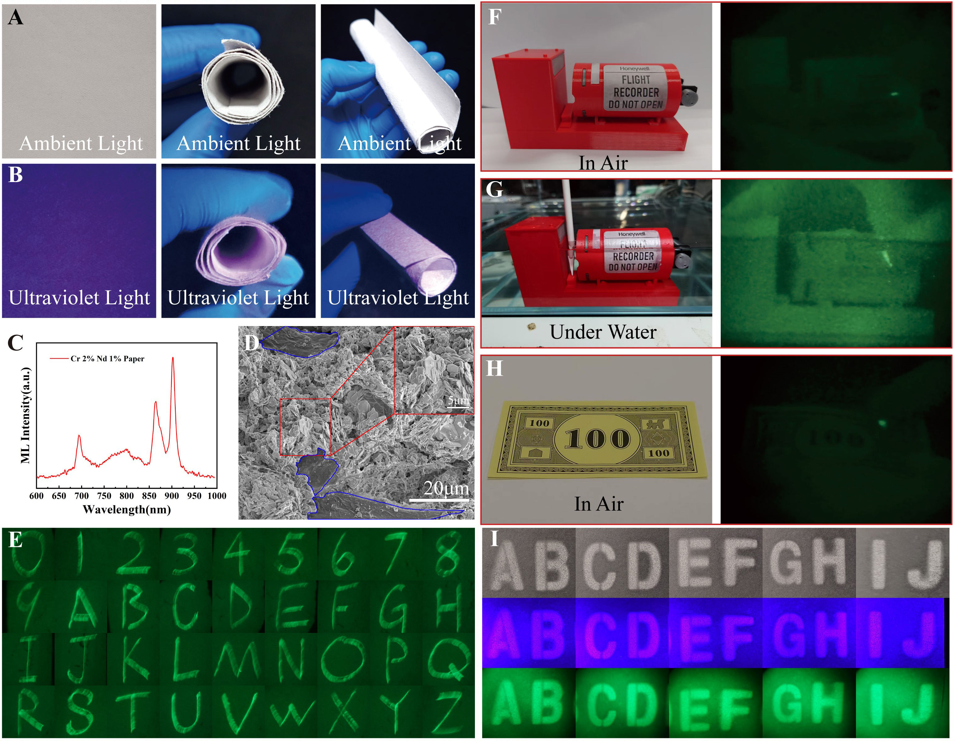
Figure 5. (A) Flexible ML paper sheet made of SrAl12O19:2%Cr3+, 1%Nd3+ under ambient light; (B) Flexible ML paper sheet made of SrAl12O19:2%Cr3+, 1%Nd3+ under UV light; (C) ML spectrum of SrAl12O19:2%Cr3+, 1%Nd3+ ML paper sheet; (D) SEM of SrAl12O19:2%Cr3+, 1%Nd3+ ML paper sheet (The area outlined in blue is paper fiber.); (E) Under night vision conditions, letters were written on SrAl12O19:2%Cr3+, 1%Nd3+ ML paper and recorded via time-lapse photography; (F) NIR ML comparison of black boxes in air atmosphere; (G) NIR ML comparison of black boxes under water condition; (H) NIR ML comparison of banknotes; (I) Printed letters with SrAl12O19:2%Cr3+, 1%Nd3+ sprayer under the condition of ambient light, UV light and night vision. ML: mechanoluminescence; SEM: scanning electron microscope; NIR: near-infrared; UV: ultraviolet.
The microstructure of the paper composite was analyzed by FESEM. As shown in Figure 5D and Supplementary Figure 22, the SEM images show the unique platelike morphology with uniform distribution of the SrAl12O19:2%Cr3+,1%Nd3+ sample within the paper fibers of the sheet. The EDS confirmed the presence of all constituent elements [Supplementary Figure 23]. Figure 5E shows a long-exposure image captured with an ONV3+ night vision device, showing the numbers 0-9 and the 26 letters of the alphabet written directly onto the NIR-ML paper sheet using a glass rod. Supplementary Video 2 demonstrates the bending and writing operations performed on the paper sheet in which the NIR ML is clearly captured under night vision device. The paper sheet presents excellent ML repeatability even after 100 sliding experiments [Supplementary Figure 24]. Therefore, this paper exhibits excellent anti-counterfeiting characteristics, as the written information remains invisible to the naked eye but detectable in the NIR spectrum using specialized equipment.
Besides, the SrAl12O19:2%Cr3+ phosphor, which exhibits composition and luminescence stability under aqueous environment [Supplementary Figures 6 and 12], shows great potential for applications in underwater NIR ML devices. Accordingly, Figure 5F-H demonstrates the application of this ML coating composed of SrAl12O19:2%Cr3+ phosphor on various substrates, including black boxes (tested both in air and underwater) and banknotes [Supplementary Videos 3-5]. The ML induced by sand and stone abrasion on the seabed-simulated black box, as well as the ML response from manually scratching the banknotes were simulated. Figure 5I also demonstrates the creation of English letter patterns employing SrAl12O19:2%Cr3+ 1%Nd3+ coating after passing through a perforated plate mask. The patterns were imaged under three conditions: ambient light, UV excitation, and through night vision device with UV excitation. When viewed through night vision device, the intense emission confirms the material’s efficient NIR ML capability. Therefore, this dual-mode luminescence - comprising both NIR PL and ML which are only detectable using night vision device under light and mechanical stimulation [Supplementary Video 6] - enables effective application in advanced anti-counterfeiting and specialized marking, significantly enhancing the complexity of information encryption. Additionally, we conducted exploratory ultrasonic testing on PDMS films mixed with SrAl12O19:2%Cr3+,1%Nd3+. Experimental results demonstrate the material’s excellent response to ultrasonic forces, offering new insights for NIR stress luminescence applications within biological organisms [Supplementary Video 7].
CONCLUSIONS
This study presents the development of a platelike SrAl12O19 NIR ML material via Cr3+ sole-doping and co-doping with rare-earth ions like Nd3+, Yb3+, Er3+. Tunable ML was achieved by modulating the Cr3+ doping concentration, which effectively alters the crystal field strength. The introduction of lanthanide rare earth ions further enabled multispectral emission within NIR-Ⅱ region. This material exhibits excellent chemical stability, outstanding self-recoverable properties, and long-term water resistance. Based on the Cr3+, Nd3+ co-doped system, NIR ML paper sheet and sprayable coating were designed and demonstrated for applications in information encryption, dynamic response, and special identification. This research provides new insights for expanding the application scenarios of NIR ML materials and advancing smart display technologies.
DECLARATIONS
Acknowledgments
The authors thank Dr. Nan Jian from Electron Microscope Center of Shenzhen University for his help in HRTEM. The authors also acknowledge researcher Mengmeng Ma for assistance from The Photonics Center of Shenzhen University. The authors also thank researcher Yingzhen Li for the assistance on XRD measurements received from the College of Physics and Optoelectronic Engineering of Shenzhen University.
Authors’ contributions
Design: Zhang, Q.; Peng, D.
Experiments: Zhang, Q.; Fang, Z.; Wu, M.
Manuscript writing: Zhang, Q.; Liu, S.; Peng, D.
Manuscript revision and supervision: Zhang, Q.; Fang, Z.; Wu, M.; Ma, Q.; Liu, Y.; Zhou, J.; Liu, S.; Peng, D.
All authors reviewed and edited the final version.
Availability of data and materials
The data that supports the findings of this study are available in the Supplementary Materials of this article. Further data are available from the corresponding author upon reasonable request.
AI and AI-assisted tools Statement
Not applicable.
Financial support and sponsorship
This work was supported by the Natural Science Foundation of China (62275170), the Guangdong Provincial Science Fund for Distinguished Young Scholars (2022B1515020054), the Key-Area Research and Development Program of Guangdong Province (2024B0101080001), Shenzhen Fundamental Research Project (JCYJ20240813141624033), Scientific Research Foundation as Phase III construction of high level University 2035 plan (0000050101).
Conflicts of interest
All authors declared that there are no conflicts of interest.
Ethical approval and consent to participate
Not applicable.
Consent for publication
Not applicable.
Copyright
© The Author(s) 2026.
Supplementary Materials
REFERENCES
1. Chang, S.; Zhang, K.; Peng, D.; Deng, Y.; Shan, C.; Dong, L. Mechanoluminescent functional devices: developments, applications and prospects. Nano. Energy. 2024, 122, 109325.
2. Schramm, S.; Weiß, D. Bioluminescence - the vibrant glow of nature and its chemical mechanisms. Chembiochem 2024, 25, e202400106.
3. Wang, J.; Yao, K.; Cui, K.; et al. Contact electrification induced multicolor self-recoverable mechanoluminescent elastomer for wearable smart light-emitting Devices. Adv. Opt. Mater. 2023, 11, 2203112.
4. Zhou, B.; Liu, J.; Huang, X.; et al. Mechanoluminescent-triboelectric bimodal sensors for self-powered sensing and intelligent control. Nano-Micro. Lett. 2023, 15, 72.
5. Shin, H. G.; Timilsina, S.; Sohn, K. S.; Kim, J. S. Digital image correlation compatible mechanoluminescent skin for structural health monitoring. Adv. Sci. 2022, 9, 2105889.
6. Dong, Y.; An, W.; Wang, Z.; Zhang, D. An artificial intelligence-assisted flexible and wearable mechanoluminescent strain sensor system. Nano-Micro. Lett. 2024, 17, 62.
7. Duan, S.; Sang, M.; Chen, H.; et al. Shear stiffening-based mechanoluminescent device for impact-thermal coupling protection and impact visualization. Adv. Funct. Mater. 2024, 34, 2411821.
8. Jiang, S.; Wu, X.; Yang, F.; Rommelfanger, N. J.; Hong, G. Activation of mechanoluminescent nanotransducers by focused ultrasound enables light delivery to deep-seated tissue in vivo. Nat. Protoc. 2023, 18, 3787-820.
9. Wu, S.; Zhou, G.; Wu, Y.; et al. Multiple defect-induced high-resolution near-infrared mechanoluminescent materials for non-destructive detection of blood glucose and lipids. Adv. Mater. 2024, 36, 2408508.
10. Li, L.; Wondraczek, L.; Li, L.; et al. CaZnOS:Nd3+ emits tissue-penetrating near-infrared light upon force loading. ACS. Appl. Mater. Interfaces. 2018, 10, 14509-16.
11. Xiong, P.; Peng, M. Near infrared mechanoluminescence from the Nd3+ doped perovskite LiNbO3:Nd3+ for stress sensors. J. Mater. Chem. C. 2019, 7, 6301-7.
12. Xiong, P.; Peng, M.; Qin, K.; Xu, F.; Xu, X. Visible to near-infrared persistent luminescence and mechanoluminescence from Pr3+‐doped LiGa5O8 for energy storage and bioimaging. Adv. Opt. Mater. 2019, 7, 1901107.
13. Chen, C.; Zhuang, Y.; Tu, D.; Wang, X.; Pan, C.; Xie, R. Creating visible-to-near-infrared mechanoluminescence in mixed-anion compounds SrZn2S2O and SrZnSO. Nano. Energy. 2020, 68, 104329.
14. Liu, Z.; Yu, X.; Peng, Q.; et al. NIR mechanoluminescence from Cr3+ activated Y3Al5O12 with intense zero phonon line. Adv. Funct. Mater. 2023, 33, 2214497.
15. Tian, B.; Zhao, L.; Wang, Y.; et al. Interfacial charge flow modulation of CaF2/CaAl12O19:Dy heterojunctions for enhanced mechanoluminescence in flexible composites. Adv. Mater. 2025, 38, e15048.
16. Zhang, Y.; Yu, Y.; Zhao, Q. A three-mode optical thermometer based on Cr3+ doped CaAl12O19 phosphors. Ceram. Int. 2025, 51, 7321-9.
17. Suo, H.; Wang, Y.; Zhang, X.; et al. A broadband near-infrared nanoemitter powered by mechanical action. Matter 2023, 6, 2935-49.
18. Liu, S.; Guo, Y.; Song, Z.; Peng, D.; Liu, Q.; Wang, F. Bright chromium-sensitized lanthanide NIR‐II mechanoluminescence in a piezoelectric oxide. Adv. Mater. 2025, 37, e06957.
19. Shao, P.; Xiong, P.; Xiao, Y.; Chen, Z.; Chen, D.; Yang, Z. Self-recoverable NIR mechanoluminescence from Cr3+ doped perovskite type aluminate. Adv. Powder. Mater. 2024, 3, 100165.
20. Wu, S.; Xiao, B.; Xiao, Y.; Shao, P.; Wang, Y.; Xiong, P. Cr3+-activated broadband near-infrared mechanoluminescence in garnet compound. Nano. Energy. 2023, 116, 108811.
21. Li, C.; Schramma, N.; Wang, Z.; et al. Ultrasensitive and robust mechanoluminescent living composites. Sci. Adv. 2023, 9, eadi8643.
22. Pei, L.; Yu, Y.; Ma, Z.; Wang, X.; Mao, Q.; Zhong, J. Molten salt synthesis of single-crystalline Sr2MgSi2O7: Eu2+, Dy3+ nanoplates: breaking the afterglow-size trade-off. Inorg. Chem. 2025, 64, 9084-92.
23. Hu, M.; Yang, W.; Tan, H.; et al. Template-free synthesis of mesoporous and crystalline transition metal oxide nanoplates with abundant surface defects. Matter 2020, 2, 1244-59.
24. Scarabelli, L.; Sun, M.; Zhuo, X.; et al. Plate-like colloidal metal nanoparticles. Chem. Rev. 2023, 123, 3493-542.
25. Tu, D.; Xu, C.; Fujio, Y. Intense red emitting mechanoluminescence from CaZnOS:Mn,Li with c-axis preferred orientation. J. Adv. Dielect. 2014, 04, 1450017.
26. Vishista, K.; Gnanam, F. Microstructural development of SrAl12O19 in alumina-strontia composites. J. Eur. Ceram. Soc. 2009, 29, 77-83.
27. Shannon, R. D. Revised effective ionic radii and systematic studies of interatomic distances in halides and chalcogenides. Acta. Cryst. A. 1976, 32, 751-67.
28. Lin, X.; Zhang, R.; Tian, X.; et al. Coordination geometry-dependent multi-band emission and atypically deep-trap-dominated NIR persistent luminescence from chromium‐doped aluminates. Adv. Opt. Mater. 2018, 6, 1701161.
29. Groppi, G.; Cristiani, C.; Forzatti, P. Preparation, characterisation and catalytic activity of pure and substituted La-hexaaluminate systems for high temperature catalytic combustion. Appl. Catal. B-Environ. 2001, 35, 137-48.
30. Machida, M.; Eguchi, K.; Arai, H. Catalytic properties of BaMAl11O19-α (M = Cr, Mn, Fe, Co, and Ni) for high-temperature catalytic combustion. J. Catal. 1989, 120, 377-86.
31. Tam, T. T. H.; Quang, N. V.; Tu, N.; et al. Promising deep‐red emitting Cr3+‐doped SrAl12O19 phosphors for plant growth LEDs. Luminescence 2024, 39, e4851.
32. Adachi, S. Review - photoluminescence properties of Cr3+-activated oxide phosphors. ECS. J. Solid. State. Sci. Technol. 2021, 10, 026001.
33. Meng, X.; Wang, Z.; Huo, X.; et al. Mechanoluminescence and photoluminescence properties of high efficiency near-infrared phosphor and its multifunctional application. Mater. Today. Chem. 2025, 48, 102893.
34. Verdun, H.; Wortman, D.; Morrison, C.; Bradshaw, J. Optical properties of Nd3+ in single crystal SrAl12O19. Opt. Mater. 1997, 7, 117-28.
35. Yan, W.; Chen, Y.; Yin, M. Quenching mechanism of Er3+ emissions in Er3+ - and Er3+/Yb3+-doped SrAl12O19 nanophosphors. J. Rare. Earths. 2011, 29, 202-6.
37. Carnall, W. T.; Fields, P. R.; Rajnak, K. Electronic energy levels in the trivalent lanthanide aquo ions. I. Pr3+, Nd3+, Pm3+, Sm3+, Dy3+, Ho3+, Er3+, and Tm3+. J. Chem. Phys. 1968, 49, 4424-42.
38. Van Pieterson, L.; Heeroma, M.; De Heer, E.; Meijerink, A. Charge transfer luminescence of Yb3+. J. Lumin. 2000, 91, 177-93.
39. Deng, Y.; Peng, D.; Shen, C. L.; et al. Energy transfer-assisted color conversion of persistent mechanoluminescence in RhB@SiO2/SrAl2O4:Eu,Dy system for multilevel information encryption. Laser. &. Photonics. Rev. 2024, 18, 2400251.
40. Wang, C.; Liu, D.; Wei, G.; et al. Enabling multimodal luminescence in a single nanoparticle for X-ray imaging encryption and anticounterfeiting. Nano. Lett. 2024, 24, 9691-9.
41. Wu, J.; Zhou, X.; Luo, J.; et al. Stretchable and self-powered mechanoluminescent triboelectric nanogenerator fibers toward wearable amphibious electro-optical sensor textiles. Adv. Sci. 2024, 11, 2401109.
Cite This Article
How to Cite
Download Citation
Export Citation File:
Type of Import
Tips on Downloading Citation
Citation Manager File Format
Type of Import
Direct Import: When the Direct Import option is selected (the default state), a dialogue box will give you the option to Save or Open the downloaded citation data. Choosing Open will either launch your citation manager or give you a choice of applications with which to use the metadata. The Save option saves the file locally for later use.
Indirect Import: When the Indirect Import option is selected, the metadata is displayed and may be copied and pasted as needed.
About This Article
Copyright

Data & Comments
Data
0


Comments
Comments must be written in English. Spam, offensive content, impersonation, and private information will not be permitted. If any comment is reported and identified as inappropriate content by OAE staff, the comment will be removed without notice. If you have any queries or need any help, please contact us at [email protected].