
fig3
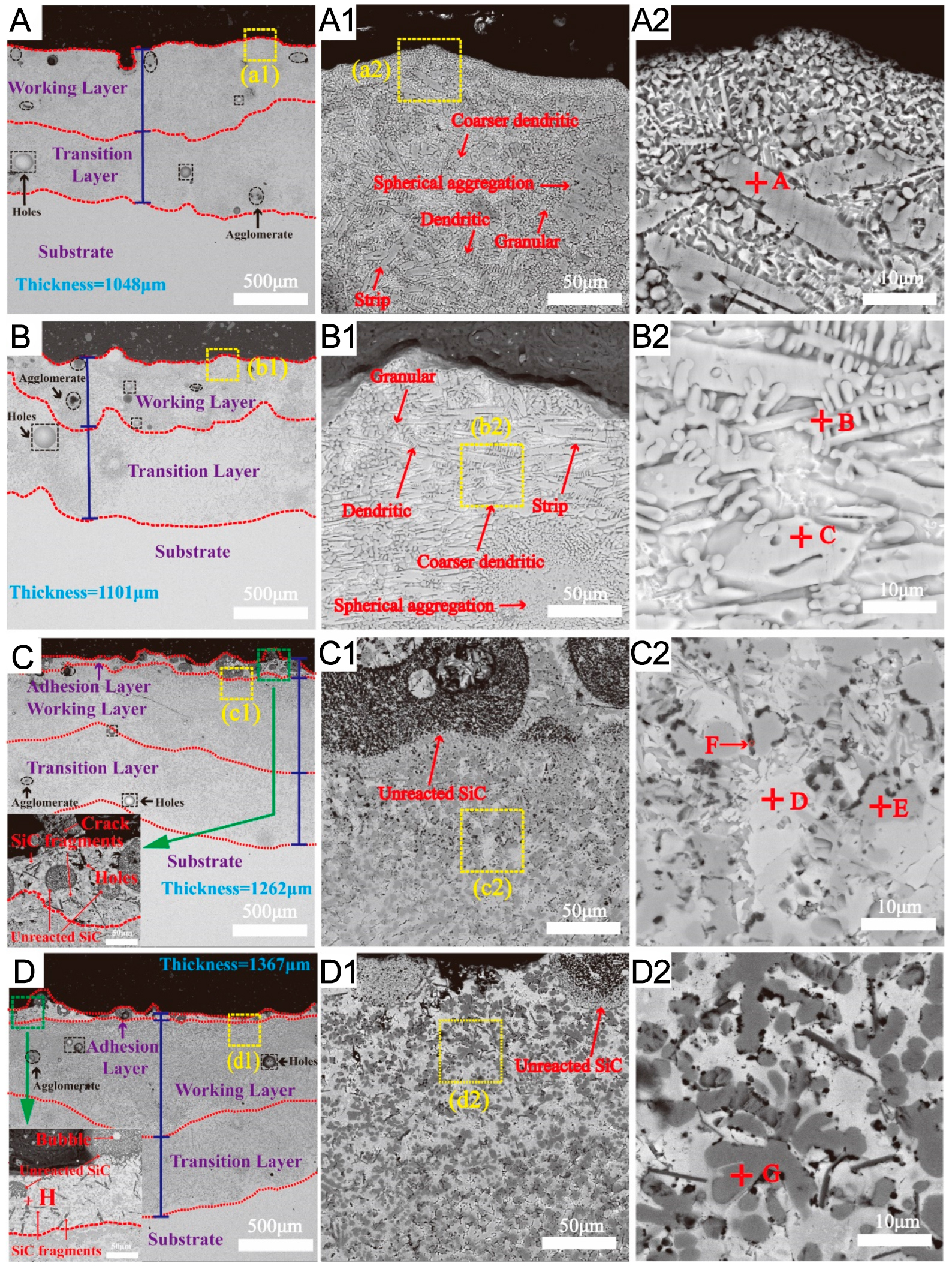
Figure 3. SEM images of the cross-sectional microstructure of the coatings: (A) WL-F0; (B) WL-F5; (C) WL-F10; (D) WL-F15; (A1-D1) Higher-magnification views of the regions in (A-D), respectively; (A2-D2) Further enlargements of (A1-D1), respectively. This figure is adapted from Ref.[51]. Licensed under CC-BY 4.0. SEM: Scanning electron microscopy.