
fig2
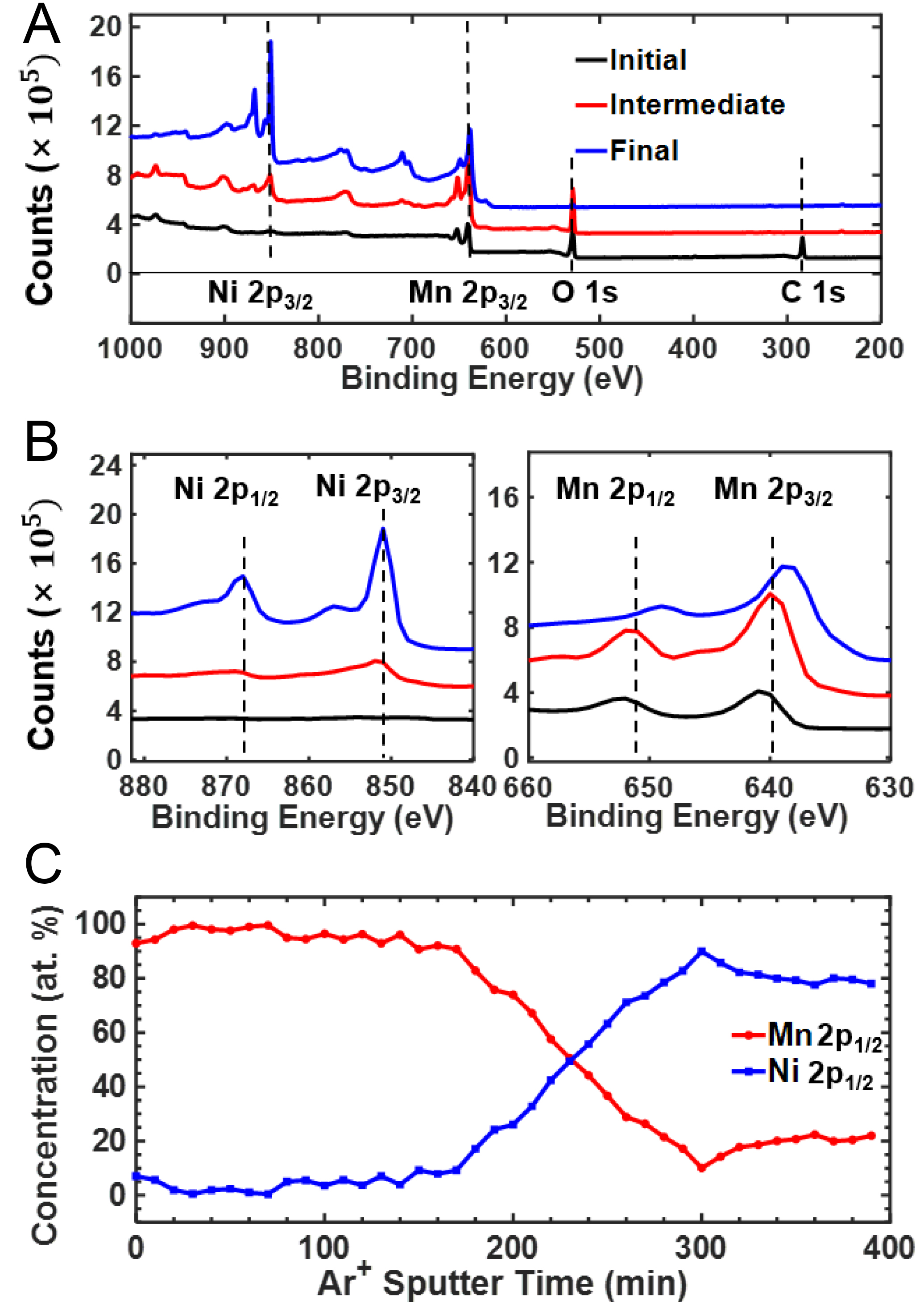
Figure 2. XPS depth profile of Ni54Mn46 oxidized for 2.8 h at 650 K. (A) Survey spectra confirming the presence of O, Ni, and Mn; (B) Ni 2p and Mn 2p core-level spectra after sequential sputtering (initial surface, intermediate, and final); (C) Relative Ni and Mn concentrations derived from the 2p1/2 components, selected to minimize interference from overlapping spectral features and Auger contributions. XPS: X-ray photoelectron spectroscopy.