


fig2
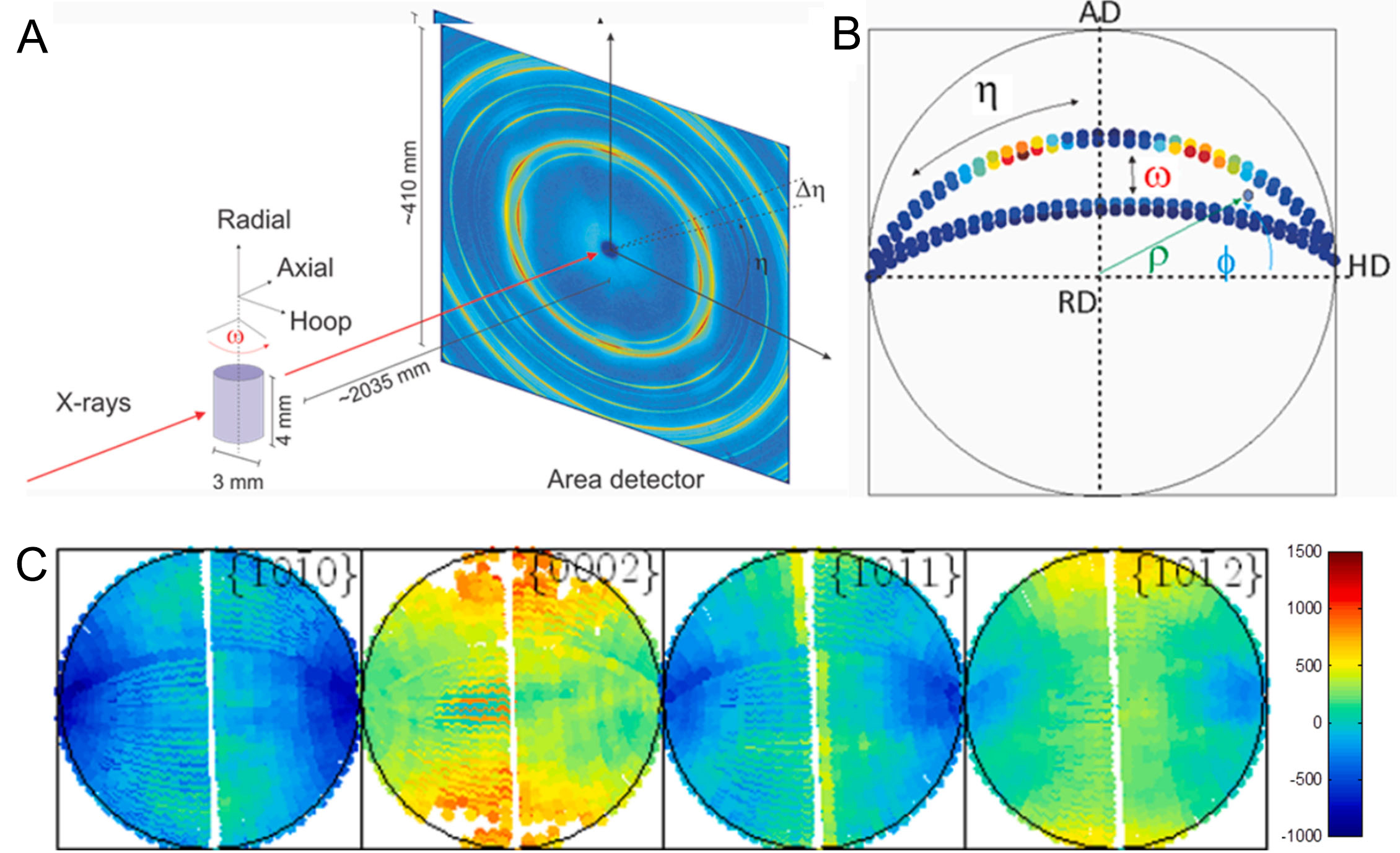
Figure 2. (A) Schematic illustration of the experimental configuration used for in situ HEXRD measurements. The sample is aligned with the axial direction (AD) parallel to the X-ray beam, the hooping direction (HD) horizontal, and the radial direction (RD) vertical. To analyze the peak profiles of grains with different orientations, the specimen rotates around the RD; (B) Construction of a stereographic projection, where each point encodes information such as peak intensity and lattice strain, defined by the azimuth angle (η) and the rotation angle (ω); (C) Experimental strain pole figures. (Reproduced from Ref.[50][50]). HEXRD: High-energy X-ray diffraction.


