fig6

Evidence of shear stress component in <InlineParagraph>Lüders</InlineParagraph> front based on an <i>in-situ</i> synchrotron high-energy X-ray diffraction experiment

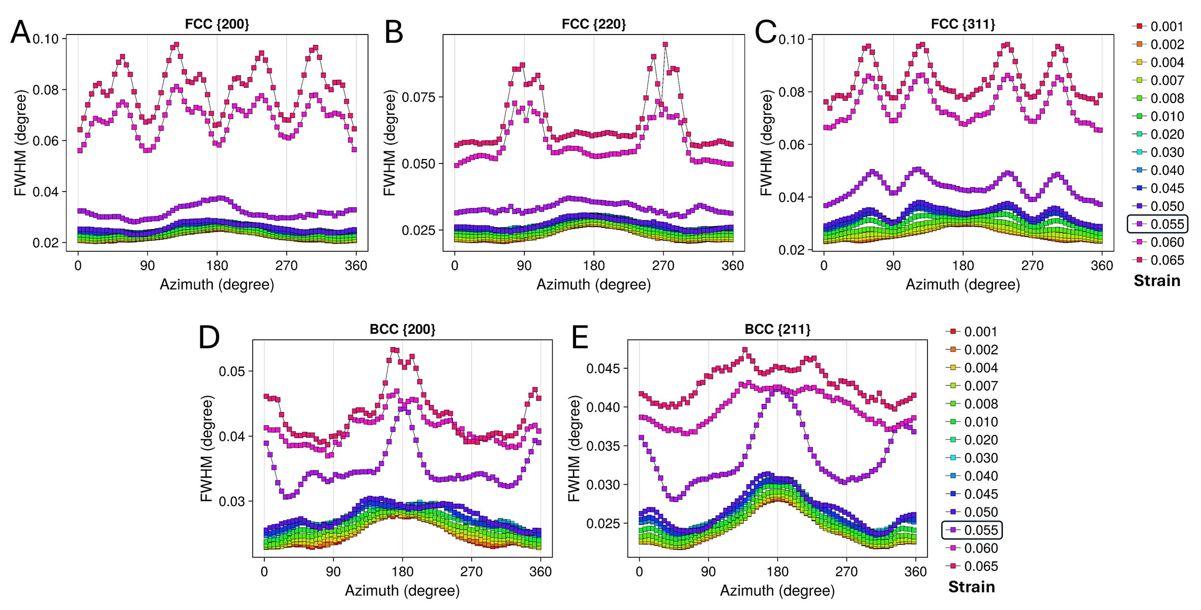
Figure 6. The peak full width at half maximum as a function of azimuth for the FCC γ and BCC α/α’ phases from a series of strain points. (A) FCC {200}. (B) FCC {220}. (C) FCC {311}. (D) BCC {200}.(E) BCC {211}. The strain-point color coding is consistent with that used in the inset of Figure 4. The boxed strain of 0.055 corresponds to the point at which the Lüders bands first interact with the X-ray. (For interpretation of the references to color in this figure legend, the reader is referred to the web version of this article).

Microstructures
ISSN 2770-2995 (Online)

Portico

All published articles are preserved here permanently:

https://www.portico.org/publishers/oae/

Portico

All published articles are preserved here permanently:

https://www.portico.org/publishers/oae/