


fig3
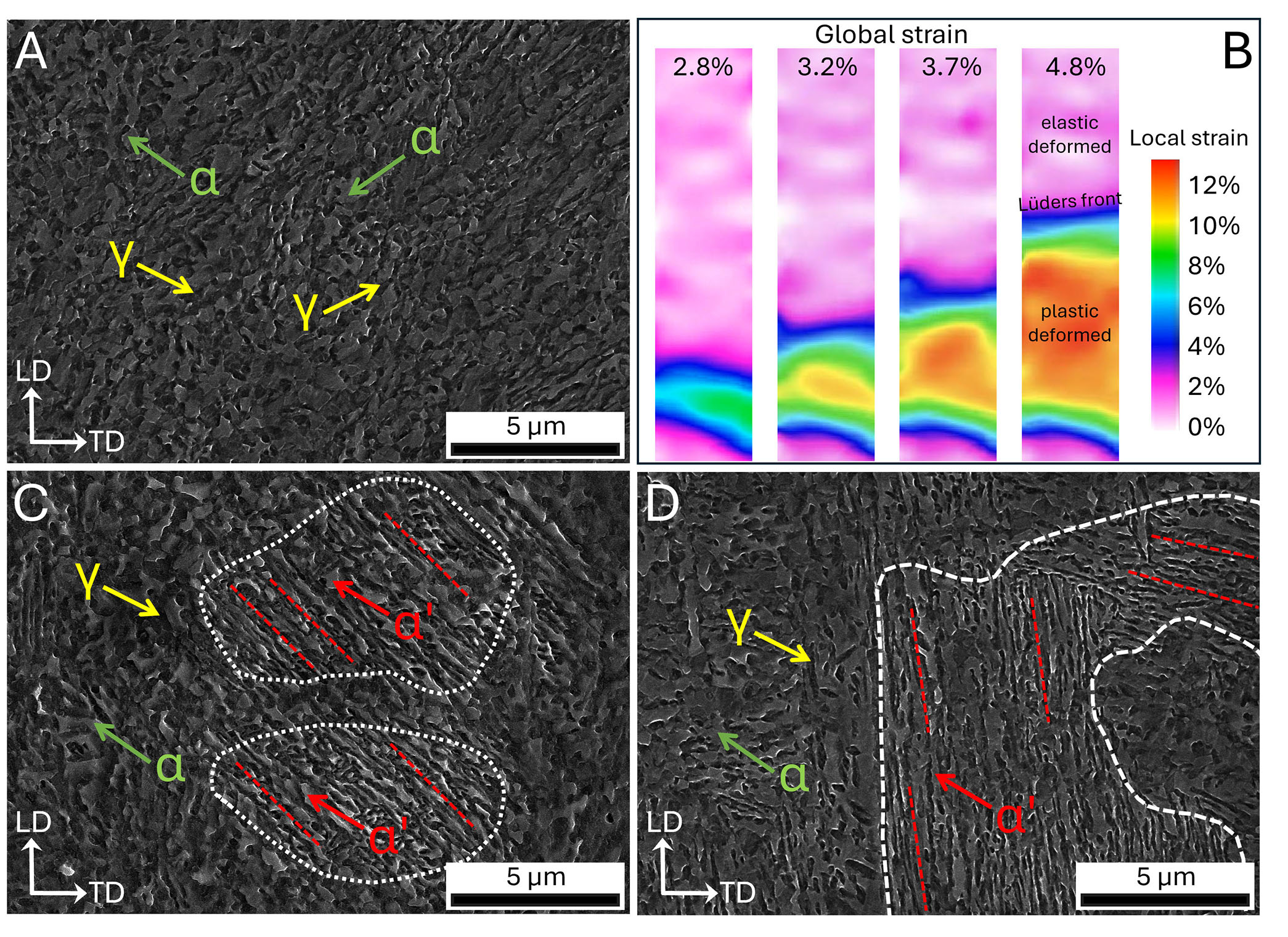
Figure 3. DIC strain field and SEM micrographs. (A) SEM micrograph of the undeformed region. (B) DIC strain field and localized strain. (C) SEM micrograph of the
Figure 3. DIC strain field and SEM micrographs. (A) SEM micrograph of the undeformed region. (B) DIC strain field and localized strain. (C) SEM micrograph of the


All published articles are preserved here permanently:
https://www.portico.org/publishers/oae/


