


fig1
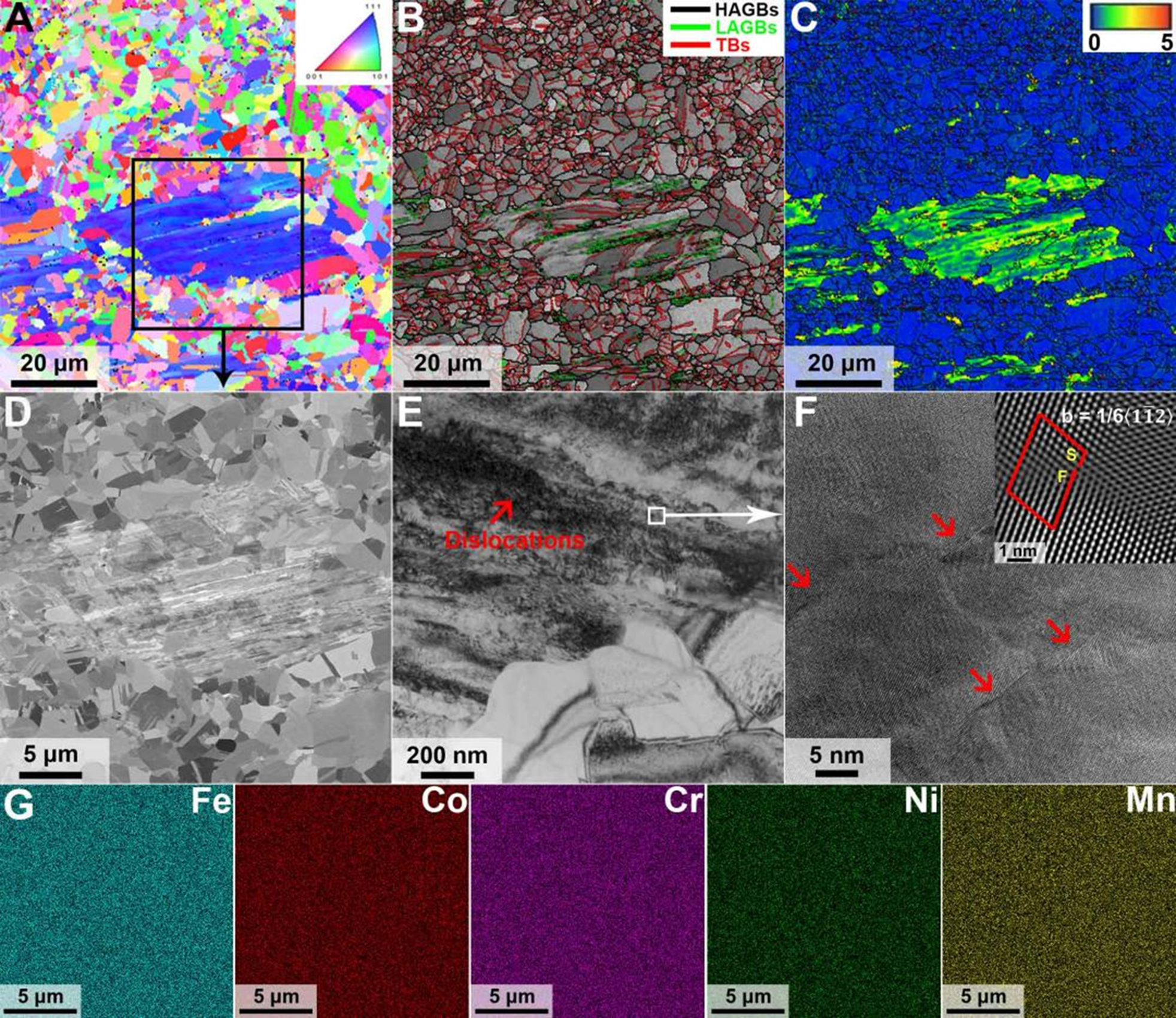
Figure 1. Initial microstructure of the HS-HEA. (A) Inverse pole figure map; (B) Boundary map overlayed with image quality map. High-angle grain boundaries, low-angle grain boundaries, and annealing twin boundaries are highlighted; (C) Kernel average misorientation. These EBSD maps reveal the FCC-structured HS-HEA consisting of both recrystallized and non-recrystallized zones; (D) ECCI image showing the microstructures of the marked zones in (A); (E) Bright-field transmission electron image revealing the non-recrystallized zones with high densities of dislocations; (F) Zoom-in TEM image and the fast Fourier transform filtered high-resolution TEM image of partial dislocation obtained from the areas marked in (E). S and F represent the starting and ending points of Burgers circuit, respectively. b, Burgers vector; (G) SEM-EDS maps of five principal elements, i.e., Fe, Co, Cr, Ni, and Mn, for the zone in (D). All elements are distributed uniformly between recrystallized and non-recrystallized zones. HEA: High-entropy alloy; HS: heterogeneous structure; EBSD: electron backscatter diffraction; FCC: face-centered cubic; ECCI: electron channeling contrast imaging; TEM: transmission electron microscopy; SEM: scanning electron microscope; EDS: energy-dispersive X-ray spectroscopy.


