


fig3
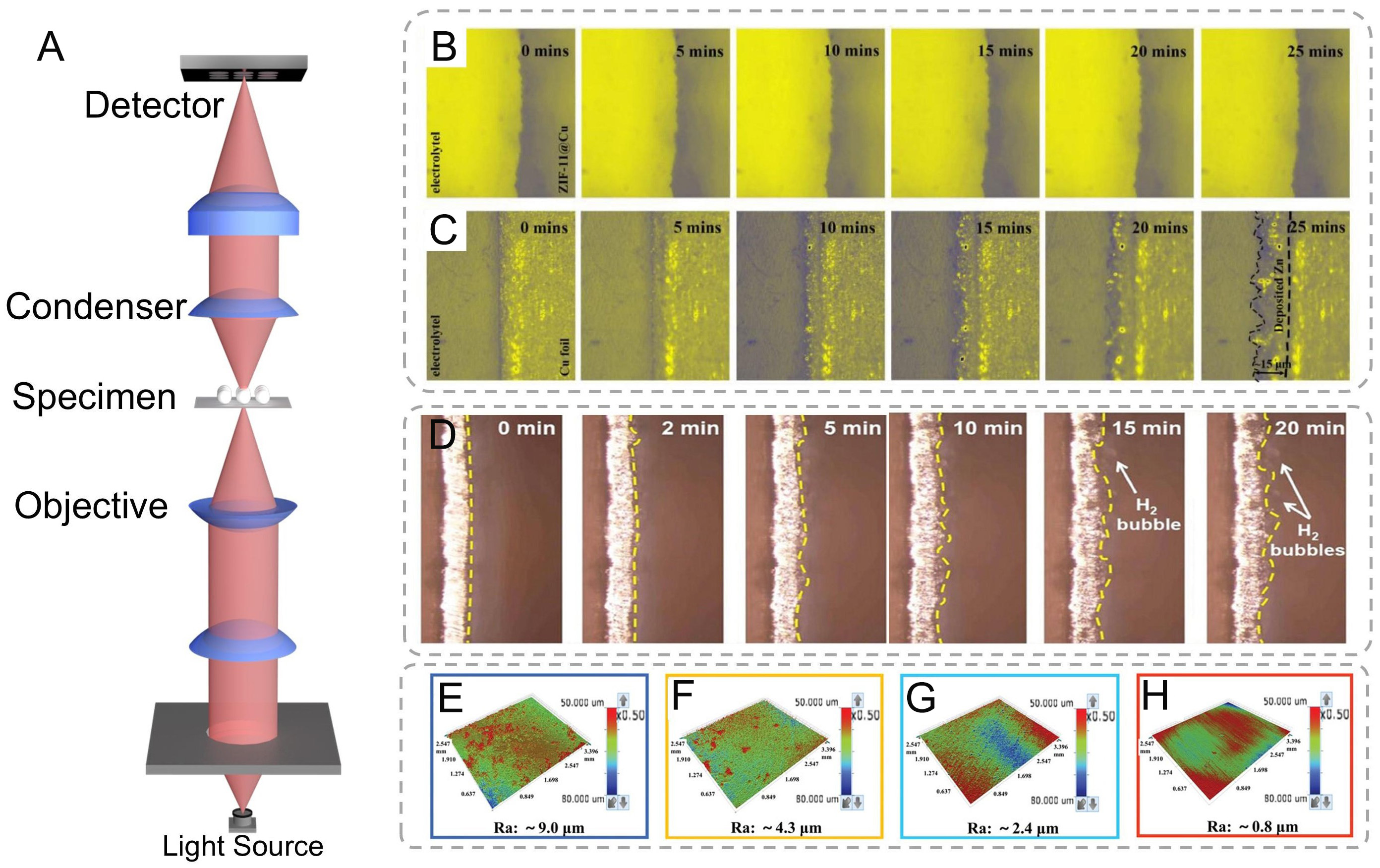
Figure 3. (A) Schematics of the optical microscope. In situ optical microscopy images of Zn deposits on (B) ZIF-11@Cu and (C) bare Cu electrode surface at 0, 5, 10, 15, 20, and 25 min. (B and C) are reproduced from Ref.[92]. with permission, Copyright 2021, Elsevier. (D) In situ operando optical microscope images showing hydrogen evolution behavior[42]. Copyright 2024, Wiley. (E-H) 3D optical images of Zn foils from Zn-Zn symmetrical batteries after cycling in 2 M ZnSO4[96]. Copyright 2022, Wiley.


