


fig5
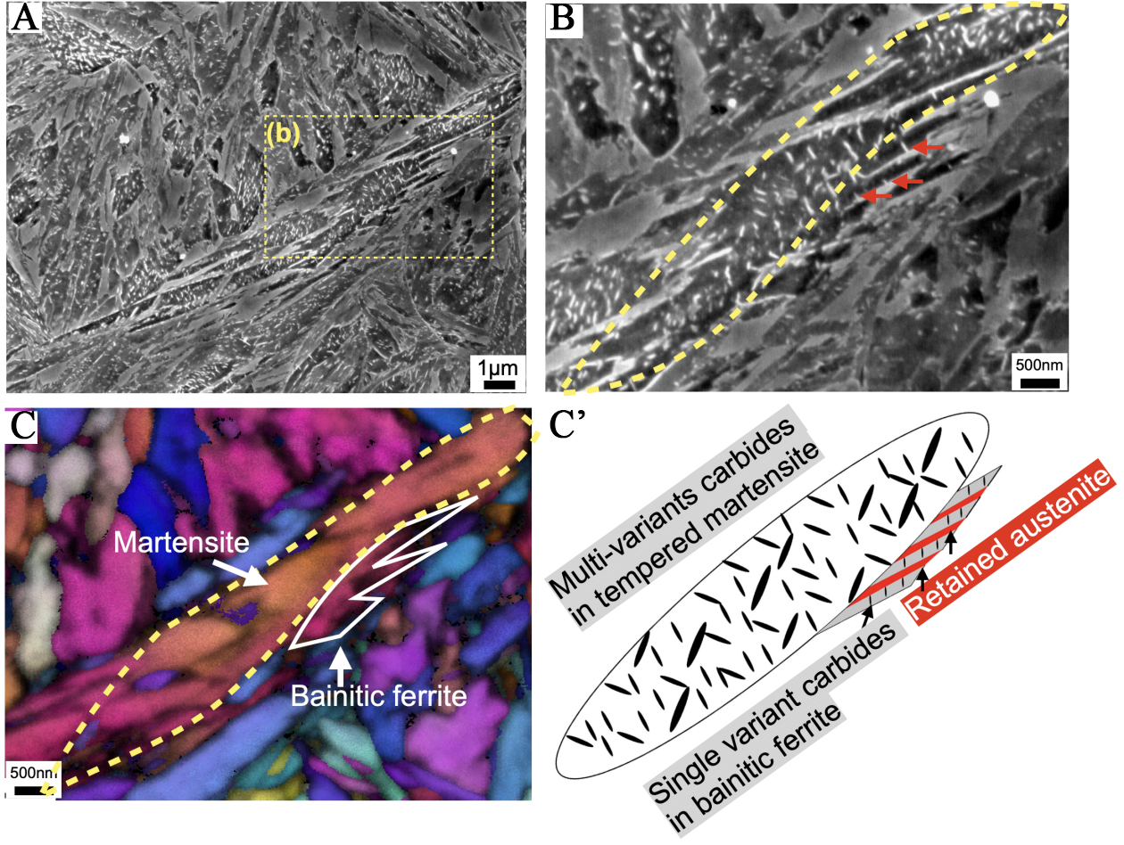
Figure 5. SEM images of specimens subjected to (A) isothermal holding at 230 °C for 90 min; (B) Enlarged SEM image and (c) EBSD IPF map of the rectangular area highlighted in (A); (C’) Schematic illustration of the transformed microstructure, consisting of tempered martensite and bainite. SEM: Scanning electron microscope; EBSD: electron backscatter diffraction; IPF: inverse pole figure.


