


fig4
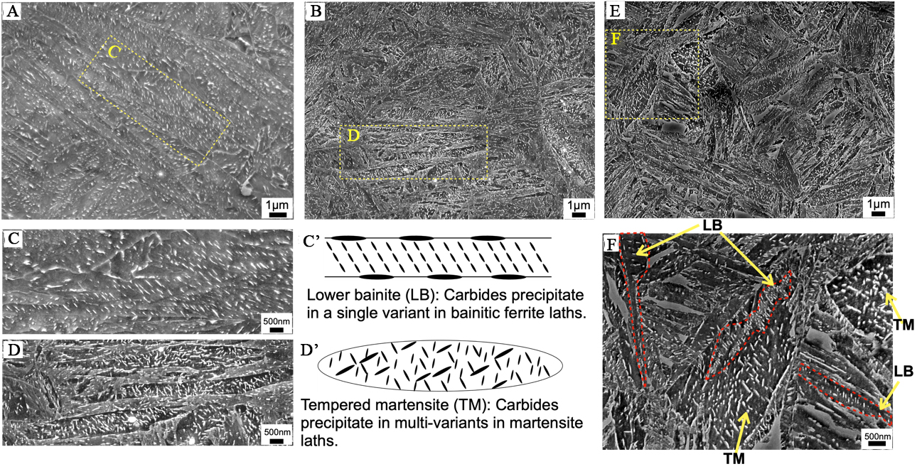
Figure 4. SEM images of specimens subjected to (A) isothermal holding at 320 °C for 90 min and (B) quenching to ambient temperature followed by tempering at 230 °C for 90 min. (C) and (D) Enlarged views of the rectangular areas in (A) and (B), highlighting typical LB and TM, respectively. Schematic illustration of (C’) LB and (D’) TM. SEM microstructures obtained by (E) isothermal holding at 250 °C for 90 min, and (F) presents the enlarged views of the rectangular areas in (E). SEM: Scanning electron microscope; LB: lower bainite; TM: tempered martensite.


