


fig5
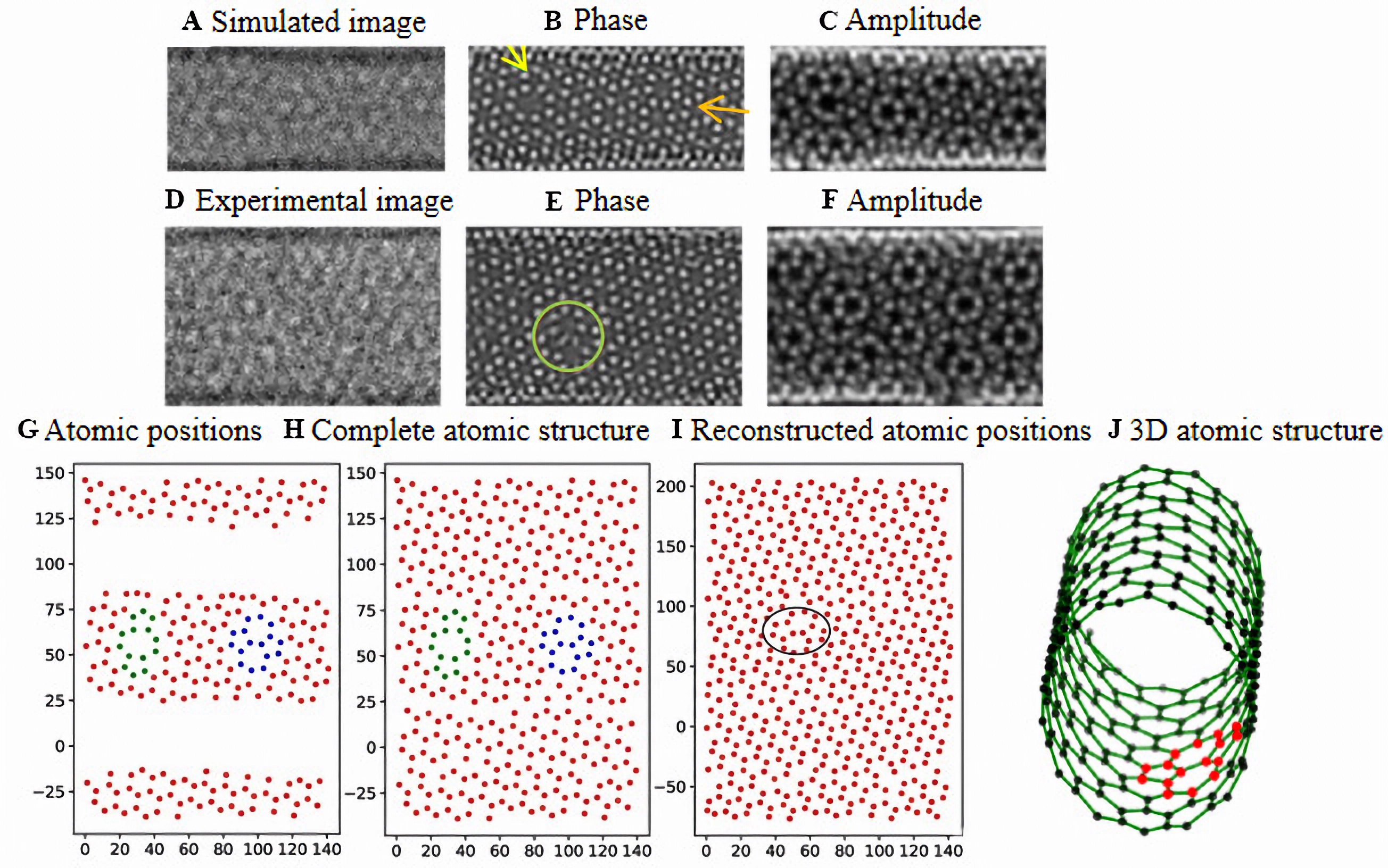
Figure 5. (A) Simulated and (B) experimental HRTEM images of an SWCNT. (C and D) Phase and (E and F) amplitude components of the wave function predicted from (C and E) simulated and (D and F) experimental images using WRGAN; (G) Atomic positions extracted from (C and E) and projected onto a 2D plane, corresponding to the simulated HRTEM image; (H) Complete atomic structure after estimating missing sidewall atoms in (G); (I) Final reconstructed atomic positions derived from (D and F) with all missing atoms interpolated. Defect sites are highlighted in (C and D) and (G-I); (J) The 3D atomic structure after cylindrical rolling from the sheet of (I). HRTEM: High-resolution transmission electron microscopy; SWCNT: single-walled carbon nanotube; WRGAN: Wave-Reconstruction Generative Adversarial Networks.


