

Epitranscriptomic landscapes in periodontitis: unraveling the regulatory roles of RNA Modifications
 0
Graphical Abstract
Abstract
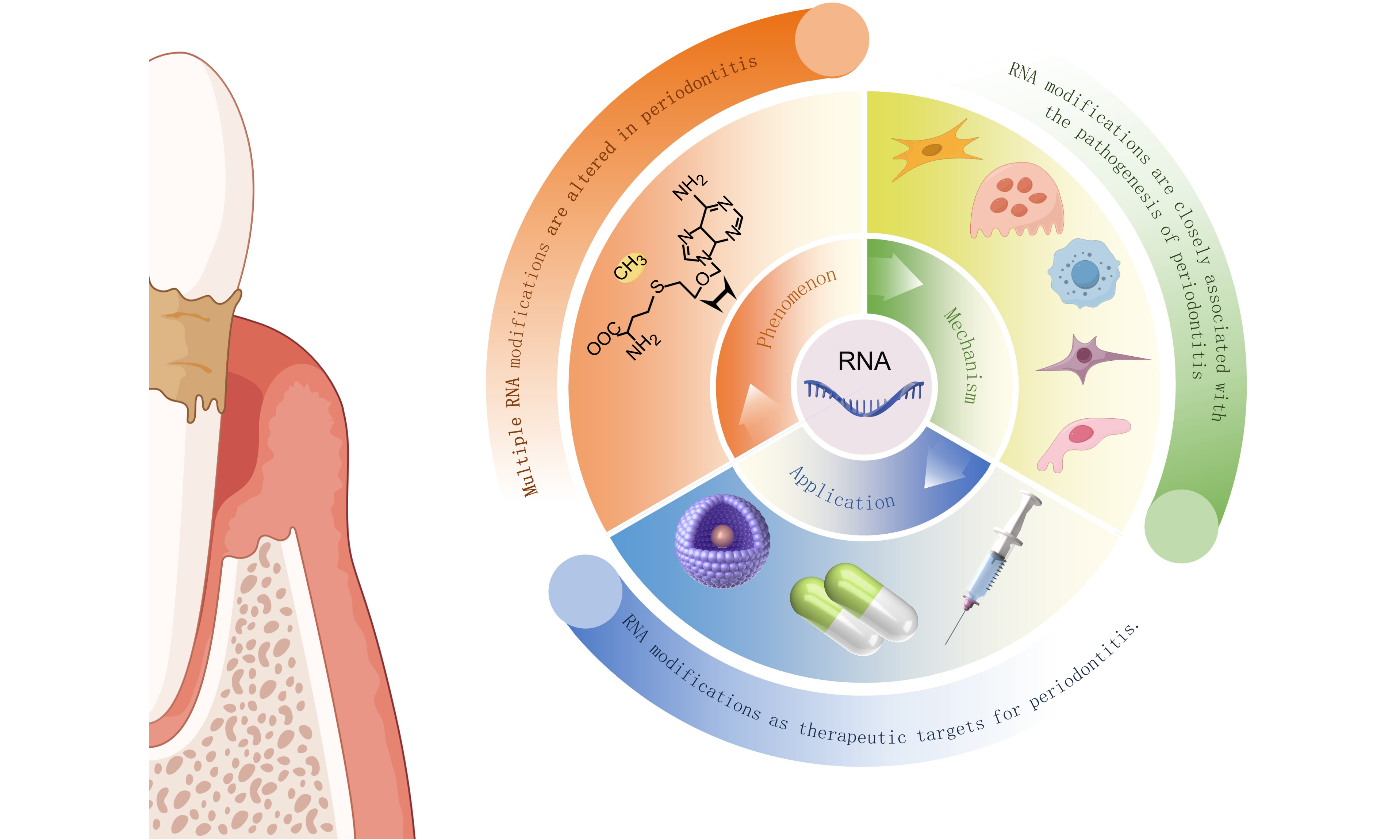
Epitranscriptomics has emerged as a rapidly expanding field investigating chemical modifications of RNA molecules that regulate gene expression without altering nucleotide sequences. Among these modifications, N6-methyladenosine (m6A) represents the most prevalent internal modification in eukaryotic messenger RNAs, with its dynamic and reversible regulation mediated by writers, erasers, and readers. Periodontitis, a chronic inflammatory disease affecting tooth-supporting tissues, involves complex interactions between microbial factors and host immune responses. Recent evidence has revealed significant roles for RNA modifications in periodontal pathogenesis, offering a molecular perspective on the disease heterogeneity influenced by host and environmental factors. This review synthesizes current understanding of how m6A and other RNA modifications contribute to periodontitis, examining their effects on key cellular processes including osteogenic differentiation, inflammatory responses, and bone remodeling. The involvement of specific modifications - including N4-acetylcytidine (ac4C), N6,2’-O-dimethyladenosine (m6Am), N7-methylguanosine (m7G), 5-methylcytosine (m5C), and N1-methyladenosine (m1A) - is also discussed. Furthermore, translational opportunities are explored, encompassing small molecule inhibitors targeting modification enzymes, repurposed pharmaceutical agents, natural compounds, and advanced biomaterials. The review highlights how epitranscriptomic mechanisms offer new perspectives on periodontal disease pathogenesis and presents emerging directions for diagnostic biomarker development and targeted therapeutic strategies.
Keywords


 Download PDF
INTRODUCTION
Periodontitis is a chronic inflammatory disease of microbial etiology that affects the tooth-supporting tissues, representing one of the most prevalent conditions worldwide and a major cause of tooth loss in adults[1,2]. The disease is characterized by a dysbiotic biofilm triggering an aberrant host inflammatory response, leading to progressive destruction of the periodontal ligament, alveolar bone resorption, and ultimately tooth loss if left untreated[3]. Beyond its oral manifestations, periodontitis has been increasingly recognized for its bidirectional associations with systemic conditions including diabetes mellitus, cardiovascular diseases, and adverse pregnancy outcomes, underscoring its significance as a public health priority[4].
The pathogenesis of periodontitis involves complex interactions between microbial factors and host immune responses, with genetic and epigenetic determinants modulating individual susceptibility and disease progression[2,3,5]. While the microbial etiology is well-established, the molecular mechanisms governing the exaggerated inflammatory response and impaired tissue regeneration in periodontitis remain incompletely understood. Recent advances in epitranscriptomics have unveiled a new layer of gene regulation through RNA modifications. Among these, N6-methyladenosine (m6A) has attracted particular attention due to its prevalence and critical regulatory roles in eukaryotic messenger RNAs and non-coding RNAs[6]. The dynamic and reversible nature of m6A modification, orchestrated by methyltransferases (writers), demethylases (erasers), and binding proteins (readers), allows for precise spatiotemporal control of RNA metabolism, including splicing, export, translation, and decay[7].
The periodontal tissues comprise a heterogeneous population of cells - including gingival fibroblasts (HGFs), periodontal ligament stem cells (PDLSCs), bone marrow mesenchymal stem cells (BMSCs), osteoblasts, osteoclasts, macrophages, and cementoblasts - that collectively maintain tissue homeostasis, mount innate and adaptive immune responses, and mediate the destruction and regeneration of the periodontium. The function and fate of these diverse cell types are profoundly influenced by the epitranscriptomic landscape, which this review explores in detail.
Beyond m6A, the epitranscriptomic landscape encompasses numerous other modifications including N4-acetylcytidine (ac4C), N6,2’-O-dimethyladenosine (m6Am), N7-methylguanosine (m7G), 5-methylcytosine (m5C), and N1-methyladenosine (m1A), each with distinct regulatory functions and implications in human diseases[8]. While research on non-m6A modifications in periodontitis remains in its infancy, preliminary studies suggest their potential involvement in inflammatory signaling and bone metabolism, warranting further investigation.
This review provides a comprehensive synthesis of current knowledge regarding RNA modifications in periodontitis, with particular emphasis on m6A modification and its regulatory mechanisms. The emerging evidence linking specific m6A regulators and target genes to periodontal inflammation, immune cell function, and alveolar bone homeostasis is discussed in detail. Additionally, the growing literature on non-m6A modifications and their contributions to periodontal pathology is examined. Finally, the translational implications of these findings are considered, and future directions for research are outlined that may ultimately lead to novel diagnostic and therapeutic strategies for periodontitis management.
It should be noted that while this review focuses primarily on periodontitis, mechanistic insights derived from apical periodontitis, osteoporosis, and peri-implant models are also discussed where direct periodontitis evidence is lacking. These related models offer valuable clues regarding the roles of RNA modifications in inflammatory bone remodeling and cell differentiation, which may inform future periodontal research. Throughout this review, we explicitly distinguish direct evidence from periodontitis models versus complementary evidence from related models to provide a transparent assessment of the current state of knowledge.
M6A MODIFICATION AND PERIODONTITIS
m6A modification: an overview
m6A represents the most abundant internal modification in mammalian mRNA, accounting for approximately 0.2%-0.6% of all adenosines in mammalian mRNA[9,10]. This modification involves the methylation of the nitrogen-6 position of adenosine, creating a molecular mark that influences multiple aspects of RNA metabolism[7]. The discovery of the first m6A demethylase, fat mass and obesity-associated protein (FTO), in 2010 fundamentally challenged the previously held notion that RNA modifications were static and irreversible, marking the emergence of epitranscriptomics as a dynamic regulatory layer[9].
The installation, removal, and recognition of m6A modifications are orchestrated by three classes of proteins collectively referred to as writers (methyltransferases), erasers (demethylases), and readers (binding proteins)[11], as presented in Figure 1. The methyltransferase complex responsible for m6A deposition, known as the writer complex, is a multicomponent assembly centered on methyltransferase-like 3 (METTL3) and methyltransferase-like 14 (METTL14) as the core catalytic subunits[12]. METTL3 functions as the catalytic subunit that transfers the methyl group from S-adenosylmethionine to the adenosine residue, while METTL14 serves a primarily supportive and structural role within the writer complex: it stabilizes the methyltransferase complex and facilitates RNA substrate binding, rather than directly catalyzing methyl transfer[12]. Additional components including Wilms tumor 1-associated protein (WTAP), KIAA1429 (also known as VIRMA), RNA-binding motif protein 15 (RBM15), and zinc finger CCCH domain-containing protein 13 (ZC3H13) contribute to the proper localization and substrate specificity of the methyltransferase complex[12]. This multicomponent nature allows for sophisticated regulation of m6A deposition in response to cellular signals and environmental cues.
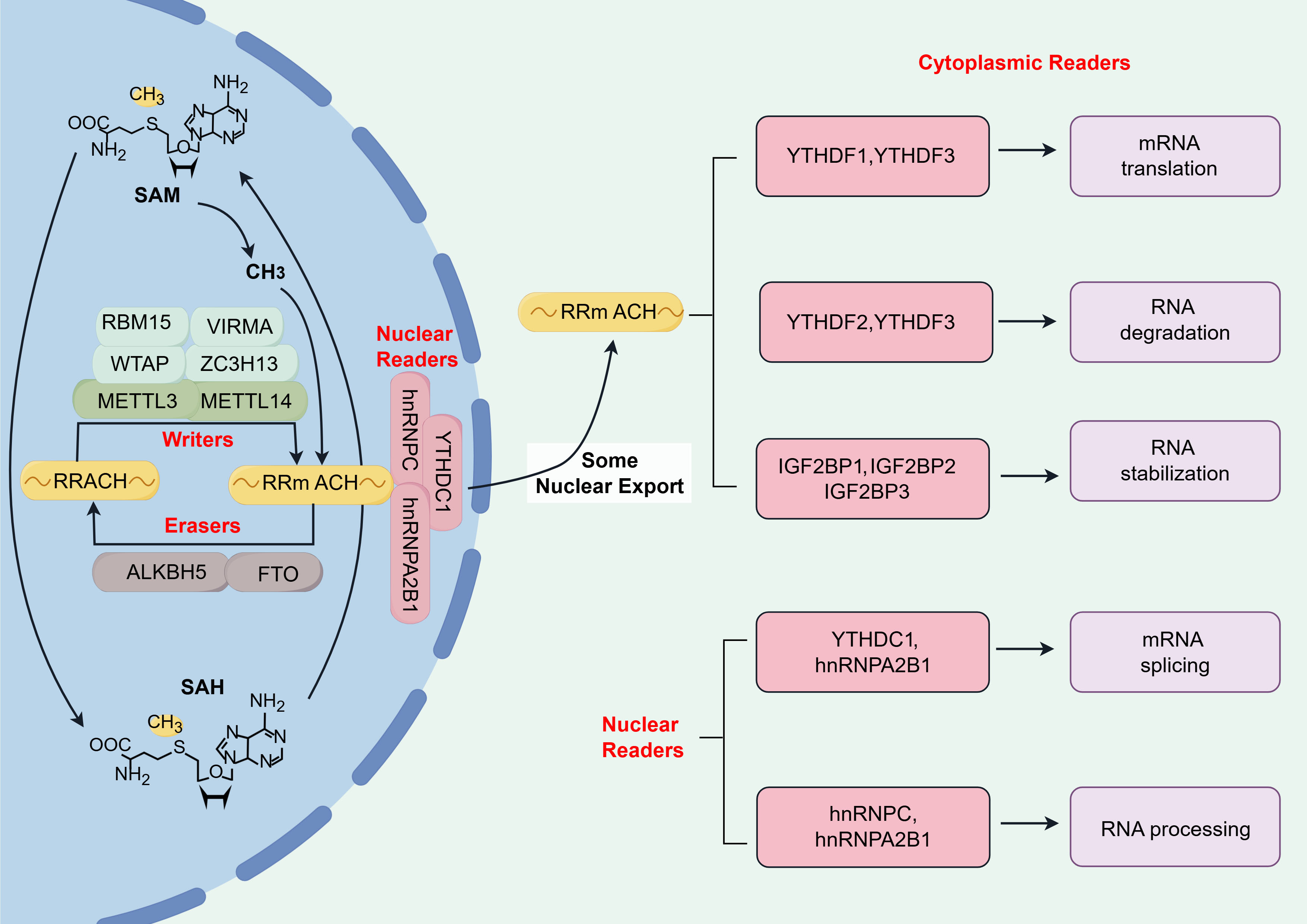
Figure 1. Overview of m6A modification. (Created with Figdraw; ID: TUTTP80f46). RBM15: RNA-binding motif protein 15; VIRMA: vir like m6A methyltransferase associated; WTAP: Wilms tumor 1-associated protein; ZC3H13: zinc finger CCCH domain-containing protein 13; METTL3: methyltransferase-like 3; METTL14: methyltransferase-like 14; ALKBH5: AlkB homolog 5; FTO: fat mass and obesity-associated protein; SAM: S-adenosylmethionine; SAH: S-adenosylhomocysteine; YTHDC1: YTH N6-methyladenosine RNA binding protein C1; hnRNPC: heterogeneous nuclear ribonucleoprotein C; hnRNPA2B1: heterogeneous nuclear ribonucleoprotein A2/B1; YTHDF: YTH N6-methyladenosine RNA binding protein F; IGF2BP: insulin-like growth factor 2 mRNA-binding protein; m6A: N6-methyladenosine.
The removal of m6A modifications is catalyzed by two primary demethylases or erasers: FTO and AlkB homolog 5 (ALKBH5)[7]. Both enzymes are Fe(II)/α-ketoglutarate-dependent dioxygenases but exhibit distinct substrate preferences and subcellular localizations[7]. FTO demonstrates broader substrate specificity, capable of demethylating m6A as well as m6Am at the cap-proximal nucleotide, whereas ALKBH5 appears more specific for internal m6A residues[7,13]. The discovery of these erasers established that m6A modification is a reversible process, enabling dynamic regulation of gene expression in response to physiological and pathological stimuli[9].
The functional consequences of m6A modification are largely mediated by reader proteins that specifically recognize and bind to methylated adenosines. The most extensively characterized readers are the YT521-B homology (YTH) domain-containing proteins, including YTH N6-methyladenosine RNA binding protein F1 (YTHDF1), YTHDF2, YTHDF3, and YTH N6-methyladenosine RNA binding protein C1 (YTHDC1)[12]. YTHDF2 primarily promotes mRNA decay by localizing bound transcripts to processing bodies, while YTHDF1 enhances translation efficiency by interacting with translation initiation machinery[12]. YTHDF3 appears to cooperate with both DF1 and DF2, modulating both translation and decay[14]. Nuclear reader YTHDC1 regulates alternative splicing and nuclear export of m6A-modified transcripts[15]. Beyond YTH domain proteins, other readers including insulin-like growth factor 2 mRNA-binding proteins (IGF2BPs) and heterogeneous nuclear ribonucleoproteins (hnRNPs) contribute to the diverse functional outcomes of m6A modification by influencing RNA stability, localization, and processing[12].
m6A has been extensively studied in cancers, where it modulates tumorigenesis and progression through regulating various oncogenes and tumor suppressors. For instance, METTL3-mediated m6A modification of pri-miR-93 promotes hepatocellular carcinoma progression via CDKN1A suppression[16], and the interplay between m6A modification and ferroptosis has emerged as a promising therapeutic avenue in cancer research[17].
In the context of the oral cavity and periodontal tissues, m6A modification has emerged as an important regulator of cellular functions essential for tissue homeostasis. PDLSCs, which play a fundamental role in periodontal regeneration, exhibit dynamic changes in m6A levels during osteogenic differentiation[18]. Similarly, HGFs and immune cells residing in the periodontium likely depend on proper m6A regulation to maintain appropriate inflammatory responses and tissue remodeling capacity[19,20]. The recognition that m6A modification governs key processes such as cell proliferation, differentiation, and inflammatory mediator production has provided a rationale for investigating its role in periodontitis pathogenesis.
m6A modification in periodontitis and related models
The role of m6A modification in periodontitis has gained increasing attention in recent years [Figure 2]. Evidence from bioinformatic analyses, clinical samples, and cellular studies collectively supports its involvement in disease pathogenesis. Genetic variants affecting m6A modification may contribute to periodontitis susceptibility. A genome-wide integration of periodontitis GWAS data with the m6AVar database identified 104 m6A-associated SNPs to be linked to periodontitis, with the leading SNP rs2723183 predicted to regulate IL-37 expression[21]. m6A modification patterns also shape the immune microenvironment of periodontitis. Analysis of 310 periodontitis samples identified 1,631 m6A phenotype-related genes and revealed three distinct m6A modification subtypes, each with unique immune cell infiltration profiles, immune reaction activities, and HLA gene expression patterns[22]. Clinical tissue samples have provided direct evidence of m6A dysregulation in periodontitis. Using m6A microarray analysis of human gingival tissues, 458 differentially methylated mRNAs and 750 differentially expressed mRNAs were identified between disease and health[23]. Two novel m6A-modified biomarkers, DNER and GNL2, were validated as downregulated in periodontitis and may serve as potential diagnostic targets[23]. At the cellular level, the impaired osteogenic differentiation of periodontal ligament cells under inflammatory conditions is linked to altered m6A methylation. MeRIP-seq and RNA-seq revealed that differentially m6A-methylated genes were primarily enriched in histone modification processes[24]. Among these, 25 histone modification genes were identified as potential regulators of osteogenic differentiation through m6A-dependent mechanisms[24].
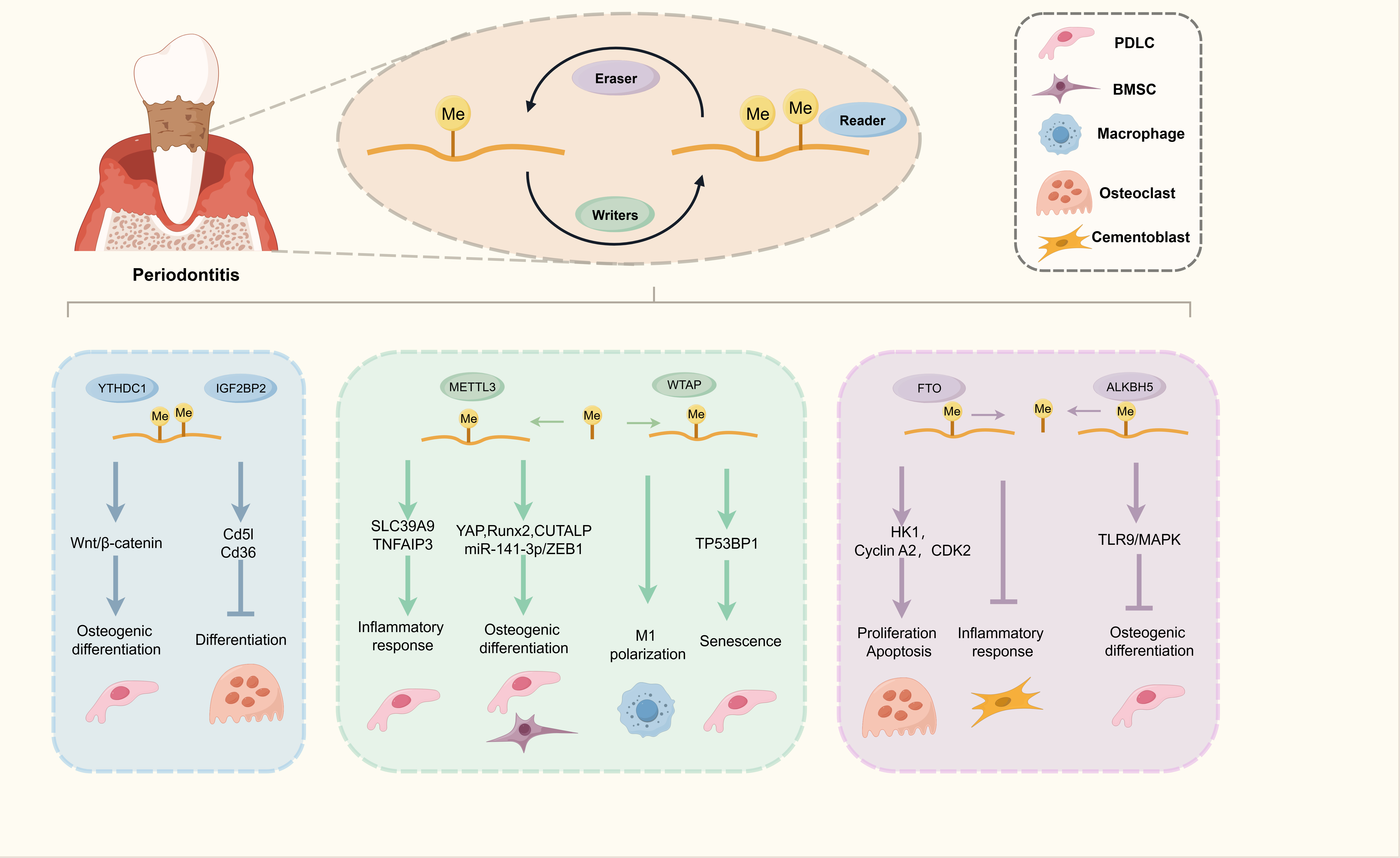
Figure 2. The regulatory landscape of m6A modification in periodontitis and related disease models. (Created with Figdraw; ID: AYWWUd8ee8). PDLC: Periodontal ligament cell; BMSC: bone marrow mesenchymal stem cell; YTHDC1: YTH N6-methyladenosine RNA binding protein C1; IGF2BP: insulin-like growth factor 2 mRNA-binding proteins; METTL3: methyltransferase-like 3; WTAP: Wilms tumor 1-associated protein; SLC39A9: solute carrier family 39 member 9; TNFAIP3: TNF alpha induced protein 3; YAP: Yes1 associated transcriptional regulator; Runx2: RUNX family transcription factor 2; TP53BP1: tumor protein p53 binding protein 1; FTO: fat mass and obesity-associated protein; ALKBH5: AlkB homolog 5; TLR9: toll-like receptor 9; MAPK: mitogen-activated protein kinase; CDK2: cyclin dependent kinase 2; HK1: hexokinase 1; ZEB1: zinc finger E-box binding homeobox 1; m6A: N6-methyladenosine.
METTL3 orchestrates osteogenic differentiation of periodontal progenitors
Among the m6A regulators, METTL3 is the most extensively studied in periodontal research. METTL3 and its mediated m6A modification play critical roles in promoting osteogenic differentiation of various periodontal progenitor cells[18,25-27]. Mechanistically, METTL3 increases the m6A methylation of Yes1 associated transcriptional regulator (YAP) mRNA in an IGF2BP1-mediated manner, thus enhancing YAP mRNA stability[18]. Furthermore, METTL3 enhances the translation efficiency of YAP by recruiting YTHDF1 and eukaryotic translation initiation factor 3 subunit A (eIF3a) to the translation initiation complex[18]. METTL3-based m6A modification also favors osteogenic differentiation of PDLSCs and BMSCs through IGF2BP1-mediated stabilization of RUNX family transcription factor 2 (Runx2) mRNA, a master transcription factor for osteogenesis[25,28]. Beyond direct regulation of osteogenic genes, METTL3 orchestrates a complex network of non-coding RNAs. Comprehensive analyses of long non-coding RNA (lncRNA) and mRNA m6A modification patterns in PDLSCs from periodontitis patients and healthy individuals have revealed dynamic epitranscriptomic remodeling[29]. METTL3-mediated m6A modification regulates osteogenic differentiation through lncRNA CUTALP in periodontal mesenchymal stem cells derived from periodontitis patients[30]. MicroRNAs are also involved in METTL3-mediated regulation of osteogenic differentiation. METTL3 facilitated the conversion of pri-miR-141 into miR-141-3p via m6A modification, resulting in increased miR-141-3p levels, which in turn suppressed zinc finger E-box binding homeobox 1 (ZEB1) expression. The miR-141-3p/ZEB1 axis mediates METTL3’s protective effects on osteogenic differentiation in PDLSCs[31].
METTL3 modulates inflammatory responses in periodontal cells
Beyond its role in osteogenic differentiation, METTL3 actively participates in the regulation of inflammatory responses. Knockdown of METTL3 or METTL14 suppresses LPS-induced interleukin-6 expression in periodontal ligament cells[32]. METTL3 and METTL14 mediate m6A modification of zinc transporter solute carrier family 39 member 9 (SLC39A9) mRNA, which subsequently influences intracellular zinc ion levels[32]. This alteration affects DNA methylation levels at the interleukin 6 (IL-6) gene promoter, establishing a mechanistic link between RNA methylation and epigenetic regulation of inflammatory genes[32]. Genetic deletion of METTL3 alleviates periodontal destruction via suppressing NLRP3 inflammasome activation[19]. The stability of TNF alpha induced protein 3 (TNFAIP3) transcript is significantly attenuated upon m6A modification[19]. When METTL3 is silenced, accumulated TNFAIP3 facilitates the ubiquitination of NIMA related kinase 7 (NEK7), subsequently impairing NLR family pyrin domain containing 3 (NLRP3) inflammasome assembly and reducing inflammatory tissue damage[19].
WTAP shapes the periodontal immune microenvironment and regulates stem cell senescence
In addition to METTL3, other components of the m6A writer complex play significant roles in periodontitis pathogenesis. WTAP expression is upregulated in periodontitis tissues[33]. Reducing WTAP expression promotes a shift from pro-inflammatory (M1) to pro-regenerative (M2) macrophage phenotype under inflammatory conditions, thereby attenuating periodontal inflammation and favoring tissue regeneration[33]. WTAP also contributes to periodontitis pathogenesis by promoting PDLSC senescence and impairing osteogenic differentiation[34]. Cellular senescence, a state of stable cell-cycle arrest triggered by replication stress or DNA damage, contributes to chronic tissue degeneration and represents a therapeutic target in various diseases[35]. WTAP promotes senescence in PDLSCs through m6A-dependent regulation of tumor protein p53 binding protein 1 (TP53BP1), leading to increased expression of senescence-associated proteins[34].
FTO and ALKBH5 as key regulators of bone remodeling and cementoblast function
The m6A erasers FTO and ALKBH5 also play important roles in periodontal pathology, particularly in bone remodeling. FTO promotes osteoclastogenesis and alveolar bone resorption through multiple mechanisms[36,37]. Evidence from apical periodontitis models suggests a role for FTO in inflammatory bone loss, where reduced m6A levels in alveolar bone and osteoclasts correlate with upregulated FTO expression[36]. Mechanistically, FTO-mediated demethylation of hexokinase 1 mRNA enhances glycolytic pathway activity, which stabilizes RANK protein through USP14-mediated deubiquitination, thereby promoting osteoclastogenesis[36]. Extending these observations to periodontitis, it is plausible that similar FTO-dependent mechanisms may contribute to alveolar bone resorption, though direct evidence in periodontitis models remains to be established. Moreover, FTO promotes the stability of S phase-related genes including Cyclin A2 and CDK2 mRNAs in a YTHDF2-dependent manner, thus modulating the proliferation and apoptosis of osteoclast precursors in inflammatory conditions[37]. Furthermore, FTO is involved in the biology of cementoblasts, the specialized cells responsible for cementum formation[38]. Cementum is a thin, mineralized tissue covering the tooth root that serves two critical functions: it anchors the periodontal ligament fibers to the tooth, providing stable tooth support, and it acts as a protective barrier against microbial invasion. The destruction of cementum during periodontitis directly compromises tooth attachment and hinders the formation of a functional new attachment following therapy. Therefore, preserving cementoblast function and promoting cementum regeneration are essential for successful periodontal treatment. Research indicates that FTO is a key regulator of the cementoblast inflammatory response. In a mouse model of apical periodontitis, Fto expression was found to be significantly downregulated[38]. Similarly, exposing cementoblast cell lines (OCCM-30) to the pro-inflammatory cytokine tumor necrosis factor-α (TNF-α) reduced Fto expression while upregulating inflammation-related genes[38]. Functional studies revealed that knocking down Fto in cementoblasts under inflammatory stress further exacerbated the expression of genes associated with inflammation and tissue degradation[38]. Notably, this effect was not due to changes in the stability of these mRNAs, suggesting that FTO’s regulatory role in cementoblasts may involve alternative mechanisms, such as influencing translation efficiency or other post-transcriptional pathways[38]. These findings highlight that the loss of FTO under inflammatory conditions contributes to cementoblast dysfunction and cementum degradation, positioning FTO as a potential therapeutic target for preserving cementum integrity and promoting periodontal regeneration. Beyond FTO, ALKBH5 also plays a role in regulating inflammation and osteogenic differentiation by activating the toll-like receptor 9 (TLR9)/ mitogen-activated protein kinase (MAPK) pathway in periodontitis[39].
YTHDC1 and IGF2BP2 mediate osteogenesis and stage-specific periodontal inflammation
Recent investigations have elucidated the critical functions of m6A readers in periodontal biology. YTHDC1 positively modulates the osteogenic capacity of PDLSCs through the Wnt/β-catenin signaling pathway[40]. PDLSCs overexpressing YTHDC1 exhibit enhanced bone formation capacity in vivo, highlighting the therapeutic potential of targeting m6A readers for bone regeneration[40]. However, it should be noted that this in vivo evidence derives from an osteoporotic bone-defect model rather than a periodontitis model; therefore, the role of YTHDC1 in periodontitis-associated bone loss remains to be directly validated.
Beyond the YTH domain-containing proteins, the IGF2BP family - comprising IGF2BP1, IGF2BP2, and IGF2BP3 - has emerged as another class of m6A readers that recognize and stabilize m6A-modified transcripts. A growing body of evidence has implicated IGF2BPs in the regulation of tumorigenesis and cancer progression. For instance, a pan-cancer analysis demonstrated that IGF2BPs are broadly upregulated across multiple cancer types, where their expression is closely associated with poor prognosis, tumor stemness, and the tumor immune microenvironment, including significant correlations with macrophage infiltration and immune checkpoint molecules[41]. In the context of periodontitis, a recent study revealed that IGF2BP2 plays a dual-phase regulatory role by ameliorating inflammation in the early stage while promoting osteoclast differentiation and bone loss in the advanced stage through direct interaction with Cd5l and Cd36 mRNA[20].
m6A modification bridges periodontitis with systemic diabetes mellitus
m6A modification not only participates in the regulation of periodontitis itself but also plays a role in its exacerbation by systemic diseases such as diabetes mellitus. Diabetes and periodontitis share a bidirectional relationship, and a meta-analysis of longitudinal cohort studies has confirmed that individuals with diabetes are more likely to develop incident periodontitis[42]. Advanced glycation end products impair BMSC osteogenesis in diabetic periodontitis via FTO-mediated m6A modification of sclerostin[43]. AGEs upregulate FTO expression, leading to decreased m6A levels on sclerostin mRNA and increased sclerostin expression, which inhibits osteogenic differentiation[43]. Hyperglycemia disrupts alveolar bone homeostasis by inducing stem cell senescence through the METTL3/Foxo3/SIRT1 axis[44]. High glucose combined with P. gingivalis LPS induces BMSC senescence, elevates m6A levels, and increases METTL3 expression. m6A hypermodification on the Foxo3 coding sequence accelerates mRNA decay through YTHDF2 recognition, disrupting the anti-aging effects of SIRT1[44]. METTL3-mediated m6A modifications of NLRP3 accelerate alveolar bone resorption through enhancing macrophage pyroptosis in diabetic periodontitis[45].
Context-dependent and opposing effects of m6A regulators: a critical synthesis
Taken together, the studies reviewed above reveal that the effects of m6A regulators in periodontitis are not uniformly beneficial or detrimental but are highly context-dependent [Figure 3]. METTL3 provides the most striking example: it promotes osteogenic differentiation of PDLSCs and BMSCs and thus appears protective for periodontal regeneration[18,25,28], yet genetic deletion of Mettl3 alleviates NLRP3-driven periodontal destruction[19], and METTL3 promotes stem cell senescence and macrophage pyroptosis under diabetic conditions[44,45]. Similarly, FTO exerts seemingly opposite effects in different compartments: it promotes osteoclastogenesis and alveolar bone resorption[36,37] while protecting cementoblasts from inflammatory dysfunction[38]. These apparent contradictions can be reconciled by considering three variables: (i) cell type - the same regulator may stabilize pro-osteogenic transcripts in mesenchymal progenitors but pro-inflammatory transcripts in immune cells; (ii) disease stage - early inflammatory and late regenerative phases impose different transcriptomic demands, as illustrated by the dual-phase role of IGF2BP2[20]; and (iii) inflammatory and metabolic context - hyperglycemia and AGEs reshape the expression of m6A regulators and redirect their substrate repertoire[43-45]. Future studies should therefore define m6A function within cell-type-, stage-, and context-resolved frameworks rather than assigning blanket “protective” or “pathogenic” labels to individual regulators [Table 1].
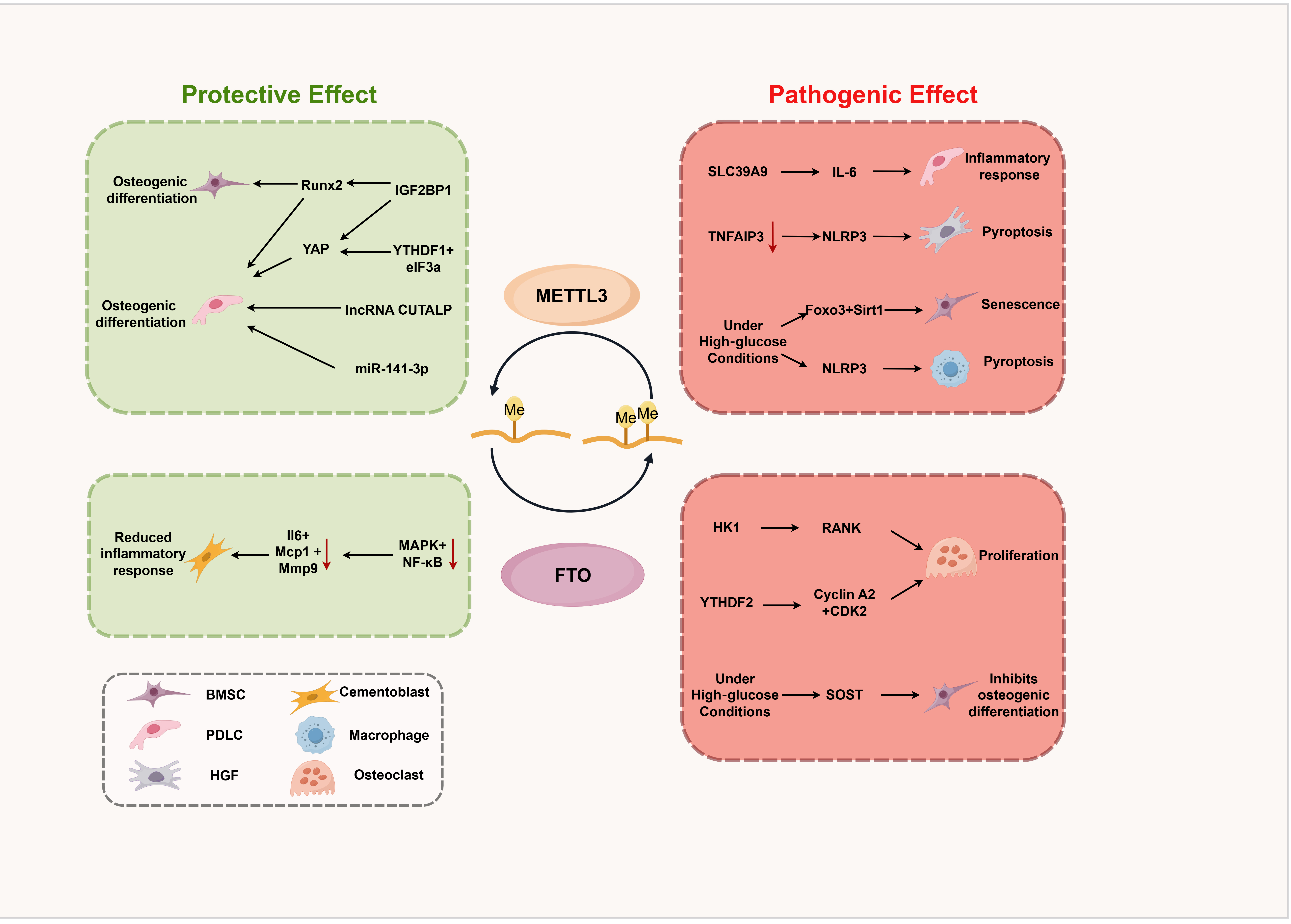
Figure 3. The m6A machinery acts as a double-edged sword in periodontitis and related conditions. (Created with Figdraw; ID: IIOTU9a2ca). Runx2: RUNX family transcription factor 2; YAP: yes1 associated transcriptional regulator; IGF2BP: insulin-like growth factor 2 mRNA-binding protein; YTHDF: YTH N6-methyladenosine RNA binding protein F; METTL3: methyltransferase-like 3; FTO: fat mass and obesity-associated protein; eIF3a: eukaryotic translation initiation factor 3 subunit A; MAPK: mitogen-activated protein kinase; NF-κ B: nuclear factor-kappa B; BMSC: bone marrow mesenchymal stem cell; PDLC: periodontal ligament cell; HGF: human gingival fibroblast; SLC39A9: solute carrier family 39 member 9; IL-6: interleukin 6; NLRP3: NLR family pyrin domain containing 3; TNFAIP3: TNF alpha induced protein 3; HK1: hexokinase 1; CDK2: cyclin dependent kinase 2; SOST: sclerostin; Mcp1: monocyte chemoattractant protein 1; Mmp9: matrix metallopeptidase 9; RANK: receptor activator of NF-κB; m6A: N6-methyladenosine.
Summary of m6A regulators implicated in periodontitis and related experimental models
| Regulator | Role | Cell type / model | Target | Affected pathway | Biological effect | References |
| METTL3 | Writer | PDLCs | YAP mRNA | IGF2BP1/YTHDF1-mediated stabilization and translation of YAP mRNA | Promotes osteogenic differentiation | [18] |
| METTL3 | Writer | PDLSCs; BMSCs | Runx2 mRNA | IGF2BP1-mediated stabilization of Runx2 mRNA | Promotes osteogenic differentiation | [25,28] |
| METTL3 | Writer | PDLSCs | lncRNA CUTALP | lncRNA CUTALP/miR-30b-3p/Runx2 axis | Promotes osteogenic differentiation | [30] |
| METTL3 | Writer | PDLSCs | miR-141-3p | miR-141-3p/ZEB1 Axis | Promotes osteogenic differentiation | [31] |
| METTL3/METTL14 | Writer | PDLCs | SLC39A9 mRNA | Zinc-regulated IL-6 promoter DNA methylation | Promotes inflammatory responses | [32] |
| METTL3 | Writer | HGFs/macrophages; periodontitis mice | TNFAIP3 mRNA | NLRP3 inflammasome activation | Promotes inflammatory bone destruction | [19] |
| METTL3 | Writer | BMSCs; diabetic periodontitis mice | Foxo3 mRNA | Foxo3/Sirt1 axis | Promotes cellular senescence; disrupts bone homeostasis | [44] |
| METTL3 | Writer | Macrophages; diabetic periodontitis mice | NLRP3 mRNA | Pyroptosis | Accelerates alveolar bone resorption | [45] |
| WTAP | Writer | Macrophages; periodontitis mice | - | M1/M2 polarization | Promotes inflammation; inhibits regeneration | [33] |
| WTAP | Writer | PDLSCs | TP53BP1 mRNA | Oxidative stress | Promotes cellular senescence; inhibits osteogenic differentiation | [34] |
| FTO | Eraser | Osteoclasts; apical periodontitis rats | HK1 mRNA | Glycolysis | Promotes osteoclastogenesis; promotes bone resorption | [36] |
| FTO | Eraser | Osteoclast precursors; periodontitis mice | Cyclin A2, CDK2 mRNAs | Cell proliferation and apoptosis | Promotes osteoclast development; promotes bone resorption | [37] |
| FTO | Eraser | Cementoblasts; apical periodontitis mice | - | p65, p38, ERK1/2, and JNK pathways | inhibits inflammatory response | [38] |
| FTO | Eraser | BMSCs; diabetic periodontitis mice | Sclerostin mRNA | Wnt signalling pathway | Inhibits osteogenic differentiation | [43] |
| ALKBH5 | Eraser | PDLSCs | TLR9 mRNA | MAPK pathway | Promotes inflammatory responses; inhibits osteogenic differentiation | [39] |
| YTHDC1 | Reader | PDLSCs; osteoporosis rats | - | Wnt/β-catenin signalling pathway | Promotes osteogenic differentiation | [40] |
| IGF2BP2 | Reader | Macrophages/osteoclasts; periodontitis mice | Cd5l, Cd36 mRNAs | - | Dual-phase: anti-inflammation early, pro-osteoclastogenesis late | [20] |
NON-M6A MODIFICATIONS IN PERIODONTITIS AND RELATED CONTEXTS
While m6A represents the most extensively studied RNA modification in periodontal research, a growing body of evidence supports the involvement of other epitranscriptomic marks in disease pathogenesis. These non-m6A modifications - including ac4C, m6Am, m5C, m7G, and m1A - exert distinct regulatory functions through their unique writer, eraser, and reader proteins, as shown in Figure 4. Recent studies have begun to unravel their contributions to periodontal inflammation, immune dysregulation, and alveolar bone remodeling.
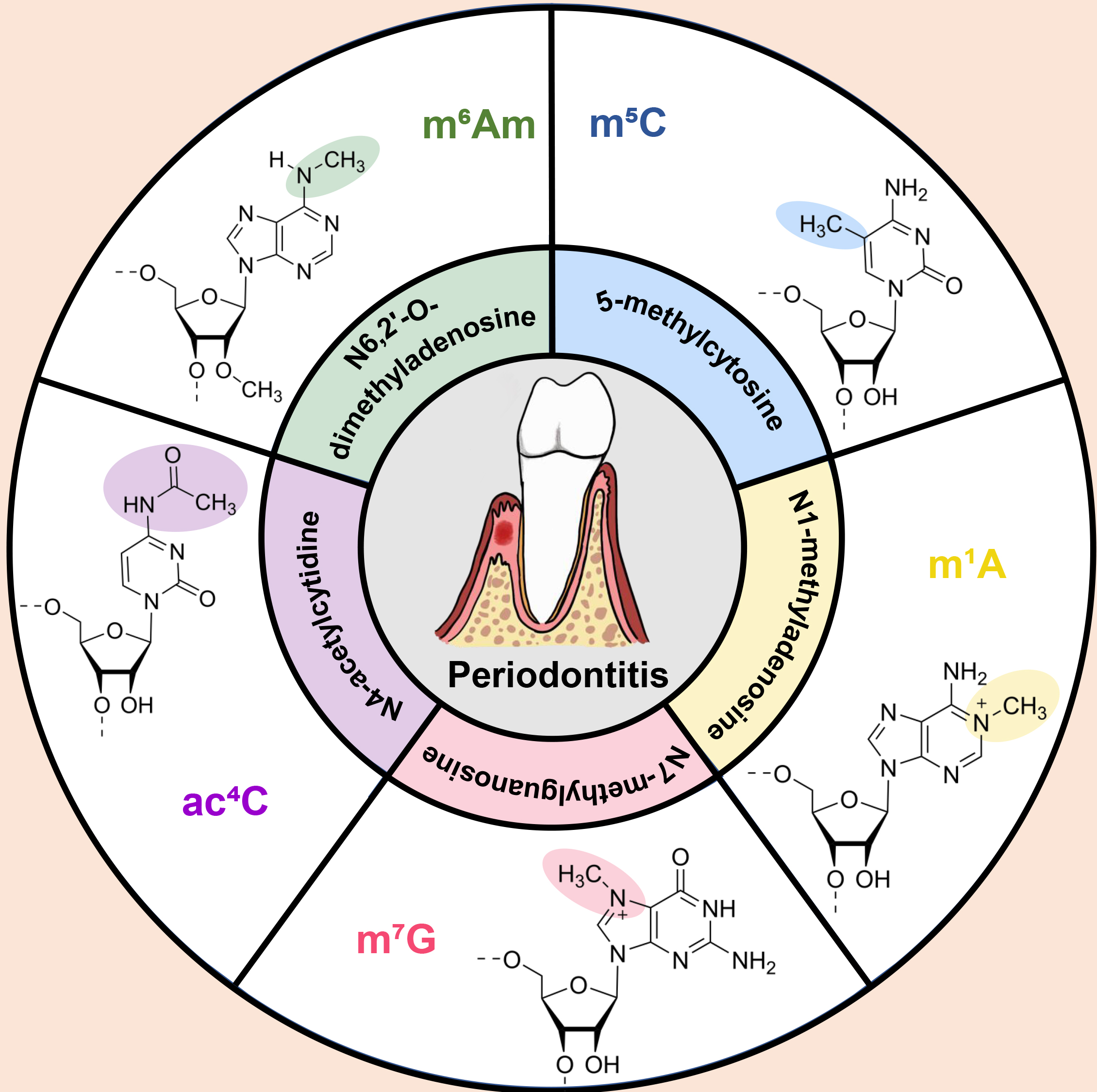
Figure 4. Overview of non-m6A modifications.
Ac4C modification in periodontitis
Among non-m6A modifications, ac4C has emerged as the most extensively studied in the periodontal field. ac4C is the only known acetylated RNA modification in eukaryotic cells, catalyzed exclusively by the N-acetyltransferase 10 (NAT10)[46]. This modification involves the transfer of an acetyl group to the N4 position of cytidine, forming ac4C that enhances mRNA stability and translation efficiency through improved base-pairing interactions[47]. Initially identified in tRNA and rRNA, ac4C has recently been mapped to mammalian mRNAs, where it is enriched within coding regions and promotes translation elongation[48].
In periodontitis, NAT10 and ac4C modification regulate osteogenic differentiation, osteoclastogenesis, and macrophage inflammatory responses. NAT10 promotes osteogenic differentiation of PDLSCs by regulating Vascular endothelial growth factor A (VEGFA)-mediated Phosphatidylinositol 3-kinase (PI3K)/ protein kinase B (AKT) signaling through ac4C modification[49]. NAT10 catalyzes ac4C modification of VEGFA mRNA, enhancing its stability and expression, which subsequently activates the PI3K/AKT pathway[49]. NAT10 also promotes osteoclastogenesis in inflammatory bone loss by catalyzing Fos mRNA ac4C modification and upregulating the MAPK signaling pathway[50]. This modification stabilizes Fos mRNA, increasing c-Fos protein expression and enhancing osteoclast differentiation[50]. Furthermore, inflammatory signaling is orchestrated by a complex interplay of regulatory layers. While our focus is on RNA modifications, it is important to recognize that protein post-translational modifications, such as ubiquitination and phosphorylation, also critically control inflammatory cascades, as exemplified in the regulation of type I interferon signaling[51]. NAT10 regulates the LPS-induced inflammatory response in macrophages through the NADPH oxidase 2 (NOX2)- reactive oxygen species (ROS)- nuclear factor-kappa B (NF-κB) pathway[52]. Mechanistically, NAT10 catalyzes ac4C modification of NOX2 mRNA and enhances its stability and expression; the resulting accumulation of reactive oxygen species then drives phosphorylation of NF-κB, switching on NF-κB-dependent inflammatory gene expression[52]. This cascade itself illustrates how chemical modifications at distinct regulatory layers act in concert: an RNA-level ac4C modification controls NOX2 abundance, whereas protein-level post-translational modifications (phosphorylation) execute the downstream inflammatory switch.
m6Am modification in periodontitis
m6Am is a unique RNA modification located at the first transcribed nucleotide adjacent to the 5’ cap structure of mRNAs[8]. This modification combines two distinct chemical marks: 2’-O-methylation of the ribose and N6-methylation of the adenosine base[53]. m6Am is installed by the methyltransferase Phosphorylated CTD interacting factor 1 (PCIF1), which specifically catalyzes N6-methylation of 2’-O-methyladenosine to generate m6Am[54]. The biological functions of m6Am include regulating mRNA stability, translation efficiency, and nuclear export[53].
Emerging evidence suggests a role for m6Am modification in periodontitis. Multiplexed LC-MS/MS quantification of salivary RNA modifications revealed significant alterations in m6Am levels in periodontitis patients compared to healthy controls[55]. Salivary m6Am may serve as a potential diagnostic biomarker for periodontal disease[55]. The m6Am methyltransferase PCIF1 regulates periodontal inflammation. Loss of PCIF1 attenuates periodontal inflammation in whole-body and myeloid lineage-specific knockout mouse models. Pcif1 deletion inhibits macrophage phagocytosis and migration through m6Am-Csf1r signaling[56].
Other non-m6A modifications: m7G, m5C, and m1A
A limited but growing body of research has also focused on the roles of m7G, m5C, and m1A modifications in periodontitis [Table 2].
Summary of non-m6A RNA modifications in periodontitis and related models
| Modification | Regulator | Experimental sample / model | Key finding | Level of evidence | References |
| ac4C | NAT10 | PDLSCs | Stabilizes VEGFA mRNA → PI3K/AKT; promotes osteogenesis | Direct | [49] |
| ac4C | NAT10 | Osteoclasts; periodontitis mice; calvarial osteolysis mice | Stabilizes Fos mRNA → MAPK; promotes osteoclastogenesis | Direct | [50] |
| ac4C | NAT10 | Macrophages; periodontitis mice | Stabilizes NOX2 mRNA → ROS-NF-κB; promotes inflammatory responses | Direct | [52] |
| m6Am | PCIF1 | Macrophages; Periodontitis mice | Stabilizes Csf1r mRNA; promotes periodontal inflammation | Direct | [56] |
| m6Am | - | Saliva of periodontitis patients | Salivary m6Am levels altered; biomarker candidate | Direct | [55] |
| m7G | METTL1 | PDLSCs | Promotes stabilization and translation of osteogenic genes; promotes osteogenesis | Direct | [59] |
| m5C | NSUN2 | Macrophages on titanium implants | Methylates IRF4; promotes PDLSC osteogenesis and HUVEC angiogenesis | Indirect (peri-implant context) | [61] |
| m1A | RRP8, ALKBH3, MAK16, and DDX18 | EndoC-βH1 cells; Gingival tissues of periodontitis patients | m1A regulators dysregulated; diagnostic biomarker candidates | Direct | [64] |
m7G is best known as the 5’ cap structure of eukaryotic mRNAs, where it protects transcripts from degradation and facilitates translation initiation[57]. Internal m7G modifications are installed primarily by the methyltransferase methyltransferase-Like-1 (METTL1)[58]. Research has found that METTL1 promotes osteogenic differentiation of PDLSCs by enhancing m7G modification and translation efficiency of osteogenic genes including RUNX2, osteocalcin (OCN), and osteopontin (OPN)[59].
m5C is a widespread RNA modification catalyzed by members of the NOP2/Sun RNA methyltransferase (NSUN) family of methyltransferases, with NSUN2 being the most extensively characterized m5C writer for mRNAs and non-coding RNAs[60]. m5C modification influences multiple aspects of RNA metabolism and has been implicated in stem cell differentiation and immune responses. Direct evidence linking m5C to periodontitis remains limited. However, periodontal-related mechanistic insights have emerged from a study investigating macrophage-titanium implant interactions. NSUN2 methylates interferon regulatory factor 4 (IRF4) to affect the capacity of macrophages attached to titanium implants[61]. This modification subsequently influences the osteogenic differentiation of PDLSCs and angiogenesis of human umbilical vein endothelial cells (HUVECs) in vitro[61]. While this study does not directly examine periodontitis, it reveals crosstalk between m5C modification, immune cells, and bone-forming cells that may be relevant to the peri-implant and periodontal microenvironment.
m1A refers to the methylation of the nitrogen-1 position of adenosine, introducing a positive charge that can alter RNA structure and protein-RNA interactions[62]. The modification is catalyzed by the tRNA methyltransferase 6 (TRMT6)/ tRNA methyltransferase 61A (TRMT61A) complex, while demethylation is mediated by AlkB homolog 3 (ALKBH3)[63]. Combined bioinformatics and experimental validation identified m1A-modified genes as potential diagnostic biomarkers in periodontitis and diabetes[64]. Specific m1A regulators such as ALKBH3 and m1A-modified transcripts are dysregulated in periodontal tissues from patients with periodontitis and diabetes, correlating with disease severity and inflammatory markers[64].
TRANSLATIONAL IMPLICATIONS AND FUTURE DIRECTIONS
The expanding understanding of RNA modifications in periodontitis offers multiple translational opportunities. Small molecule inhibitors targeting RNA modification enzymes have shown promise in preclinical studies. Two distinct FTO inhibitors have shown therapeutic potential in inflammatory bone loss. Dac51, an FTO inhibitor, exhibited inhibitory activity on osteoclastogenesis[36]. Notably, FB23-2, another FTO inhibitor, was also shown to suppress osteoclast-related gene expression, osteoclast formation, and bone resorption ability in cultured cells, and systemic treatment with FB23-2 significantly reduced alveolar bone loss in mice with experimental periodontitis[37]. For NAT10, the specific inhibitor Remodelin has shown consistent protective effects[50,52]. Administration of Remodelin reduced macrophage infiltration and bone resorption in ligature-induced periodontitis mice[52]. Furthermore, Remodelin treatment remarkably impeded ligature-induced alveolar bone loss and lipopolysaccharide-induced inflammatory calvarial osteolysis, highlighting the therapeutic potential of targeting ac4C modification in inflammatory bone diseases[50].
Beyond direct enzyme inhibitors, repurposing existing drugs and exploring natural compounds have emerged as attractive strategies. Ropinirole, a dopamine agonist primarily used in Parkinson’s disease treatment, has recently been found to suppress LPS-induced periodontal inflammation by inhibiting NAT10 in an ac4C-dependent manner, thereby exerting therapeutic effects in periodontitis[65]. This drug repurposing approach leverages existing safety data and could accelerate clinical translation. Natural small molecule compounds have also shown promise. Coptisine chloride, an isoquinoline alkaloid derived from traditional medicinal plants, was identified as a novel METTL3 inhibitor that exerts therapeutic effects on periodontitis by alleviating NLRP3 inflammasome activation and reducing alveolar bone loss[19]. These findings highlight the potential of natural products as sources of epitranscriptome-modulating agents.
Advanced biomaterials and drug delivery systems represent another frontier for translation, with several innovative platforms demonstrating therapeutic efficacy through modulation of RNA modifications. An intelligent pH-responsive MXene-based nanosystem (MPDM) was engineered to deliver METTL3/14 modulators for m6A-mediated osteogenic reprogramming; this system exhibited therapeutic efficacy in both in vitro and in vivo models of periodontitis, demonstrating the potential of combining nanomaterial engineering with epitranscriptomic modulation[66]. A biosafe injectable hydrogel drug-controlled delivery system was constructed as a bone-enhancing factory and loaded with quercetin, a natural flavonoid with antioxidant properties. Quercetin was demonstrated to reduce oxidative stress in orofacial mesenchymal stem cells (OMSCs) and promote osteogenic differentiation through molecular mechanisms involving m6A modification of Per1, ultimately enhancing periodontal bone repair in vivo[67]. Curcumin-carbon dots (CUR-CD), another nanomaterial-based approach, were shown to suppress periodontitis through downregulation of METTL3-mediated m6A modification of IRE1α[68]. These biomaterial-based approaches offer the advantage of localized delivery and sustained release, overcoming the limitations of systemic drug administration and enabling targeted modulation of epitranscriptomic pathways in periodontal tissues.
Despite these promising advances, several practical hurdles must be overcome before epitranscriptomic therapies can reach periodontal clinics. A major challenge is the cell-type specificity and context-dependent effects of RNA modification regulators. As discussed above, the same regulator can exert opposing effects in different periodontal cell populations. This functional pleiotropy implies that systemic inhibition may cause unintended on-target effects in non-target tissues. Therefore, targeting decisions must consider cell type, disease stage, delivery mode, dosage, etc. Another barrier is the current lack of human validation, as most evidence derives from murine models and in vitro systems that do not fully recapitulate human periodontal disease complexity. Rigorous pharmacokinetic, pharmacodynamic, and safety studies are needed before human trials. Methodological bottlenecks also constrain the field’s progress. Current epitranscriptomic detection techniques, including antibody-based approaches such as MeRIP-seq, are limited by antibody specificity, resolution, and the inability to quantify stoichiometry at single-base resolution. Emerging technologies such as direct RNA sequencing and mass spectrometry-based quantification offer improved resolution but remain costly and technically demanding[55,69,70]. The development of more accessible, quantitative, and cell-type-specific epitranscriptomic profiling methods is urgently needed.
Future research should prioritize several key areas. First, single-cell epitranscriptomic approaches (e.g., scRNA-seq combined with m6A detection) would resolve modification dynamics at the individual cell level, revealing cell-type-specific regulatory networks obscured by bulk analyses. Second, current research has focused almost exclusively on the downstream consequences of RNA modifications, whereas the upstream signals—how periodontal pathogens and their virulence factors trigger RNA modification changes in host cells - remain poorly defined. Elucidating these pathogen-driven mechanisms would reveal novel intervention points. Third, it remains unknown whether epitranscriptomic profiles correlate with distinct disease stages, rapid progression, or treatment response - this should be a priority for future clinical cohort studies. Fourth, salivary RNA-modification biomarkers hold promise for non-invasive disease monitoring and personalized treatment, but their clinical application requires systematic evaluation and standardization of pre-analytical variables, including collection timing, storage conditions, RNA degradation, and salivary flow rate.
CONCLUSION
The emerging field of epitranscriptomics has revealed RNA modifications as critical regulators of gene expression with profound implications for periodontitis pathogenesis. Current evidence suggests that dysregulation of m6A machinery, particularly METTL3 and FTO, contributes to periodontitis through effects on osteogenic differentiation, osteoclastogenesis, and inflammatory responses. The identification of ac4C modification as a regulator of both bone remodeling and immune function highlights the importance of non-m6A marks. Emerging evidence for m6Am, m7G, m5C, and m1A modifications expands the epitranscriptomic landscape relevant to periodontal disease. Translational implications are emerging, with small molecule inhibitors, repurposed drugs, natural compounds, and advanced biomaterials showing promise in preclinical studies. Integration of epitranscriptomic approaches with conventional periodontal therapy holds promise for improving patient outcomes through personalized, mechanism-based treatments.
DECLARATIONS
Acknowledgments
The authors appreciate Figdraw (www.figdraw.com ID: TYAIR47735) for providing convenience for drawing the Graphical Abstract.
Author contributions
Conceptualization: Guo Y, Ma L, Huang X
Data curation: Guo Y
Formal analysis: Guo Y
Methodology: Guo Y
Writing - original draft: Guo Y, Liu H
Writing - review & editing: Guo Y, Liu H, Ma L, Huang X, Cao Z
Funding acquisition: Ma L, Huang X, Cao Z
All authors have read and agreed to the published version of the manuscript.
Availability of data and materials
Data sharing is not applicable to this article, as no new data were created or analyzed in this study.
AI and AI-assisted tools statement
The authors used AI-assisted tools (Kimi Work, Moonshot AI; version 3.2.4, released 2026-08-31) to improve English language expression during manuscript preparation. All AI-generated outputs were critically reviewed and revised by the authors, who assume full responsibility for the manuscript’s content.
Financial support and sponsorship
None.
Conflicts of interest
All authors declared that there are no conflicts of interest.
Ethical approval and consent to participate
Not applicable.
Consent for publication
Not applicable.
Copyright
© The Author(s) 2026.
REFERENCES
1. Feng Y, Guo X, Yang Y, Qiu W, Chen Z, Fang F. Noncoding RNAs in periodontitis: progress and perspectives (Review). Int J Mol Med. 2025;56:1-17.
2. Larsson L, Kavanagh NM, Nguyen TVN, Castilho RM, Berglundh T, Giannobile WV. Influence of epigenetics on periodontitis and peri-implantitis pathogenesis. Periodontol 2000. 2022;90:125-37.
3. Hathaway-Schrader JD, Novince CM. Maintaining homeostatic control of periodontal bone tissue. Periodontol 2000. 2021;86:157-87.
4. Villoria GEM, Fischer RG, Tinoco EMB, Meyle J, Loos BG. Periodontal disease: a systemic condition. Periodontol 2000. 2024;96:7-19.
5. Balta MG, Papathanasiou E, Blix IJ, Van Dyke TE. Host modulation and treatment of periodontal disease. J Dent Res. 2021;100:798-809.
6. Larsson L, Giraldo-Osorno PM, Garaicoa-Pazmino C, Giannobile WV, Asa'ad F. DNA and RNA methylation in periodontal and peri-implant diseases. J Dent Res. 2025;104:131-9.
8. Sun H, Li K, Liu C, Yi C. Regulation and functions of non-m6A mRNA modifications. Nat Rev Mol Cell Biol. 2023;24:714-31.
9. Roundtree IA, Evans ME, Pan T, He C. Dynamic RNA modifications in gene expression regulation. Cell. 2017;169:1187-200.
10. Zhang L, Wei J, Zou Z, He C. RNA modification systems as therapeutic targets. Nat Rev Drug Discov. 2026;25:59-78.
11. Lin LC, Liu ZY, Yang JJ, Zhao JY, Tao H. m6A epitranscriptomic modification in diabetic microvascular complications. Trends Pharmacol Sci. ;2023:991-1008.
12. Feng G, Wu Y, Hu Y, et al. Small molecule inhibitors targeting m6A regulators. J Hematol Oncol. 2024;17:30.
13. Oerum S, Meynier V, Catala M, Tisné C. A comprehensive review of m6A/m6Am RNA methyltransferase structures. Nucleic Acids Res. 2021;49:7239-55.
14. Shi H, Wang X, Lu Z, et al. YTHDF3 facilitates translation and decay of N6-methyladenosine-modified RNA. Cell Res. 2017;27:315-28.
15. Roundtree IA, Luo GZ, Zhang Z, et al. YTHDC1 mediates nuclear export of N6-methyladenosine methylated mRNAs. Elife. 2017;6:e31311.
16. Wang Y, Zhang X, Yang F, Shang J, Yang Q. METTL3-mediated m6A modification of pri-miR-93 promotes hepatocellular carcinoma progression via CDKN1A suppression. Cancer Genet. 2025;296-297:31-40.
17. Roshan M K, Pal R, Kumari S, Kumar S, Muthuswamy S. The interplay of N6-methyladenosine and ferroptosis in cancer: a promising therapeutic avenue. Cancer Genet. 2025;296-297:15-24.
18. Sun X, Meng X, Piao Y, Dong S, Dong Q. METTL3 promotes the osteogenic differentiation of human periodontal ligament cells by increasing YAP activity via IGF2BP1 and YTHDF1-mediated m6A modification. J Periodontal Res. 2024;59:1017-30.
19. Zhou X, Yang X, Huang S, et al. Inhibition of METTL3 alleviates NLRP3 inflammasome activation via increasing ubiquitination of NEK7. Adv Sci (Weinh). 2024;11:e2308786.
20. Ma XX, Zhou XY, Feng MG, et al. Dual role of IGF2BP2 in osteoimmunomodulation during periodontitis. J Dent Res. 2024;103:208-17.
21. Lin W, Xu H, Wu Y, Wang J, Yuan Q. In silico genome-wide identification of m6A-associated SNPs as potential functional variants for periodontitis. J Cell Physiol. 2020;235:900-8.
22. Zhang X, Zhang S, Yan X, et al. m6A regulator-mediated RNA methylation modification patterns are involved in immune microenvironment regulation of periodontitis. J Cell Mol Med. 2021;25:3634-45.
23. Wang Z, Chen H, Peng L, He Y, Wei J, Zhang X. DNER and GNL2 are differentially m6A methylated in periodontitis in comparison with periodontal health revealed by m6A microarray of human gingival tissue and transcriptomic analysis. J Periodontal Res. 2023;58:529-43.
24. Zou X, Liu C, Wu X, Yuan Z, Yan F. Changes in N6-methyladenosine RNA methylomes of human periodontal ligament cells in response to inflammatory conditions. J Periodontal Res. 2023;58:444-55.
25. Sun X, Meng X, Piao Y, Dong S, Dong Q. METTL3 promotes osteogenic differentiation of human periodontal ligament stem cells through IGF2BP1-mediated regulation of Runx2 stability. Int J Med Sci. 2024;21:664-73.
26. Huang J, Guo C, Wang Y, Zhou Y. Role of N6-adenosine-methyltransferase subunits METTL3 and METTL14 in the biological properties of periodontal ligament cells. Tissue Cell. 2023;82:102081.
27. Yao C, Xin H, Liu S, et al. Role of METTL3-mediated m6A modification in osteogenic differentiation of periodontal ligament stem cells extracted from adult periodontal ligaments ex-vivo. Cell Mol Biol (Noisy-le-grand). 2023;69:23-8.
28. Zhou S, Zhang G, Wang K, Yang Z, Tan Y. METTL3 potentiates osteogenic differentiation of bone marrow mesenchymal stem cells via IGF2BP1/m6A/RUNX2. Oral Dis. 2024;30:1313-21.
29. Zhang X, Liu J, Gao J, et al. N6-methyladenosine promotes osteogenic differentiation of PDLSCs from periodontitis patients. Oral Dis. 2024;30:1322-36.
30. Chen X, Qin Y, Wang X, et al. METTL3-mediated m6A modification regulates the osteogenic differentiation through LncRNA CUTALP in periodontal mesenchymal stem cells of periodontitis patients. Stem Cells Int. 2024;2024:3361794.
31. Li W, Alimujiang A. METTL3 promotes osteogenic differentiation of human periodontal ligament stem cells under the inflammatory microenvironment through the miR-141-3p/ZEB1 axis. Cell Biochem Biophys. 2025;83:1771-83.
32. Huang J, Wang Y, Zhou Y. METTL3 and METTL14 regulate IL-6 expression via RNA m6A modification of zinc transporter SLC39A9 and DNA methylation of IL-6 in periodontal ligament cells. Biochim Biophys Acta Mol Cell Res. 2024;1871:119605.
33. Li Y, Yang Y, Niu Y, et al. The role of WTAP in regulating macrophage-mediated osteoimmune responses and tissue regeneration in periodontitis. Front Immunol. 2024;15:1423378.
34. Xiong M, Wang T, Liu Y. WTAP Contributes to periodontitis pathogenesis by promoting PDLSC senescence and impairing osteogenic differentiation via m6A-dependent regulation of TP53BP1. Immun Inflamm Dis. 2026;14:e70335.
35. Ray SK, Mukherjee S. Exploring replication stress and cellular senescence as key targets in novel cancer therapies. Cancer Genet. 2025;298-299:78-87.
36. Qian Y, Wu J, Yang W, et al. FTO-associated osteoclastogenesis promotes alveolar bone resorption in apical periodontitis male rat via the HK1/USP14/RANK pathway. Nat Commun. 2025;16:1519.
37. He J, Zhao Y, Zhang Y, Zhang Z, Li D, Xu Q. FTO regulates osteoclast development by modulating the proliferation and apoptosis of osteoclast precursors in inflammatory conditions. Cell Signal. 2024;117:111098.
38. Li B, Du M, Sun Q, Cao Z, He H. m6 A demethylase Fto regulates the TNF-α-induced inflammatory response in cementoblasts. Oral Dis. 2023;29:2806-15.
39. Zhang Y, Li J, Chen M, Zhang N, Yu X, Wen Y. ALKBH5 promotes inflammation and inhibits osteogenic differentiation by activating the TLR9/MAPK pathway in human periodontal ligament stem cells. Cytotechnology. 2026;78:21.
40. Tan D, Li Q, Chen Z, et al. YTHDC1 Modulates the osteogenic capacity of hPDLSCs via Wnt/β-catenin signalling pathway for the treatment of bone defects in osteoporosis rats. Cell Prolif. 2025;58:e70020.
41. Shao W, Zhao H, Zhang S, et al. A pan-cancer landscape of IGF2BPs and their association with prognosis, stemness and tumor immune microenvironment. Front Oncol. 2022;12:1049183.
42. Botelho J, Singh S, Varenne B, et al. Oral health and diabetes: a systematic review and meta-analysis. Lancet Public Health. 2026;11:e555-66.
43. Zhou J, Zhu Y, Ai D, et al. Advanced glycation end products impair bone marrow mesenchymal stem cells osteogenesis in periodontitis with diabetes via FTO-mediated N6-methyladenosine modification of sclerostin. J Transl Med. 2023;21:781.
44. Zhou J, Li X, Xiao L, Zhu Y, Yang Y, Gao X. Hyperglycemia disrupts alveolar bone homeostasis by inducing stem cell senescence through Mettl3/Foxo3/Sirt1 axis in diabetic periodontitis. Life Sci. 2026;388:124215.
45. Yang Q, Xiao J, Liu Y, et al. METTL3-mediated m6A modifications of NLRP3 accelerate alveolar bone resorption through enhancing macrophage pyroptosis. Cell Signal. 2025;127:111572.
46. Wubulikasimu Z, Zhao H, Mao F, Zhao X. Emerging roles of RNA N4-acetylcytidine modification in reproductive health. Protein Cell. 2025;16:458-77.
47. Zhang Y, Lei Y, Dong Y, et al. Emerging roles of RNA ac4C modification and NAT10 in mammalian development and human diseases. Pharmacol Ther. 2024;253:108576.
48. Ouyang W, Huang Z, Wan K, Nie T, Chen H, Yao H. RNA ac4C modification in cancer: unraveling multifaceted roles and promising therapeutic horizons. Cancer Lett. 2024;601:217159.
49. Cui Z, Xu Y, Wu P, et al. NAT10 promotes osteogenic differentiation of periodontal ligament stem cells by regulating VEGFA-mediated PI3K/AKT signaling pathway through ac4C modification. Odontology. 2023;111:870-82.
50. Yang R, Yu W, Lin L, et al. NAT10 promotes osteoclastogenesis in inflammatory bone loss by catalyzing Fos mRNA ac4C modification and upregulating MAPK signaling pathway. J Adv Res. 2025;72:303-17.
51. Chen X, Cheng Q, Gong F, Zheng H. Protein post-translational modification-mediated control of type I interferon cascade signaling. Medicine Bulletin. 2025;1:37-59.
52. Zhang Z, Zhang Y, Cai Y, et al. NAT10 regulates the LPS-induced inflammatory response via the NOX2-ROS-NF-κB pathway in macrophages. Biochim Biophys Acta Mol Cell Res. 2023;1870:119521.
53. Jin H, Shi Z, Zhou T, Xie S. Regulation of m6Am RNA modification and its implications in human diseases. J Mol Cell Biol. 2024;16:mjae012.
54. Liu WW, Zheng SQ, Li T, et al. RNA modifications in cellular metabolism: implications for metabolism-targeted therapy and immunotherapy. Signal Transduct Target Ther. 2024;9:70.
55. Vignon M, Bastide A, Attina A, et al. Multiplexed LC-MS/MS quantification of salivary RNA modifications in periodontitis. J Periodontal Res. 2023;58:959-67.
56. Song W, Liu L, Liang H, et al. m6Am methyltransferase PCIF1 regulates periodontal inflammation. J Dent Res. 2024;103:1130-40.
57. Luo Y, Yao Y, Wu P, Zi X, Sun N, He J. The potential role of N7-methylguanosine (m7G) in cancer. J Hematol Oncol. 2022;15:63.
58. Wu Q, Fu X, Liu G, He X, Li Y, Ou C. N7-methylguanosine modification in cancers: from mechanisms to therapeutic potential. J Hematol Oncol. 2025;18:12.
59. Zhao C, Li K, Wu A. METTL1-mediated m7G modification regulates osteogenic differentiation of human periodontal ligament stem cells. Clinics (Sao Paulo). 2025;80:100813.
60. Mao Z, Tian Y, Wu L, Zhang Y. Epitranscriptomic mechanisms and implications of RNA m5C modification in cancer. Theranostics. 2025;15:8404-28.
61. Xiao L, Wu D, Zhang T, He C, Guo X, Yang H. NSUN2 methylates IRF4 to affect the capacity of macrophages attached to titanium implant on osteogenic differentiation of PDLSCs and angiogenesis of HUVECs in vitro. BMC Oral Health. 2024;24:1371.
62. Liu Y, Zhang S, Gao X, Ru Y, Gu X, Hu X. Research progress of N1-methyladenosine RNA modification in cancer. Cell Commun Signal. 2024;22:79.
63. Wang X, Ma X, Chen S, et al. Harnessing m1A modification: a new frontier in cancer immunotherapy. Front Immunol. 2024;15:1517604.
64. Wang K, Wang A, Qiu L, Xu L, Wei J. Combined bioinformatics and experimental validation identifies m1A-modified genes as potential diagnostic biomarkers in periodontitis and diabetes. Arch Oral Biol. 2025;180:106428.
65. Liao H, Ma H, Meng H, Kang N, Wang L. Ropinirole suppresses LPS-induced periodontal inflammation by inhibiting the NAT10 in an ac4C-dependent manner. BMC Oral Health. 2024;24:510.
66. Chen X, Lin Z, Lei H, et al. An Intelligent MXene nanoplatform for bone regeneration in periodontitis through synergistic scavenging pathogenic cfDNA and ROS. Adv Healthc Mater. 2026;15:e05749.
67. Zhu H, Cai C, Yu Y, et al. Quercetin-loaded bioglass injectable hydrogel promotes m6A alteration of Per1 to alleviate oxidative stress for periodontal bone defects. Adv Sci (Weinh). 2024;11:e2403412.
68. Li L, Wang Y, Gao L, Wu S, Jin Y. Curcumin-carbon dots suppress periodontitis via regulating METTL3/IRE1α signaling. Crit Rev Eukaryot Gene Expr. 2025;35:25-35.
69. Liu C, Sun H, Yi Y, et al. Absolute quantification of single-base m6A methylation in the mammalian transcriptome using GLORI. Nat Biotechnol. 2023;41:355-66.
Cite This Article
 Open Access
How to Cite
Guo Y, Liu H, Ma L, Huang X, Cao Z. Epitranscriptomic landscapes in periodontitis: unraveling the regulatory roles of RNA Modifications. J Transl Genet Genom. 2026;10:574-91. https://dx.doi.org/10.20517/jtgg.2026.31
Download Citation
If you have the appropriate software installed, you can download article citation data to the citation manager of your choice. Simply select your manager software from the list below and click on download.
Export Citation File
Type of Import
Tips on Downloading Citation
Citation Manager File Format
Type of Import
Direct Import: When the Direct Import option is selected (the default state), a dialogue box will give you the option to Save or Open the downloaded citation data. Choosing Open will either launch your citation manager or give you a choice of applications with which to use the metadata. The Save option saves the file locally for later use.
Indirect Import: When the Indirect Import option is selected, the metadata is displayed and may be copied and pasted as needed.
About This Article
Copyright

Data & Comments
Data
0










Comments
Comments must be written in English. Spam, offensive content, impersonation, and private information will not be permitted. If any comment is reported and identified as inappropriate content by OAE staff, the comment will be removed without notice. If you have any queries or need any help, please contact us at [email protected].