fig5
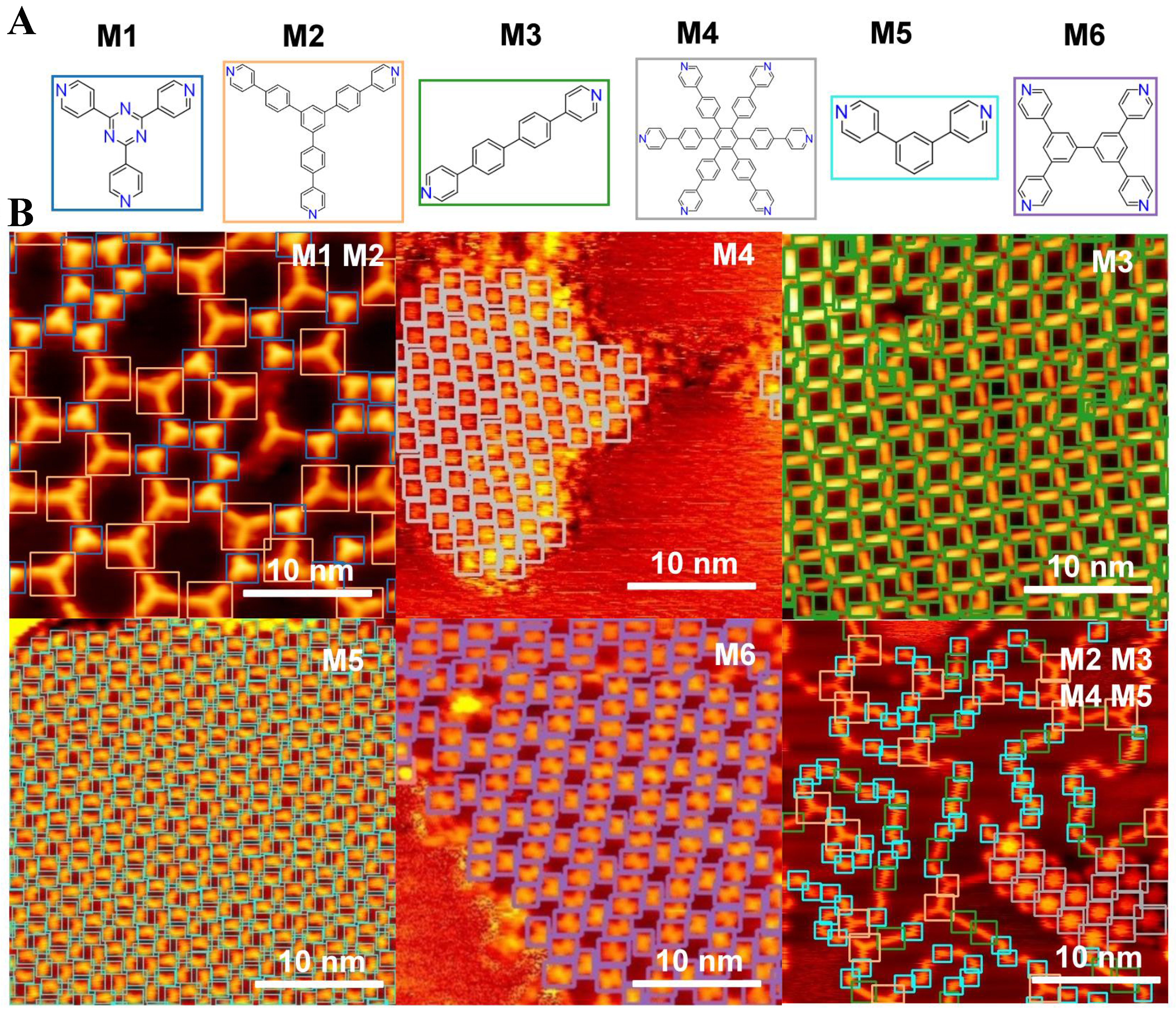
Figure 5. STM molecular images and prediction results. (A) Chemical structures of six molecular categories (M1-M6); (B) STM images and prediction results obtained using the well-trained model. The detected molecules are highlighted with color-coded markers: blue squares denote molecule M1, orange squares denote M2, green squares denote M3, grey squares denote M4, cyan squares denote M5, and purple squares denote M6. STM: Scanning tunneling microscopy.