


fig13
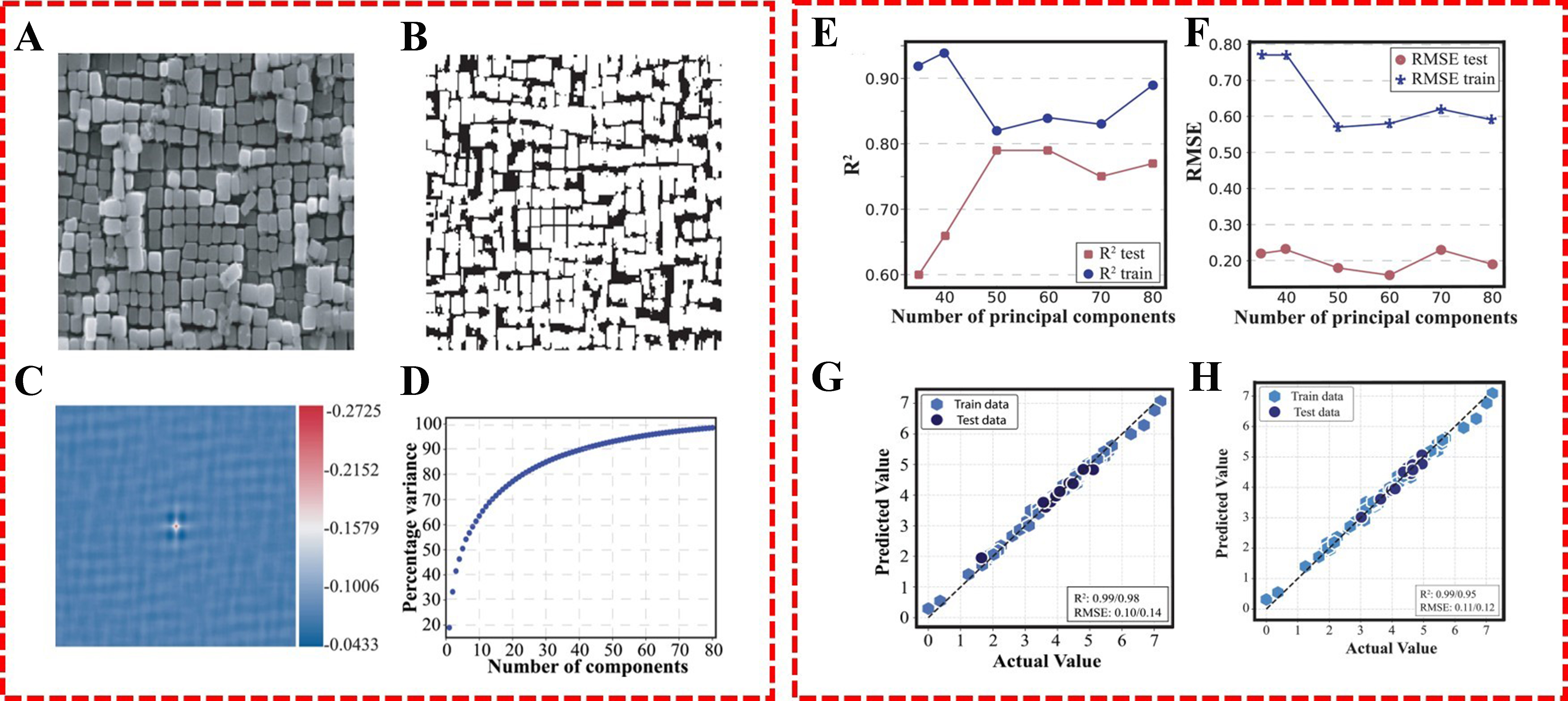
Figure 13. (A) Original SEM images; (B) Phase-separated binary images; (C) 2D point correlation functions; (D) Percentage variance of binarized micrograph captured as a function of number of principal components; (E and F) Microstructure and composition-based SVR predictions; (G) GPR fitting; (H) LASSO feature-reduced model fitting[85]. SEM: Scanning electron microscopy; SVR: support vector regressor; GPR: gaussian process regression; LASSO: least absolute shrinkage and selection operator.