
fig6
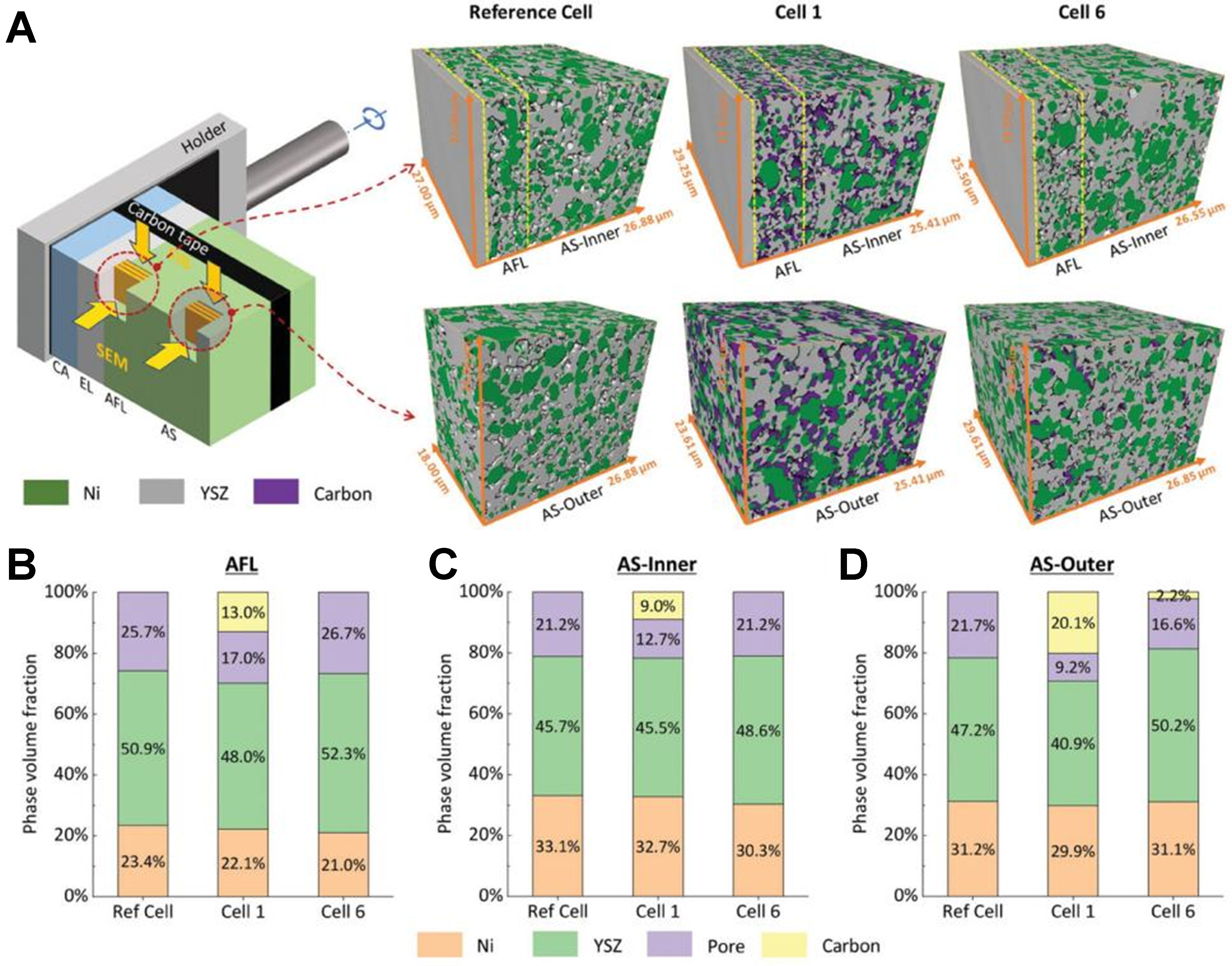
Figure 6. (A) Illustration of continuous slicing and imaging using orthogonal FIB-SEM, along with the 3D reconstructed volumes of the reference cell; Volume fractions of Ni, YSZ, pore, and carbon phases in the three cells are shown for (B) AFL, (C) AS-Inner, and (D) AS-Outer (Reprinted from Ref.[54] under open access license of CC BY-NC 4.0). FIB-SEM: Focused ion beam-scanning electron microscopy; 3D: three-dimensional; YSZ: yttria-stabilized zirconia; AFL: anode functional layer; AS: anode support; CA: cathode; EL: electrode.