

A synopsis of the guidelines for diagnosis and treatment of fulminant myocarditis
 0

Graphical Abstract
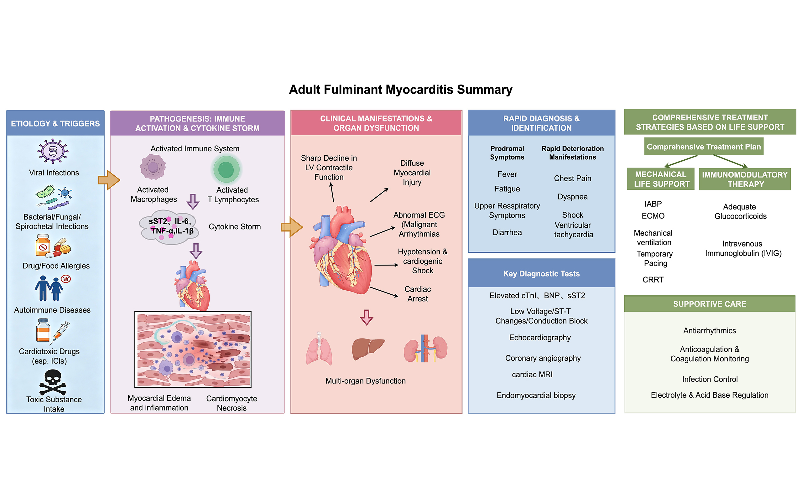
Abstract
Fulminant myocarditis (FM) represents the most severe subtype of myocarditis, associated with a high in-hospital mortality. For the clinical management of FM, the “life support-based comprehensive treatment regimen” is recommended, which places emphasis on suppressing the core pathophysiological mechanisms underlying FM, namely the excessive activation of innate immune responses and the inflammatory storm. Mechanical circulatory support and immunomodulatory therapy with adequate doses of both glucocorticoids and immunoglobulins constitute the two foundational pillars of this regimen. Notably, the treatment regimen underscores the principle of “ultra-early recognition, ultra-early diagnosis, ultra-early prediction, and ultra-early rescue”. As the first clinical guideline for the diagnosis and treatment of adult FM, this document provides evidence-based and effective therapeutic approaches, which are crucial for significantly reducing mortality and improving long-term prognosis in affected patients.
Keywords


 Download PDF
INTRODUCTION
Myocarditis refers to inflammatory injury of the myocardium, characterized by inflammatory cell infiltration and cardiac dysfunction. Fulminant myocarditis (FM) represents the most severe phenotype, with acute onset and rapid deterioration. Patients with FM often develop hemodynamic instability, severe arrhythmias, multiple organ failure, and even sudden cardiac death. Authorized by the Chinese Society of Cardiology, the Chinese Expert Consensus on the Diagnosis and Treatment of Fulminant Myocarditis was first published in 2017[1]. This document has since been updated and formally upgraded into the Chinese Society of Cardiology Guidelines on the Diagnosis and Treatment of Adult Fulminant Myocarditis in 2024 (hereinafter referred to as the “2024 Chinese Guidelines”)[2,3]. In comparison with its preceding consensus, the 2024 Chinese Guidelines incorporate several novel sections, covering epidemiology, pathological classification, management and follow-up during the convalescent phase, complication mitigation, the establishment of a rapid response team for FM and healthcare system construction. Furthermore, the guidelines integrate the latest advances in the pathophysiological mechanisms of FM and provide a more detailed exposition of diagnostic workup and therapeutic regimens. In line with international standards, recommendations are graded into four classes (I, IIa, IIb and III) and three levels of evidence (A, B, C)[4,5]. This synopsis presents a condensed version of the guidelines, aiming to help readers rapidly grasp the core contents of the guidelines, especially for popularization of the key clinical concepts in primary healthcare units.
EPIDEMIOLOGY OF FM
Accurate epidemiological data on the incidence of FM remain limited. This is largely because FM is sporadic and progresses extremely rapidly, which frequently results in underdiagnosis or misdiagnosis. Consequently, direct estimation of FM incidence via population-based epidemiological surveys is challenging. To date, available data were primarily derived from fatal cases of acute myocarditis. Based on these estimates, the average incidence of FM is approximately 9-23 per 1 million person-years[6-9]. When extrapolated to the Chinese population, this corresponds to an estimated annual caseload of 12,600 to 32,200 cases. Nevertheless, this figure is likely to be a marked underestimate due to the underdiagnosis and misdiagnosis of FM, underscoring the urgent need for dedicated epidemiological investigations to ascertain the true disease burden.
Previous studies reported an in-hospital mortality of 45%-55.6% among patients with FM treated in tertiary medical centers[7,10,11]. FM affects individuals from adolescence to old age. In a cohort of 344 patients with histologically confirmed FM, the median age was 54 years, and older age was associated with poorer 90-day outcomes (death or heart transplantation)[12]. In another study comprising 220 FM patients, the mean age among non-survivors was 52.63 years, compared with a median age of 35.00 years among survivors[13], further supporting the prognostic implications of age.
ETIOLOGY, PATHOPHYSIOLOGY AND PATHOLOGY OF FM
The etiology of FM parallels that of acute non-fulminant myocarditis, comprising infectious and non-infectious causes. Viral infection is the primary etiology, while non-infectious factors mainly include autoimmune diseases and drug toxicity.
The pathophysiology of FM is complex. Excessive activation of the innate immune responses and rapid release of inflammatory mediators, which is referred to as “cytokine storm”, are considered the core mechanisms underlying FM. Myocardial injury occurs via two major pathways: direct viral cytotoxicity to cardiomyocytes, and inflammatory damage induced by the cytokine storm [Figure 1][14,15].
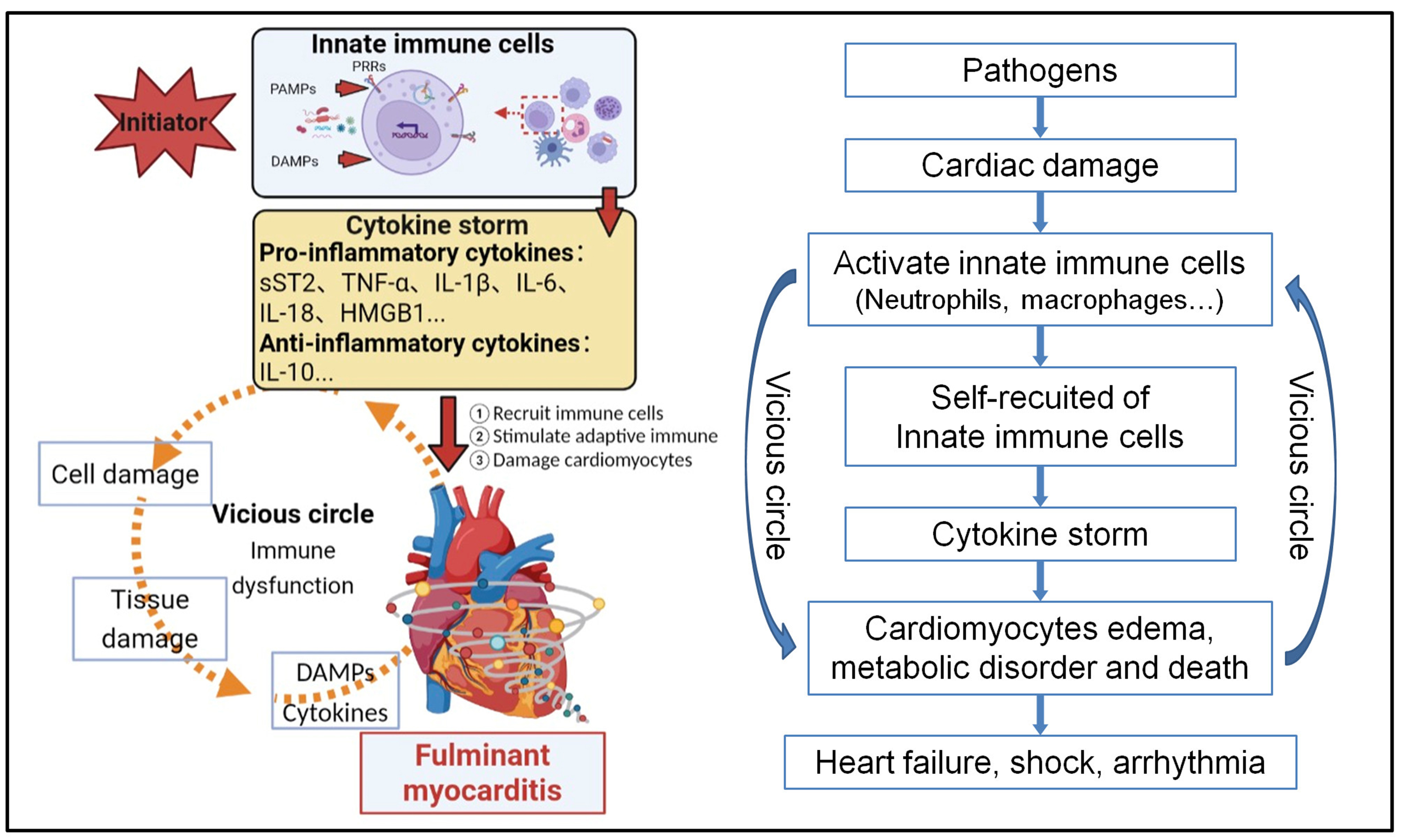
Figure 1. A summary of the pathophysiological mechanisms underlying FM. FM is initiated by viral or other causative agents that invade and damage cardiomyocytes, thereby recruiting neutrophils to the site of injury. The activated neutrophils subsequently promote the infiltration of monocytes and macrophages, which in turn trigger PAMP- or DAMP-mediated activation of innate immune cells. This activation precipitates a robust cytokine storm, inflicting further injury on myocardial tissue. The ensuing tissue destruction amplifies DAMPs-driven immune overactivation, establishing a self-perpetuating vicious cycle that ultimately culminates in the overt clinical syndrome of FM. PAMPs: Pathogen-associated molecular patterns; DAMPs: damage-associated molecular patterns; PRRs: pattern recognition receptors; sST-2: soluble growth stimulation expressed gene 2; TNF-α: tumor necrosis factor-α; IL: interleukin; HMGB1: high mobility group box 1; FM: fulminant myocarditis. The drawing part was created with BioRender (https://biorender.com/6yp4kqp).
According to established quantitative immunohistological criteria, myocarditis is defined as ≥ 14 infiltrating leukocytes/mm2 on myocardial biopsy[16]. FM typically demonstrates extensive inflammatory cell infiltration and cardiomyocyte necrosis, with inflammatory cell counts exceeding 50/mm2[17].
Notably, pathological findings do not always correlate with clinical severity, and some patients diagnosed with FM show only mild histological changes. Therefore, FM remains primarily a clinical diagnosis.
CLINICAL EVALUATION AND DIAGNOSIS OF FM
Early symptoms of FM are usually nonspecific, often presenting as cold-like prodromal symptoms lasting 3-5 days. FM is mainly characterized by hemodynamic instability resulting from severe myocardial injury, and some patients may develop coma or even sudden death due to malignant arrhythmias[18,19]. Physical signs are also nonspecific, but FM patients typically exhibit hypotension, dyspnea or respiratory depression, and abnormal heart rate or rhythm, indicating hemodynamic collapse[20-22]. However, fatigue and poor appetite are common.
For patients with suspected FM based on prodromal symptoms, laboratory examinations should be performed [Table 1]. Elevated myocardial injury biomarkers, including high-sensitivity cardiac troponin I (hs-cTnI) or cardiac troponin I or T (cTnI or cTnT), and B-type natriuretic peptide (BNP) or N-terminal pro-B-type natriuretic peptide (NT-proBNP), reflect the presence and severity of myocardial injury[4,23]. In addition, increased leukocyte count and neutrophil percentage generally indicate infection and systemic inflammation. Liver and renal function tests, electrolyte panels, and acid-base balance help evaluate multiple organ dysfunctions. The severity of hepatic and renal impairment, especially secondary injury, strongly affects the prognosis of patients with FM. Maintenance of electrolyte and acid-base balance, particularly serum potassium, is critical[24]. Arterial blood gas analysis and blood lactate levels reflect tissue perfusion and oxygenation, and should be dynamically monitored. Coagulation function should also be regularly assessed to identify and correct disseminated intravascular coagulation (DIC), especially in patients receiving intra-aortic balloon pump (IABP) and/or extracorporeal membrane oxygenation (ECMO)[25].
The recommendation for laboratory examinations for patients with FM[3] (see evidence in Supplementary Table 1)
| Recommendations | Classa | Levelb |
| Blood routine examination is recommended every 1-2 days to track dynamic hematological changes | I | C |
| Dynamic monitoring of hs-cTnI or cTnI is recommended as the biomarker for the assessment of myocardial injury[23,56] | I | B |
| Serial measurement of plasma NT-proBNP or BNP levels is recommended to assess the severity of cardiac dysfunction and predict outcomes[57,58] | I | B |
| Serial assessments of liver/renal function, arterial blood gases, and blood lactate levels are recommended for disease severity stratification and prognostic evaluation | I | C |
| Comprehensive coagulation monitoring (including PT, PTA, APTT, TT, INR, FIB, D-dimer, and platelet count) is recommended routinely[59,60] | I | A |
| In patients with suspected FM, dynamic monitoring of inflammatory cytokines is recommended to aid diagnosis and to assess therapeutic response and prognosis[61,62] | I | B |
| Serial measurements of sST2 should be considered in patients with suspected FM[26,63,64] | IIa | C |
| Procalcitonin levels may be considered to guide antibiotic therapy and to evaluate its clinical efficacy[65-67] | IIb | A |
C-reactive protein is the most widely used marker of systemic inflammation but lacks specificity. High-throughput plasma inflammatory factor profiling in early FM has identified significant alterations in 39 out of 122 factors (28 increased and 11 decreased)[26]. Notably, the following factors were significantly elevated: soluble ST2 (sST2), plasminogen activator inhibitor-1 (PAI-1), sialic acid-binding immunoglobulin-like lectin-5 (Siglec-5), CD163, interleukin-4 (IL-4), IL-17B, and vascular endothelial growth factor-C (VEGF-C). Among these, sST2 level showed particular prominence and correlated well with clinical severity. Conversely, cytotoxic T-lymphocyte-associated protein 4 (CTLA4) was markedly reduced. Levels of these factors gradually tend to normalize with effective treatment and clinical improvement. Classic pro-inflammatory cytokines such as interleukins and tumor necrosis factor (TNF)‑α were also increased. Therefore, routine and dynamic monitoring of plasma inflammatory factors is recommended in patients with suspected FM, as it aids in diagnosis, therapeutic evaluation, and prognostic assessment.
Special investigations for FM include electrocardiography (ECG), echocardiography, chest computed tomography (CT) or X-ray, cardiac magnetic resonance imaging (CMR), and endomyocardial biopsy (EMB). Histopathological analysis based on EMB remains the gold standard for diagnosing myocarditis [Figure 2A and B]. In recent years, with advances in echocardiography and CMR, EMB is less frequently used in uncomplicated myocarditis. Several limitations restrict its broader clinical application: its diagnostic yield is highly dependent on the expertise of the pathologist, and as an invasive procedure, it carries inherent risks, including cardiac perforation with pericardial tamponade, atrioventricular block, chordae tendineae injury, and puncture site bleeding. Nevertheless, EMB is strongly recommended in patients with FM presenting with shock, acute heart failure, or severe arrhythmias, particularly when a specific etiology is suspected, such as immune checkpoint inhibitor (ICI)‑associated myocarditis, autoimmune diseases, or peripheral eosinophilia. In such cases, EMB helps clarify the etiology, pathological subtype, and extent of myocardial injury, thereby providing crucial guidance for clinical management[27].
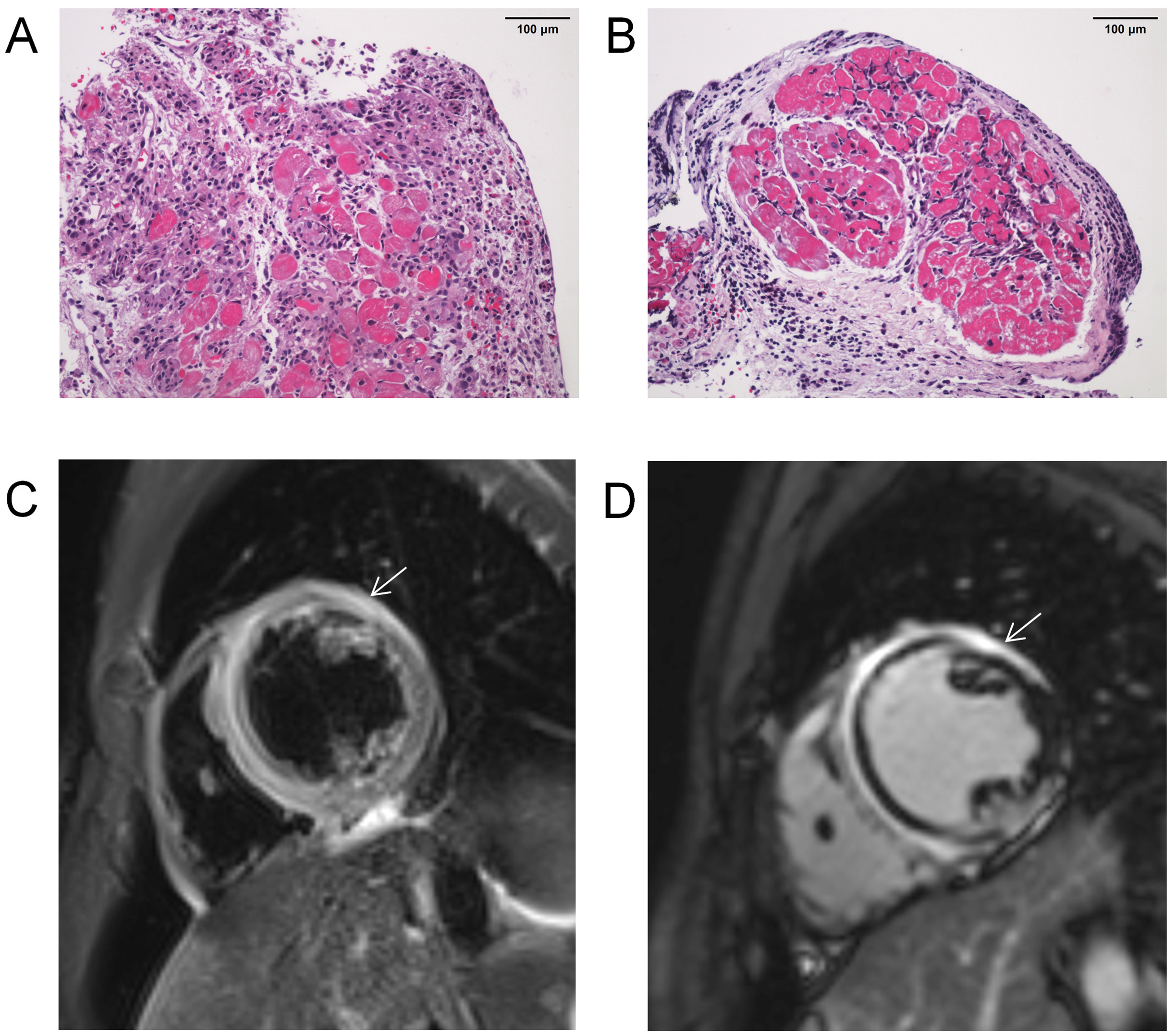
Figure 2. Representative pathological and CMR images from cases with FM. (A and B) Hematoxylin and eosin (H&E) staining of EMB specimens showing cardiac inflammatory infiltration (200×), scale bar = 100 μm; (C) T2-weighted imaging demonstrating marked myocardial edema (white arrow); (D) LGE imaging revealing myocardial injury (white arrow). CMR: Cardiac magnetic resonance imaging; LGE: late gadolinium enhancement; FM: fulminant myocarditis.
ECG shows high sensitivity but low specificity in the diagnosis of FM, and serial examinations are needed to detect dynamic changes[28]. Echocardiography allows comprehensive evaluation of cardiac structure, function and hemodynamics. Typical findings in FM include significant left ventricular systolic and diastolic dysfunctions, manifested by diffuse left ventricular hypokinesia and regional wall motion abnormalities, which may gradually improve with appropriate treatment[29,30]. Compared with acute myocarditis, FM demonstrates a more severe reduction in left ventricular longitudinal strain, which may precede overt cardiac dysfunction and thus facilitate early diagnosis[31].
CMR reveals imaging features closely resembling pathological changes in myocarditis, including myocardial edema, hyperemia, capillary leakage, necrosis, and fibrosis[32,33]. Quantitative assessment of myocardial injury can be achieved using T1 mapping, T2 mapping, and extracellular volume (ECV). The combination of CMR with elevated troponin levels has gradually emerged as a non-invasive tool for the clinical diagnosis of myocarditis, especially in low-risk patients[30]. According to current diagnostic criteria, CMR-based diagnosis of myocarditis requires at least one of the following: (i) Evidence of myocardial edema on T2 imaging, including increased T2 signal on T2-weighted imaging or elevated T2 mapping values; (ii) Evidence of myocardial injury on T1 imaging, including late gadolinium enhancement (LGE), increased T1 mapping values, or elevated ECV[32] [Figure 2C and D]. In addition, fluorodeoxyglucose uptake reflects metabolically active inflammatory cells in acute myocarditis, and fluorodeoxyglucose positron emission tomography (FDG-PET) is useful to detect disease activity when clinically indicated[34,35].
Based on the above-mentioned clinical evaluations, the diagnosis of FM is established at three levels: clinical diagnosis, pathological diagnosis, and etiological diagnosis [Figure 3]. Patients with prodromal symptoms, abnormal ECG, elevated biomarkers of myocardial injury and inflammation, and abnormal echocardiographic findings are considered to have a clinical diagnosis of FM. After exclusion of myocardial infarction by coronary angiography (CAG), CMR or EMB should be performed as early as possible to confirm the diagnosis. Finally, etiological investigations include pathogenic microorganism detection, autoimmune antibody testing, and toxicology screening.
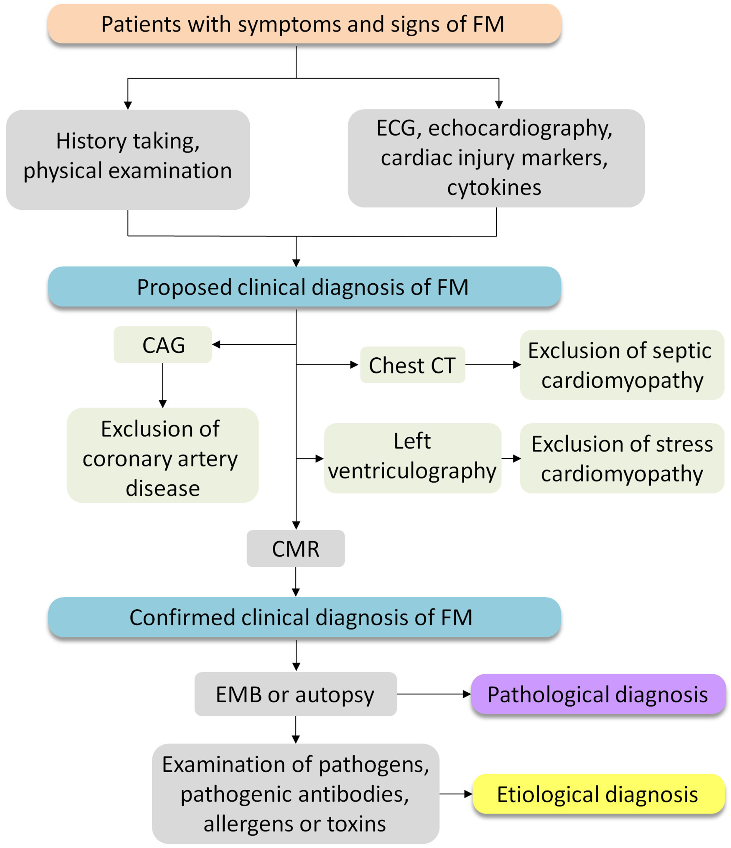
Figure 3. Diagnostic algorithm for FM[3]. Electrocardiography (ECG), echocardiography, cardiac troponin I (cTnI), N-terminal pro-B-type natriuretic peptide (NT-proBNP), and blood cytokine testing should be performed immediately; coronary angiography (CAG) is required to rule out coronary artery disease. Left ventriculography may be used to exclude stress cardiomyopathy, and chest computed tomography (CT) to rule out septic cardiomyopathy due to severe infection, when clinically indicated. Cardiac magnetic resonance imaging (CMR) should be scheduled at an appropriate time based on the patient’s clinical status. EMB: Endomyocardial biopsy; FM: fulminant myocarditis.
TREATMENT OF FM IN THE ACUTE PHASE
Once FM is diagnosed or suspected, it is recommended to start standardized treatment immediately, known as “life support-based comprehensive treatment regimen” (hereafter referred to as the “comprehensive treatment regimen”; Figure 4). To facilitate efficient implementation, hospitals are advised to establish a rapid response team and a standardized, concise, and practical clinical pathway led by the cardiovascular team. Pre-hospital emergency care, emergency intensive care, imaging and pathology services, cardiovascular surgery, and a multidisciplinary treatment support team should be integrated into the rapid response system to ensure standardized delivery of the regimen and to improve the outcomes of patients with FM[3].
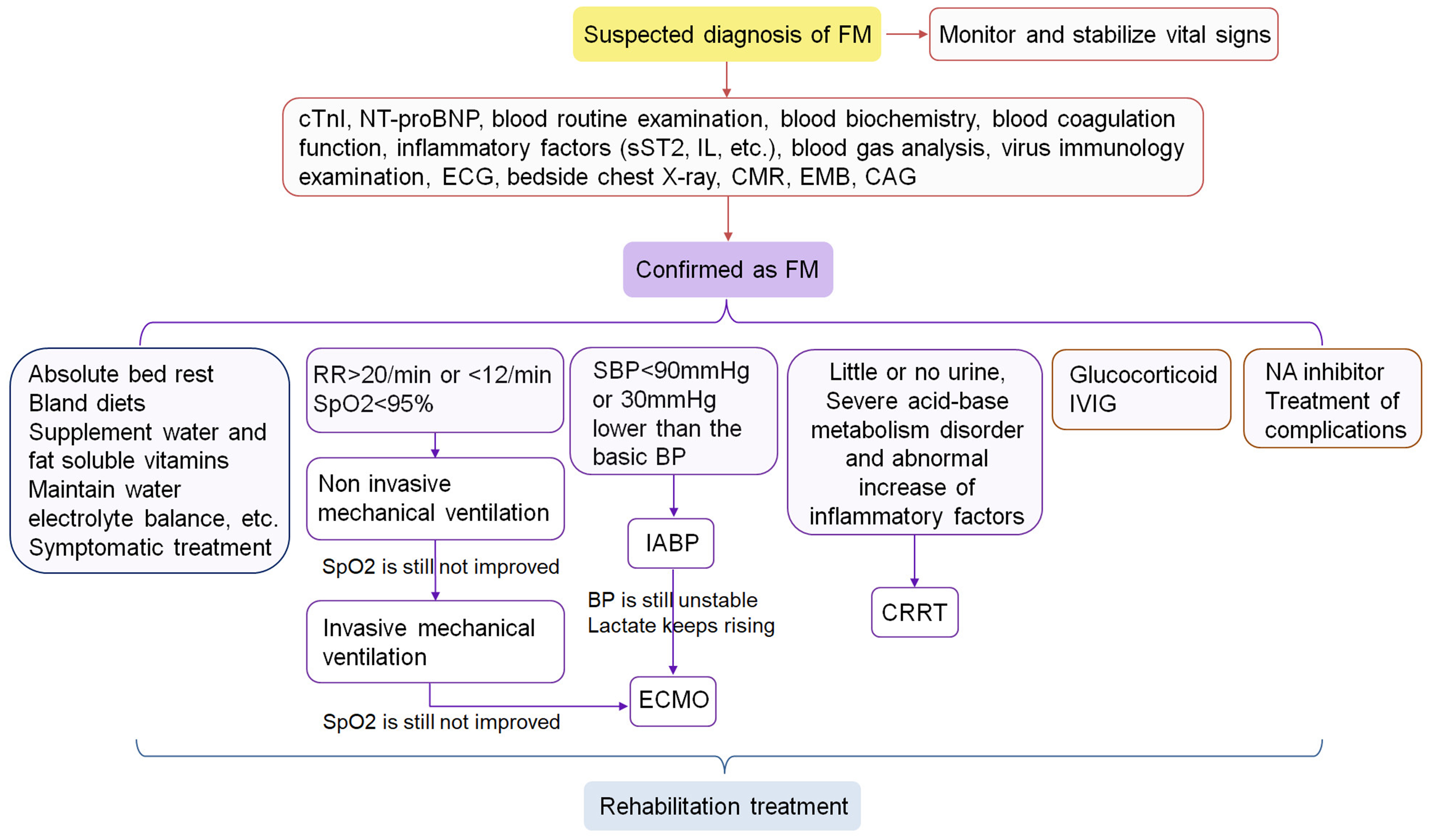
Figure 4. The treatment of FM in the acute phase[3]. NT-proBNP: N-terminal pro-B-type natriuretic peptide; cTnI: cardiac troponin I; sST2: soluble growth stimulating expression gene 2 protein; IL: interleukin; ECG: electrocardiogram; CMR: cardiac magnetic resonance imaging; EMB: endomyocardial biopsy; CAG: coronary angiography; RR: respiratory rate; SpO2: blood oxygen saturation; SBP: systolic blood pressure; BP: blood pressure; IABP: intra-aortic balloon pump; ECMO: extracorporeal membrane oxygenation; CRRT: continuous renal replacement therapy; IVIG: intravenous immunoglobulin; NA: neuraminidase; FM: fulminant myocarditis.
The comprehensive treatment regimen is guided by the principle of “ultra-early recognition, ultra-early diagnosis, ultra-early prediction, and ultra-early rescue”. The primary therapeutic goal in FM is to promptly reverse acute circulatory failure and cardiogenic shock. Key recommendations of treatment are summarized in Table 2.
Treatment of FM in the acute phase[3] (see evidence in Supplementary Table 2)
| Recommendations | Classa | Levelb |
| MCS | ||
| For patients with hemodynamic instability, early initiation of IABP is recommended[68-70] | I | A |
| Immediate initiation of ECMO is recommended when an IABP is insufficient to rectify the hemodynamic instability[71-73] | I | A |
| The Impella device, either alone or in combination with ECMO, should be considered for circulatory support in patients with FM[47,74] | IIa | C |
| Tandem heart, as a biventricular assist device, should be considered for circulatory support. In patients with predominant or concurrent right heart dysfunction, Impella RP (right heart assist device) should also be considered | IIa | C |
| Immunomodulatory Therapy | ||
| After admission, intravenous methylprednisolone at a dosage of 3-8 mg/kg per day is recommended to be started as early as possible. After 3-5 consecutive days, the dose should be reduced by half, and subsequently maintained at 20-40 mg per day for 1-3 months[75-77] | I | A |
| Early administration of IVIG at a dose of 10-20 g per day is recommended. After 3-5 days, the dose should be halved to 5-10 g per day for additional 3 days, with a total cumulative dose of approximately 2 g/kg[78-80] | I | A |
| Other pharmacotherapy | ||
| Oseltamivir phosphate capsule (75 mg orally, twice daily) should be considered, and peramivir may be used as an alternative. The course of treatment is typically 3-5 days[37,81] | IIa | C |
| Maintenance of mean arterial pressure within 60-65 mmHg is recommended to minimize the dosage and duration of vasoactive drugs, and to facilitate early referral of patients for MCS[82-84] | I | A |
| In patients on MCS, inotropic agents (levosimendan, milrinone, and cedilanid) should be considered to assist in weaning from the support devices[85,86] | IIa | C |
| Proton pump inhibitors should be considered to prevent stress ulcer and gastrointestinal bleeding[87,88] | IIa | C |
| Broad spectrum antibiotics should be considered for infection prophylaxis during and after MCS[41,42] | IIa | C |
| Routine use of antifungals is not recommended[89] | III | C |
| Other supportive treatment | ||
| Mechanical ventilation should be considered as an adjunct in acute left heart failure to improve pulmonary function, reduce the patient’s work of breathing, and decrease cardiac workload[4] | IIa | C |
| Early application of CRRT should be considered in patients with FM complicated by organ dysfunction, particularly when renal dysfunction is present[4,90] | IIa | C |
| Treatment of arrhythmia[91] | ||
| In patients with tachyarrhythmia complicated by hemodynamic instability, immediate defibrillation or synchronized electrical cardioversion should be considered. Early use of extracorporeal life support therapy should be considered when arrhythmia cannot be terminated | IIa | C |
| In patients who are relatively hemodynamically stable, antiarrhythmic drugs with negative inotropic and negative chronotropic effects (such as adrenergic β-receptor blockers or non-dihydropyridine calcium channel antagonists) are not recommended | III | C |
| In patients with bradycardia, temporary pacemaker implantation should be considered. If temporary pacing is unavailable, drugs that raise heart rate, such as isoproterenol or atropine, may be considered as an alternative[92] | IIa | C |
| In patients with bradycardia, implantation of a permanent pacemaker is not recommended in the acute phase. ICD is not recommended in patients with ventricular tachycardia in the acute phase or following disease recovery | III | C |
| In patients whose clinical condition has been stable for more than 2 weeks but in whom conduction block persists without recovery, implantation of a permanent pacemaker should be considered | IIa | C |
The core components of the comprehensive treatment regimen include: (i) mechanical circulatory support (MCS). This strategy prioritizes “resting the failing heart” over elevating blood pressure with inotropes or vasoactive agents. Accordingly, MCS devices such as IABP, ECMO and Impella are recommended to maintain hemodynamic stability and adequate organ perfusion. Vasoactive drugs are generally not recommended unless MCS is unavailable. Inotropic agents are also not recommended except for temporary use during MCS; (ii) Immunomodulatory therapy. It primarily consists of early, combined, and sufficient doses of glucocorticoids and intravenous immunoglobulin (IVIG). Immunosuppressive therapy with cytotoxic agents is not recommended; (iii) Antiviral therapy. For patients with influenza virus infection, neuraminidase inhibitors are recommended. Emerging evidence suggests that neuraminidase inhibitors may also attenuate myocardial injury through mechanisms beyond direct antiviral effects[36,37]; (iv) Other supportive treatment. It includes mechanical ventilation, continuous renal replacement therapy (CRRT), and temporary pacemaker implantation when clinically indicated.
Management of complications is also critical to optimizing therapeutic efficacy and improving prognosis in patients with FM. Arrhythmias occur in 20%-30% of FM patients. The therapeutic objective is to prevent and correct hemodynamic instability caused by arrhythmias. For tachyarrhythmias, electrical cardioversion may be used; for severe bradycardia or high-grade conduction block, temporary pacing is indicated[38]. Lethal complications such as DIC and systemic capillary leak syndrome (SCLS) should be prevented through early recognition and prompt intervention to reverse cardiogenic shock and control the cytokine storm[39,40].
It is important to note that FM is not primarily caused by bacterial infection; therefore, antibiotics are not indicated for the treatment of the underlying disease. However, broad-spectrum antibiotics are recommended for infection prophylaxis during and after MCS. If infection develops, aggressive and adequate antimicrobial therapy should be initiated promptly, followed by step-down de-escalation as early as possible to minimize microcirculatory injury[41,42].
CONVALESCENT MANAGEMENT AND LONG-TERM FOLLOW-UP OF FM
Following standardized acute-phase treatment, the majority of patients with FM achieve clinical recovery. However, some patients may exhibit persistent myocardial inflammation and edema of varying degrees, which can lead to left ventricular dilation, arrhythmias, cardiac dysfunction, or even inflammatory cardiomyopathy[43]. Therefore, rigorous and systematic clinical assessment is mandatory both before discharge and during long-term follow-up.
Post-discharge patients can be stratified into three categories: healed myocarditis, healing myocarditis, and persistent myocarditis[44]. Oral corticosteroids are effective for patients with chronic inflammatory edema and persistently elevated myocardial injury biomarkers. For patients with pathologically confirmed giant cell myocarditis (GCM), eosinophilic myocarditis (EM), cardiac sarcoidosis, or ICI-associated myocarditis, corticosteroids or other immunosuppressive agents may be administered[45].
All patients in the convalescent phase are recommended to receive angiotensin-converting enzyme inhibitors (ACEI), β-blockers, and trimetazidine[4]. Moderate-to-severe physical activity is contraindicated within the first 3-6 months after discharge. Prior to initiating cardiac rehabilitation, a comprehensive clinical re-evaluation is essential. For patients with healed myocarditis, exercise-based cardiac rehabilitation may be initiated no earlier than 6 months after the acute onset, starting with low-intensity training[44,46] [Table 3].
Convalescent management for patients with FM[3] (see evidence in Supplementary Table 3)
| Recommendations | Classa | Levelb |
| Follow-up assessment | ||
| Patients with FM require regular and comprehensive evaluation after discharge, encompassing clinical manifestations, biomarkers, resting ECG, 24-hour Holter, echocardiography and CMR, as clinically indicated | I | C |
| Exercise and cardiac rehabilitation | ||
| Moderate-to-high intensity exercise is not recommended in all patients with FM within 3-6 months following discharge[93-95] | III | B |
| Patients categorized as having “healing myocarditis” and ”healed myocarditis” may benefit from cardiac exercise rehabilitation training[44] | I | C |
| Exercise training for cardiac rehabilitation is not recommended in patients with active myocarditis in the acute phase, defined by the presence of typical symptoms, elevated biomarker levels, and abnormal echocardiographic findings[46] | III | C |
| Immunomodulatory therapy in the convalescent phase | ||
| In patients with GCM, EM, cardiac sarcoidosis, or ICI-associated myocarditis, corticosteroids or other immunosuppressive agents are recommended after discharge[96-98] | I | B |
| In patients with chronic lymphocytic myocarditis without evidence of viral infection as suggested by EMB, corticosteroid and immunosuppressive therapy are recommended to improve cardiac function[99] | I | B |
| In patients with persistently elevated myocardial injury biomarkers or CMR suggestive myocardial edema after discharge, corticosteroid therapy should be considered, as it may ameliorate myocardial inflammation[100,101] | IIa | C |
CONCLUSION
Multiple international societies have issued guidance documents on the management of myocarditis and/or FM. Table 4 summarizes key recommendations from the 2024 Chinese Guidelines[3], the 2020 American Heart Association (AHA) Scientific Statement[19], the 2024 American College of Cardiology (ACC) Expert Consensus[47], and the 2025 European Society of Cardiology (ESC) Guidelines[48]. These guidance documents demonstrate general consensus on the definition of FM, pathological diagnostic criteria, laboratory markers, diagnostic workflows, and the role of MCS. A previous review on the diagnosis and management of FM similarly aligns with these guidelines on the aforementioned aspects[49]. However, that review places particular emphasis on the life-saving role of MCS in FM management and elaborates on the efficacy, advantages and limitations of various life-support devices including Impella, VA-ECMO, ECMELLA (VA-ECMO + Impella), PROPELLA (prolonged Impella support), BIPELLA [left-sided Impella circulatory pump (CP) combined with right-sided Impella right pump (RP)], and device escalation strategies in deteriorating myocarditis cases, thereby supplementing the current guidelines with detailed MCS-related insights. By contrast, the guidelines offer more comprehensive and systematic coverage. Notably, substantial controversies persist regarding pharmacotherapy between the Chinese Guidelines and other international guidance documents.
Comparison of recommendations between current guidelines on myocarditis and FM
| 2024 Chinese guidelines[3] | 2020 AHA scientific statement[19] | 2024 ACC expert consensus[47] | 2025 ESC guidelines[48] | |
| The subject | FM | FM | Myocarditis | Myocarditis and pericarditis |
| Definition of FM | FM refers to acute diffuse inflammatory damage of myocardium with hemodynamic abnormalities | FM is defined a sudden and severe inflammation of the myocardium resulting in myocyte necrosis, edema, and cardiogenic shock | A 4-stage classification of myocarditis is proposed FM is comparable with stage D, which is defined as advanced myocarditis as evidenced by hemodynamic instability requiring inotropes/vasopressors or TCS, or electrical instability requiring intervention | FM is defined as myocarditis with acute onset (≤ 4 weeks) and hemodynamically unstable patients requiring inotropes or MCS |
| Recommendations of EMB | EMB is actively recommended for FM, especially when a specific etiology is clinically suspected, such as the use of ICIs, autoimmune diseases, peripheral blood eosinophilia, to clarify the etiology, the pathological type, the status of myocardial injury and the guidance to clinical treatment | EMB is recommended for most patients presenting with FM | Most stage D patients should undergo EMB | EMB is recommended in patients with high-risk myocarditis, and/or hemodynamic instability, and/or in patients with intermediate-risk myocarditis not responding to conventional therapy in order to detect a specific histologic subtype and to assess the presence of viral genome for treatment |
| CAG | Depending on the clinical manifestations, it is recommended to perform CAG as early as possible to rule out acute myocardial infarction, regardless of whether the patient has high-risk factors for coronary atherosclerosis | No specific recommendation for CAG | Under the circumstances of an acute rise in troponin, myocardial infarction needs to be excluded by CAG or absence of ischemic LGE on CMR | Depending on the initial clinical assessment, ACS should be ruled out by coronary CT or invasive CAG depending on likelihood of obstructive coronary artery disease |
| MCS | MCS is the preferred therapeutic strategy in patients with a diagnosis of FM Immediate use of IABP is recommended in patients with FM who present with early manifestations of shock. Addition of ECMO is recommended when IABP is insufficient to correct circulatory disturbance. In patients with severe circulatory dysfunction, severe LV dysmotility or cardiac arrest, a combination of ECMO and IABP should be performed immediately | Nearly all patients with FM will require temporary MCS | Stage D patients who develop electrical or hemodynamic instability and require TCS or advanced therapies The TCS devices used most often include IABP, intravascular axial flow device (Impella), or VA-ECMO | Temporary MCS should be performed early and in a timely manner in patients with FM with refractory cardiogenic shock Among temporary MCS, ECMO represents the most frequently applied or recommended approach |
| Immunomodulatory therapy | The immunomodulatory therapy for FM comprises very early use of adequate doses of glucocorticoids and adequate doses of immunoglobulins Cytotoxic immunosuppressant is not recommended in the treatment of FM | Immunomodulatory therapy is considered according to the type of myocarditis: · Lymphocytic myocarditis: steroids may be helpful but still controversial · GCM: high-dose steroids combined with immunosuppressants (such as cyclosporine or azathioprine) · EM: high-dose corticosteroids · ICI-associated myocarditis: high-dose corticosteroids. Antithymocyte globulin or IVIG for unstable patients who do not respond to corticosteroids. Tacrolimus or mycophenolate mofetil for stable patients who do not respond to corticosteroids | Immunosuppressive therapy is used in these conditions: · EM · GCM · Granulomatous myocarditis (sarcoid) · ICI-associated myocarditis · Other autoimmune-related conditions Immunosuppressive therapy typically includes high-dose steroids. For GCM, cyclosporine or tacrolimus should be added IVIG can be considered in the setting of inflammatory, antibody-mediated, or autoimmune disorders | Corticosteroids should be considered in patients with fulminant, non-infectious forms of myocarditis to stabilize the patients For GCM, immunosuppressive therapy typically involves corticosteroids and one or two additional immunosuppressive agents (cyclosporine, azathioprine or mycophenolate mofetil) |
| Vasoactive drugs | The use of vasoactive drugs is not recommended for patients with FM Only when MCS is unavailable, vasoactive drugs may be administered temporarily to maintain the mean arterial pressure in patients with FM. The referral of patients to a tertiary center and early use of MCS is recommended to minimize the dose and duration of vasoactive drug | It mentioned nearly all patients with FM will require vasoactive drug support, and no detail was discussed | No specific description | It mentioned FM is a cause of cardiogenic shock, and should be treated accordingly with inotropic or vasopressor support, and no detail was discussed |
| Inotropic drugs | The use of inotropic drugs is not recommended in the acute phase of FM In patients on MCS, inotropic agents should be considered to assist in weaning from the support devices | No specific description | Intravenous inotropic agents may be administered for evidence of inadequate cardiac output | It mentioned FM is a cause of cardiogenic shock, and should be treated accordingly with inotropic or vasopressor support, and no detail was discussed |
| Management of arrhythmias | For tachyarrhythmias that affect hemodynamics, cardioversion should be administered immediately, along with drugs to prevent recurrence For hemodynamically stable arrhythmias, appropriate treatment and anti-arrhythmic drugs are selected based on clinical symptoms, cardiac functional status, and the nature of the arrhythmia Temporary pacemakers should be installed immediately in patients with marked bradycardia or conduction block | No specific description | The treatment of arrhythmia in myocarditis should be in keeping with current guidelines for arrhythmia, and the detail was not discussed in this expert consensus | Temporary transvenous external pacing should be considered in patients with high-degree conduction disorders GCM is associated with a high risk of life-threatening VA and ICD implantation can be considered |
| Advanced HF referral | The patients with FM should be handled in a medical center with a rapid response team for FM. Once the diagnosis of FM is made in an institution without processing capability, the treatment with drugs should be immediately started to maintain vital signs, allowing quick referral of patients to the tertiary centers | For institutions without advanced HF surgical and medical management capabilities, consideration for transfer of patients with FM from the emergency department directly to a tertiary referral hospital is warranted | The patients with any of the following should be referred to an advanced HF center with a multidisciplinary myocarditis team: severe ventricular dysfunction; life-threatening arrhythmia; cardiogenic shock; refractory HF; fulminant onset; and most cases of HF/DCM with associated systemic immune-mediated disease; or suspected giant cell, eosinophilic, or sarcoid myocarditis | It is crucial to be aware of the severe condition and, when needed, to refer patients with FM to tertiary referral centers that can institute temporary MCS and perform early EMB |
| Follow-up | Long-term follow-up should be conducted in all patients with FM after discharge. The follow-up contents include clinical symptoms and signs, echocardiography, ECG, cTnI, NT-proBNP, inflammatory indicators such as cytokines, CMR, and EMB if necessary in order to promptly detect abnormalities of cardiac structure and function and to prevent the occurrence of complications | No specific description | Longitudinal follow-up for stage D patients are typically conducted in 2 time points after discharge: 2-4 weeks: echocardiogram, biomarkers (hs-troponin, BNP and CRP) and ECG 6 months: CMR | Follow-up is recommended in all patients with myocarditis, with clinical assessment, ECG, Holter-ECG registration, exercise test (if no signs of ongoing inflammation), echocardiography, and CMR within at least 6 months after the index hospitalization |
Previous guidelines generally recommended managing acute heart failure in FM in accordance with standard heart failure guidelines, utilizing vasoactive or inotropic agents[19,47,48]. In contrast, the Chinese Guidelines exercise extreme restraint in the use of these drugs. Vasoactive agents are generally not recommended; they may be used only temporarily to maintain mean arterial pressure when MCS is unavailable, and should be discontinued once MCS is initiated. Inotropic agents are likewise not recommended, except as short-term adjunctive therapy to facilitate successful weaning from devices in patients receiving MCS[3].
The Chinese Guidelines emphasize that the cornerstone of pharmacological therapy for improving heart failure lies in controlling overactivated inflammation through the combination of glucocorticoids and IVIG, irrespective of pathological subtypes. Other guidelines, however, hold that immunomodulatory therapy should be primarily reserved for specific subtypes of myocarditis (e.g., GCM or ICI-associated myocarditis). Historically, the use of glucocorticoids in infectious myocarditis has been controversial, primarily due to concerns regarding potential exacerbation of viral infection. However, research findings have revealed that dexamethasone administration in a mouse model of viral myocarditis reduced myocardial viral titers rather than promoting viral replication, which may result from a significant increase in interferon production[37,50]. Thus, glucocorticoids exert immunomodulatory effects alongside multiple protective roles, including anti-shock activity, stabilization of lysosomal membrane structure, mitigation of toxin-induced damage, and reduction of myocardial depressant factor release. The early initiation of adequate glucocorticoid therapy is therefore deemed essential for the management of FM.
FM is predominantly a clinical diagnosis. The disease progresses rapidly during the acute phase, and pathological diagnosis is usually not obtainable in a timely manner. Circulating levels of inflammatory factors and sST2 can identify whether patients are in a state of excessive immune activation and cytokine storm, which is of great guiding significance for the initiation of immunomodulatory therapy. Notably, sST2 exhibits high sensitivity for distinguishing FM from non-FM conditions, surpassing that of NT-proBNP and cTnI. Through quantitative analysis, it was determined that patients presenting with relevant clinical manifestations and an sST2 level > 58.39 ng·mL-1 should be considered for a suspected diagnosis of FM[26]. Additionally, dynamic changes in plasma inflammatory factors and sST2 levels serve as reliable indicators of therapeutic response and clinical improvement, and are also important for guiding dose adjustments of immunomodulatory agents. Therefore, these guidelines recommend dynamic monitoring of inflammatory factors and sST2 in FM patients. Given variations in local healthcare systems, technical and cost-related feasibility issues may affect the implementation of dynamic monitoring for these biomarkers. Regions may reasonably set the frequency and intervals of monitoring to strike an optimal balance between cost control and therapeutic management.
In previous reports, the in-hospital mortality rate among patients with FM ranged from 45%-55.6%[7,10,11]. Two multicenter studies have evaluated the treatment regimen recommended by the Chinese Guidelines. Among 169 FM patients, 81 receiving the “life support-based comprehensive regimen” had an in-hospital mortality of 3.7%, compared with 46.6% in those not receiving this regimen[37]. In another study comprising 138 FM patients, 96 treated with the recommended protocol showed an in-hospital mortality of 4.2%, versus 52.4% in patients not treated with the protocol[20]. These findings demonstrate the efficacy of the comprehensive treatment regimen. Nevertheless, both studies were retrospective in design, and all enrolled patients were diagnosed clinically without pathological confirmation. Therefore, further clinical trials on FM based on pathological or CMR-confirmed diagnoses are essential to verify therapeutic efficacy.
Notably, the therapies specified in this regimen are primarily delivered in qualified medical centers. In clinical practice, timely access of FM patients to such centers is critical for effective rescue. The healthcare system for FM management consists of three core components: referral centers, primary-care units, and connecting networks. Referral centers should establish rapid response teams for FM and be equipped with life-support devices to admit patients and deliver standardized treatment per the protocol. Primary-care units should prioritize capabilities in recognizing, diagnosing, and providing emergency management for FM, so as to stabilize patients and secure opportunities for timely referral. The connecting networks focus on regular training for primary-care providers; when FM patients present, they enable information exchange and online guidance between referral centers and local units, and allow prompt initiation of referral to minimize transit-related delays. Given regional disparities in healthcare system models and available resources, the number, distribution, and density of referral centers, primary-care units, and supporting networks vary across regions, leading to divergent response efficiencies. Provided that each region maintains the integrity of these three components according to local conditions and ensures unobstructed referral pathways, patients can be transferred to qualified centers as early as possible to maximize their chances of receiving standardized treatment.
The regimen proposed in the Chinese Guidelines still has limitations. Notably, for acute-phase management, the regimen is not stratified according to pathological subtypes, and no dedicated strategies have been established for special subtypes such as GCM and ICI-associated myocarditis. Given the abrupt onset and rapid progression of FM, most existing clinical studies focus on myocarditis in general, with FM included merely as a subgroup. There are few clinical trials specifically targeting FM, featuring small sample sizes, and substantial practical obstacles remain to the design and implementation of randomized controlled trials. Continuous accumulation of clinical evidence will therefore be required going forward. Notably, there are substantial discrepancies between the Chinese Guidelines and other international guidelines regarding the scope of application for immunomodulatory therapy, and further clinical evidence is needed to underpin the respective recommendations.
Current immunomodulatory therapies exert broad-spectrum effects through non-selective interference with inflammation. Developing targeted therapeutic approaches based on specific inflammatory pathways and molecules may represent a novel strategy for precise immunomodulation in the future (e.g., sST2-neutralizing antibodies[51]). Elevated levels of S100A8/A9 have also been associated with cardiac dysfunction and disease severity in FM patients[52], and nanoparticles delivering S100A9-targeting siRNA have been shown to block neutrophil infiltration and improve cardiac function in a mouse model[53]. Among FM patients who survive the acute phase, follow-up data indicate that approximately one-quarter of patients still present symptoms such as cardiac enlargement, heart failure, and arrhythmias one year after discharge[54]. To date, limited interventions have been established to improve the long-term outcomes of these patients, highlighting the need to advance our understanding of the chronic disease trajectory of FM. It was reported that hydroxychloroquine combined with prednisolone therapy significantly improved the prognosis and heart function in patients with chronic inflammatory cardiomyopathy[55]. In summary, these issues require further investigation, and the life support regimen will be refined and updated in light of future research findings.
DECLARATIONS
Acknowledgments
The authors acknowledge the use of Figdraw (www.figdraw.com) for creating the Graphic Abstract in this review (permission ID number TTWYOdf4f4).
Authors’ contributions
Designed the review and revised the manuscript: Wang DW
Wrote the manuscript: Wen Z, Shu H, Jiang J, Li C, Fan J, Ran X
Revised the manuscript: Wen Z, He Z
Availability of data and materials
Not applicable.
AI and AI-assisted tools statement
Not applicable.
Financial support and sponsorship
This work was supported by the National Natural Science Foundation of China (No. 82070395 to Wen Z; Nos. 82330010 and 82241034 to Wang DW), and the Scientific Research Project of Hubei Province (No. 2024CSA067 to Wang DW). The funding bodies had no role in the design, information collection, and writing of this paper.
Conflicts of interest
Wang DW is a Consulting Editor of The Journal of Cardiovascular Aging and a Guest Editor of the Special Issue “Fulminant Myocarditis: Mechanisms, Diagnostics, and Life Support Strategies”. Wang DW was not involved in any stage of the editorial process for this manuscript, including reviewer selection, manuscript handling, or decision-making. The other authors declare that they have no conflicts of interest.
Ethical approval and consent to participate
Not applicable.
Consent for publication
Not applicable.
Copyright
© The Author(s) 2026.
Supplementary Materials
REFERENCES
1. Section of Precision Medical of Chinese Society of Cardiology of Chinese Medical Association, Editorial Board of Chinese Journal of Cardiology, Working Group on Adult Myocarditis. Chinese expert consensus statement on clinical diagnosis and treatment of fulminant myocarditis in adults. Chin J Cardiovasc Dis. 2017;45:742-52.
2. Chinese Society of Cardiology, Chinese Medical Association; Editorial Board of Chinese Journal of Cardiology. Chinese guideline on the diagnosis and treatment of fulminant myocarditis in adults. Chin J Cardiovasc Dis. 2024;52:10-33.
3. Jiang J, Shu H, Wang DW, et al.; Chinese Society of Cardiology. Chinese Society of Cardiology guidelines on the diagnosis and treatment of adult fulminant myocarditis. Sci China Life Sci. 2024;67:913-39.
4. McDonagh TA, Metra M, Adamo M, et al.; ESC Scientific Document Group. 2021 ESC Guidelines for the diagnosis and treatment of acute and chronic heart failure: developed by the Task Force for the diagnosis and treatment of acute and chronic heart failure of the European Society of Cardiology (ESC) With the special contribution of the Heart Failure Association (HFA) of the ESC. Eur Heart J. 2021;42:3599-726.
5. Heidenreich PA, Bozkurt B, Aguilar D, et al.; ACC/AHA Joint Committee Members. 2022 AHA/ACC/HFSA guideline for the management of heart failure: A Report of the American College of Cardiology/American Heart Association Joint Committee on clinical practice guidelines. Circulation. 2022;145:e895-e1032.
6. Kytö V, Saraste A, Voipio-Pulkki LM, Saukko P. Incidence of fatal myocarditis: a population-based study in Finland. Am J Epidemiol. 2007;165:570-4.
7. Lee CH, Tsai WC, Hsu CH, Liu PY, Lin LJ, Chen JH. Predictive factors of a fulminant course in acute myocarditis. Int J Cardiol. 2006;109:142-5.
8. Dai H, Lotan D, Much AA, et al. Global, regional, and national burden of myocarditis and cardiomyopathy, 1990-2017. Front Cardiovasc Med. 2021;8:610989.
9. McCarthy RE 3rd, Boehmer JP, Hruban RH, et al. Long-term outcome of fulminant myocarditis as compared with acute (nonfulminant) myocarditis. N Engl J Med. 2000;342:690-5.
10. Sawamura A, Okumura T, Ito M, et al.; CHANGE PUMP Investigators. Prognostic value of electrocardiography in patients with fulminant myocarditis supported by percutaneous venoarterial extracorporeal membrane oxygenation- analysis from the CHANGE PUMP study. Circ J. 2018;82:2089-95.
11. Hung Y, Lin WH, Lin CS, et al. The prognostic role of QTc interval in acute myocarditis. Acta Cardiol Sin. 2016;32:223-30.
12. Kanaoka K, Onoue K, Terasaki S, et al.; Japanese Registry of Fulminant Myocarditis Investigators. features and outcomes of histologically proven myocarditis with fulminant presentation. Circulation. 2022;146:1425-33.
13. Liu L, Yang X, Gu Y, Jiang T, Xu J, Xu M. Predictive value of the age, creatinine, and ejection fraction (ACEF) score in patients with acute fulminant myocarditis. Front Physiol. 2021;12:596548.
14. Hang W, Chen C, Seubert JM, Wang DW. Fulminant myocarditis: a comprehensive review from etiology to treatments and outcomes. Signal Transduct Target Ther. 2020;5:287.
15. Li H, Zhang M, Zhao Q, et al. Self-recruited neutrophils trigger over-activated innate immune response and phenotypic change of cardiomyocytes in fulminant viral myocarditis. Cell Discov. 2023;9:103.
16. Maisch B, Portig I, Ristic A, Hufnagel G, Pankuweit S. Definition of inflammatory cardiomyopathy (myocarditis): on the way to consensus. A status report. Herz. 2000;25:200-9.
17. Maisch B, Ruppert V, Pankuweit S. Management of fulminant myocarditis: a diagnosis in search of its etiology but with therapeutic options. Curr Heart Fail Rep. 2014;11:166-77.
18. Ammirati E, Veronese G, Brambatti M, et al. Fulminant versus acute nonfulminant myocarditis in patients with left ventricular systolic dysfunction. J Am Coll Cardiol. 2019;74:299-311.
19. Kociol RD, Cooper LT, Fang JC, et al.; American Heart Association Heart Failure and Transplantation Committee of the Council on Clinical Cardiology. Recognition and initial management of fulminant myocarditis: a scientific statement from the American Heart Association. Circulation. 2020;141:e69-92.
20. Zhou N, Zhao Y, Jiang J, et al. Impact of mechanical circulatory support and immunomodulation therapy on outcome of patients with fulminant myocarditis: Chinese registry of fulminant myocarditis. Signal Transduct Target Ther. 2021;6:350.
21. Caforio AL, Calabrese F, Angelini A, et al. A prospective study of biopsy-proven myocarditis: prognostic relevance of clinical and aetiopathogenetic features at diagnosis. Eur Heart J. 2007;28:1326-33.
22. JCS Joint Working Group. Guidelines for diagnosis and treatment of myocarditis (JCS 2009): digest version. Circ J. 2011;75:734-43.
23. Liu C, Wang Z, Chen K, et al. The absolute and relative changes in high-sensitivity cardiac troponin I are associated with the in-hospital mortality of patients with fulminant myocarditis. BMC Cardiovasc Disord. 2021;21:571.
24. Palaka E, Grandy S, Darlington O, McEwan P, van Doornewaard A. Associations between serum potassium and adverse clinical outcomes: a systematic literature review. Int J Clin Pract. 2020;74:e13421.
25. Guglin M, Zucker MJ, Bazan VM, et al. Venoarterial ECMO for adults: JACC Scientific Expert Panel. J Am Coll Cardiol. 2019;73:698-716.
26. Wang J, He M, Li H, et al. Soluble ST2 is a sensitive and specific biomarker for fulminant myocarditis. J Am Heart Assoc. 2022;11:e024417.
28. Dai MY, Yan YC, Wang LY, Zhao CX, Wang DW, Jiang JG. Characteristics of electrocardiogram findings in fulminant myocarditis. J Cardiovasc Dev Dis. 2023;10:280.
29. Zuo H, Li R, Ma F, et al. Temporal echocardiography findings in patients with fulminant myocarditis: beyond ejection fraction decline. Front Med. 2020;14:284-92.
30. Ammirati E, Veronese G, Cipriani M, et al. Acute and fulminant myocarditis: a pragmatic clinical approach to diagnosis and treatment. Curr Cardiol Rep. 2018;20:114.
31. Zuo H, Li H, Li R, et al. Myocardial strain features by 2D-STE during the course of fulminant myocarditis: correlation with characteristics by CMR and clinical implications. Medicine. 2021;100:e25050.
32. Ferreira VM, Schulz-Menger J, Holmvang G, et al. Cardiovascular magnetic resonance in nonischemic myocardial inflammation: expert recommendations. J Am Coll Cardiol. 2018;72:3158-76.
33. Friedrich MG, Sechtem U, Schulz-Menger J, et al.; International Consensus Group on Cardiovascular Magnetic Resonance in Myocarditis. Cardiovascular magnetic resonance in myocarditis: a JACC white paper. J Am Coll Cardiol. 2009;53:1475-87.
34. Peretto G, Busnardo E, Ferro P, et al. Clinical applications of FDG-PET scan in arrhythmic myocarditis. JACC Cardiovasc Imaging. 2022;15:1771-80.
35. Archibald SJ, Holland JP, Korde A, Martins AF, Shuhendler AJ, Scott PJH. Combining nuclear medicine with other modalities: future prospect for multimodality imaging. Mol Imaging. 2024;23:15353508241245265.
36. Li C, Zhao M, Xiao L, et al. Prognostic value of elevated levels of plasma N-acetylneuraminic acid in patients with heart failure. Circ Heart Fail. 2021;14:e008459.
37. Li S, Xu S, Li C, et al. A life support-based comprehensive treatment regimen dramatically lowers the in-hospital mortality of patients with fulminant myocarditis: a multiple center study. Sci China Life Sci. 2019;62:369-80.
38. Camm AJ, Lip GY, De Caterina R, et al.; ESC Committee for Practice Guidelines (CPG). 2012 focused update of the ESC Guidelines for the management of atrial fibrillation: an update of the 2010 ESC Guidelines for the management of atrial fibrillation. Developed with the special contribution of the European Heart Rhythm Association. Eur Heart J. 2012;33:2719-47.
39. Thrombosis and Hemostasis Group, Hematology Society of Chinese Medical Association. Consensus of Chinese experts on diagnosis of disseminated intravascular coagulation (version 2017). Chin J Hematol. 2017;38:361-3.
40. Heradstveit BE, Guttormsen AB, Langørgen J, et al. Capillary leakage in post-cardiac arrest survivors during therapeutic hypothermia - a prospective, randomised study. Scand J Trauma Resusc Emerg Med. 2010;18:29.
41. Pieri M, Agracheva N, Fumagalli L, et al. Infections occurring in adult patients receiving mechanical circulatory support: the two-year experience of an Italian National Referral Tertiary Care Center. Med Intensiva. 2013;37:468-75.
42. Aslam S, Cowger J, Shah P, et al. The International Society for Heart and Lung Transplantation (ISHLT): 2024 infection definitions for durable and acute mechanical circulatory support devices. J Heart Lung Transplant. 2024;43:1039-50.
43. Tschöpe C, Ammirati E, Bozkurt B, et al. Myocarditis and inflammatory cardiomyopathy: current evidence and future directions. Nat Rev Cardiol. 2021;18:169-93.
44. Schwaab B, Kindermann I, Bjarnason-Wehrens B, et al. Viral myocarditis: a forbidden indication for cardiac rehabilitation? Eur J Prev Cardiol. 2022;29:2064-8.
45. Ammirati E, Veronese G, Bottiroli M, et al. Update on acute myocarditis. Trends Cardiovasc Med. 2021;31:370-9.
46. Kerneis M, Ammirati E, Delmas C, et al. Clinical management of acute myocarditis in daily practice: an expert practical view. Eur Heart J Acute Cardiovasc Care. 2025;14:420-31.
47. Drazner MH, Bozkurt B, Cooper LT, et al.; Writing Committee. 2024 ACC expert consensus decision pathway on strategies and criteria for the diagnosis and management of myocarditis: a report of the American College of Cardiology Solution Set Oversight Committee. J Am Coll Cardiol. 2025;85:391-431.
48. Schulz-Menger J, Collini V, Gröschel J, et al.; ESC Scientific Document Group. 2025 ESC Guidelines for the management of myocarditis and pericarditis. Eur Heart J. 2025;46:3952-4041.
49. D’Ettore N, Eghbalzadeh K, Oezkur M, Bertoldi LF, Bossard M, Pappalardo F. Diagnosis and management of patients with fulminant myocarditis. Eur Heart J Suppl. 2025;27:iv23-30.
50. Zhou Z, Zhang M, Zhao C, et al. Epoxyeicosatrienoic acids prevent cardiac dysfunction in viral myocarditis via interferon type I signaling. Circ Res. 2023;133:772-88.
51. Zhuang Y, Zhang Z, Li H, et al. Soluble ST2 drives fulminant myocarditis progression via the IGF2R-YY1 mitochondrial axis. Eur Heart J. 2026;47:5572-89.
52. He W, Wu J, Wang D, et al. Plasma proteomics identifies S100A8/A9 as a novel biomarker and therapeutic target for fulminant myocarditis. J Adv Res. 2026;81:717-30.
53. Jiang Y, Wang Z, Chen P, et al. Engineered neutrophil membrane nanosystem for targeted siRNA therapy in myocardial ischemia-reperfusion injury. J Nanobiotechnology. 2025;24:99.
54. Jiang JG, Liu C, Cui GL, et al. Long term prognosis of fulminant myocarditis and predictors related to impaired cardiac function post discharge. Chin J Cardiovasc Dis. 2022;50:263-9.
55. He W, Cui G, Chen J, et al. The efficacy and safety of hydroxychloroquine in patients with chronic inflammatory cardiomyopathy: a multicenter randomized study (HYPIC trial). BMC Med. 2025;23:467.
56. Artico J, Shiwani H, Moon JC, et al. Myocardial involvement after hospitalization for COVID-19 complicated by troponin elevation: a prospective, multicenter, observational study. Circulation. 2023;147:364-74.
57. Ukena C, Kindermann M, Mahfoud F, et al. Diagnostic and prognostic validity of different biomarkers in patients with suspected myocarditis. Clin Res Cardiol. 2014;103:743-51.
58. Ekström K, Lehtonen J, Kandolin R, Räisänen-Sokolowski A, Salmenkivi K, Kupari M. Long-term outcome and its predictors in giant cell myocarditis. Eur J Heart Fail. 2016;18:1452-8.
59. Saracoglu A, Fawzy I, Saracoglu KT, Abdallah BM, Arif M, Schmidt M. Point of care guided coagulation management in adult patients on ECMO: a systematic review and meta-analysis. J Crit Care. 2024;83:154830.
60. Zhu Y, Lan MJ, Liang JS, et al. Assessing venous thrombotic risks in extracorporeal membrane oxygenation-supported patients: a systematic review and meta-analysis. Clin Appl Thromb Hemost. 2024;30:10760296241279293.
61. Cain BS, Meldrum DR, Dinarello CA, et al. Tumor necrosis factor-alpha and interleukin-1beta synergistically depress human myocardial function. Crit Care Med. 1999;27:1309-18.
62. Wang ZH, Liao YH, Yuan J, et al. Continued elevation of plasma IL-4 and IL-17 predicts the progression from VMC to DCM. Dis Markers. 2020;2020:9385472.
63. Coronado MJ, Bruno KA, Blauwet LA, et al. Elevated sera sST2 is associated with heart failure in men ≤ 50 years old with myocarditis. J Am Heart Assoc. 2019;8:e008968.
64. Hou W, Shi T, Li Y, Li W, Xu M, Peng F. Soluble suppression of tumorigenicity 2 associated with fulminant myocarditis in children: a retrospective observational study. Medicine. 2023;102:e34784.
65. Daubin C, Valette X, Thiollière F, et al.; BPCTrea Study Group. Procalcitonin algorithm to guide initial antibiotic therapy in acute exacerbations of COPD admitted to the ICU: a randomized multicenter study. Intensive Care Med. 2018;44:428-37.
66. Huang DT, Yealy DM, Filbin MR, et al.; ProACT Investigators. Procalcitonin-guided use of antibiotics for lower respiratory tract infection. N Engl J Med. 2018;379:236-49.
67. Kyriazopoulou E, Liaskou-Antoniou L, Adamis G, et al. Procalcitonin to reduce long-term infection-associated adverse events in sepsis. A randomized trial. Am J Respir Crit Care Med. 2021;203:202-10.
68. den Uil CA, Van Mieghem NM, Bastos MB, et al. Primary intra-aortic balloon support versus inotropes for decompensated heart failure and low output: a randomised trial. EuroIntervention. 2019;15:586-93.
69. Deppe AC, Weber C, Liakopoulos OJ, et al. Preoperative intra-aortic balloon pump use in high-risk patients prior to coronary artery bypass graft surgery decreases the risk for morbidity and mortality-a meta-analysis of 9,212 patients. J Card Surg. 2017;32:177-85.
70. Chu S, Sun P, Zhang Y, et al. Intra-aortic balloon pump on in-hospital outcomes of cardiogenic shock: findings from a nationwide registry, China. ESC Heart Fail. 2021;8:3286-94.
71. Duan J, Shi Y, Luo G, Peng Y, Duan B, Zhang Z. Short-term efficacy and safety of different mechanical hemodynamic support devices for cardiogenic shock or high-risk pci: a network meta-analysis of thirty-seven trials. Shock. 2021;55:5-13.
72. Benenati S, Toma M, Canale C, et al. Mechanical circulatory support in patients with cardiogenic shock not secondary to cardiotomy: a network meta-analysis. Heart Fail Rev. 2022;27:927-34.
73. Low CJW, Ling RR, Lau MPXL, et al. Mechanical circulatory support for cardiogenic shock: a network meta-analysis of randomized controlled trials and propensity score-matched studies. Intensive Care Med. 2024;50:209-21.
74. Schrage B, Becher PM, Bernhardt A, et al. Left ventricular unloading is associated with lower mortality in patients with cardiogenic shock treated with venoarterial extracorporeal membrane oxygenation: results from an international, multicenter cohort study. Circulation. 2020;142:2095-106.
75. Lu C, Qin F, Yan Y, Liu T, Li J, Chen H. Immunosuppressive treatment for myocarditis: a meta-analysis of randomized controlled trials. J Cardiovasc Med. 2016;17:631-7.
76. Aziz KU, Patel N, Sadullah T, Tasneem H, Thawerani H, Talpur S. Acute viral myocarditis: role of immunosuppression: a prospective randomised study. Cardiol Young. 2010;20:509-15.
77. Kanyo I, An A, Haque K, Nellis ME. Comparative effectiveness of intravenous immunoglobulin vs steroids in the treatment of pediatric acute (Non-COVID) myocarditis: a pediatric health information systems database study. J Pediatr. 2026;294:115065.
78. Huang X, Sun Y, Su G, Li Y, Shuai X. Intravenous immunoglobulin therapy for acute myocarditis in children and adults. Int Heart J. 2019;60:359-65.
79. Pan B, Sun P, Pei R, Lin F, Cao H. Efficacy of IVIG therapy for patients with sepsis: a systematic review and meta-analysis. J Transl Med. 2023;21:765.
80. Kishimoto C, Shioji K, Hashimoto T, et al. Therapy with immunoglobulin in patients with acute myocarditis and cardiomyopathy: analysis of leukocyte balance. Heart Vessels. 2014;29:336-42.
81. Lee JS, Lee MS, Park Y, Lee J, Joo EJ, Eom JS. Clinical effectiveness of intravenous peramivir versus oseltamivir for the treatment of influenza in hospitalized patients. Infect Drug Resist. 2020;13:1479-84.
82. Lamontagne F, Meade MO, Hébert PC, et al.; Canadian Critical Care Trials Group. Higher versus lower blood pressure targets for vasopressor therapy in shock: a multicentre pilot randomized controlled trial. Intensive Care Med. 2016;42:542-50.
83. Lamontagne F, Richards-Belle A, Thomas K, et al.; 65 trial investigators. Effect of reduced exposure to vasopressors on 90-day mortality in older critically Ill patients with vasodilatory hypotension: a randomized clinical trial. JAMA. 2020;323:938-49.
84. Asfar P, Meziani F, Hamel JF, et al.; SEPSISPAM Investigators. High versus low blood-pressure target in patients with septic shock. N Engl J Med. 2014;370:1583-93.
85. Vally S, Ferdynus C, Persichini R, et al. Impact of levosimendan on weaning from peripheral venoarterial extracorporeal membrane oxygenation in intensive care unit. Ann Intensive Care. 2019;9:24.
86. Kim M, Seong SW, Song PS, et al. Inodilators may improve the in-hospital mortality of patients with cardiogenic shock undergoing veno-arterial extracorporeal membrane oxygenation. J Clin Med. 2022;11:4958.
87. Pahuja M, Mony S, Adegbala O, et al. Incidence and outcomes of gastrointestinal bleeding in patients with percutaneous mechanical circulatory support devices. Am J Cardiol. 2022;174:76-83.
88. Hickman AW, Lonardo NW, Mone MC, et al. Acid suppression to prevent gastrointestinal bleeding in patients with ventricular assist devices. J Surg Res. 2019;234:96-102.
89. Evans L, Rhodes A, Alhazzani W, et al. Surviving sepsis campaign: international guidelines for management of sepsis and septic shock 2021. Intensive Care Med. 2021;47:1181-247.
90. Xie Y, Chen J, Xu J, et al. Early goal-directed renal replacement therapy in acute decompensated heart failure patients with cardiorenal syndrome. Blood Purif. 2022;51:251-9.
91. Glikson M, Nielsen JC, Kronborg MB, et al. 2021 ESC Guidelines on cardiac pacing and cardiac resynchronization therapy. Europace. 2022;24:71-164.
92. Wang J, Kong F, Wang Y, et al. Pacing therapy for immune checkpoint inhibitors-associated atrioventricular block: a single-center cohort study. BMC Cardiovasc Disord. 2025;25:319.
93. Patriki D, Baltensperger N, Berg J, et al. A prospective pilot study to identify a myocarditis cohort who may safely resume sports activities 3 months after diagnosis. J Cardiovasc Transl Res. 2021;14:670-3.
94. Maron BJ, Udelson JE, Bonow RO, et al. Eligibility and disqualification recommendations for competitive athletes with cardiovascular abnormalities: task force 3: hypertrophic cardiomyopathy, arrhythmogenic right ventricular cardiomyopathy and other cardiomyopathies, and myocarditis: a scientific statement from the American Heart Association and American College of Cardiology. Circulation. 2015;132:e273-80.
95. Berg J, Kottwitz J, Baltensperger N, et al. Cardiac magnetic resonance imaging in myocarditis reveals persistent disease activity despite normalization of cardiac enzymes and inflammatory parameters at 3-month follow-up. Circ Heart Fail. 2017;10:e004262.
96. Morimoto R, Unno K, Fujita N, et al. Prospective analysis of immunosuppressive therapy in cardiac sarcoidosis with fluorodeoxyglucose myocardial accumulation: the PRESTIGE study. JACC Cardiovasc Imaging. 2024;17:45-58.
97. Kandolin R, Lehtonen J, Salmenkivi K, Räisänen-Sokolowski A, Lommi J, Kupari M. Diagnosis, treatment, and outcome of giant-cell myocarditis in the era of combined immunosuppression. Circ Heart Fail. 2013;6:15-22.
98. Brambatti M, Matassini MV, Adler ED, Klingel K, Camici PG, Ammirati E. Eosinophilic myocarditis: characteristics, treatment, and outcomes. J Am Coll Cardiol. 2017;70:2363-75.
99. Frustaci A, Russo MA, Chimenti C. Randomized study on the efficacy of immunosuppressive therapy in patients with virus-negative inflammatory cardiomyopathy: the TIMIC study. Eur Heart J. 2009;30:1995-2002.
100. Escher F, Kühl U, Lassner D, et al. Long-term outcome of patients with virus-negative chronic myocarditis or inflammatory cardiomyopathy after immunosuppressive therapy. Clin Res Cardiol. 2016;105:1011-20.
Cite This Article
 Open Access
How to Cite
Wen Z, He Z, Shu H, Jiang J, Li C, Fan J, Ran X, Wang DW. A synopsis of the guidelines for diagnosis and treatment of fulminant myocarditis. J Cardiovasc Aging. 2026;6:52. https://dx.doi.org/10.20517/jca.2026.49
Download Citation
If you have the appropriate software installed, you can download article citation data to the citation manager of your choice. Simply select your manager software from the list below and click on download.
Export Citation File
Type of Import
Tips on Downloading Citation
Citation Manager File Format
Type of Import
Direct Import: When the Direct Import option is selected (the default state), a dialogue box will give you the option to Save or Open the downloaded citation data. Choosing Open will either launch your citation manager or give you a choice of applications with which to use the metadata. The Save option saves the file locally for later use.
Indirect Import: When the Indirect Import option is selected, the metadata is displayed and may be copied and pasted as needed.
About This Article
Special Topic
Copyright

Data & Comments
Data
0










Comments
Comments must be written in English. Spam, offensive content, impersonation, and private information will not be permitted. If any comment is reported and identified as inappropriate content by OAE staff, the comment will be removed without notice. If you have any queries or need any help, please contact us at [email protected].