

fig1
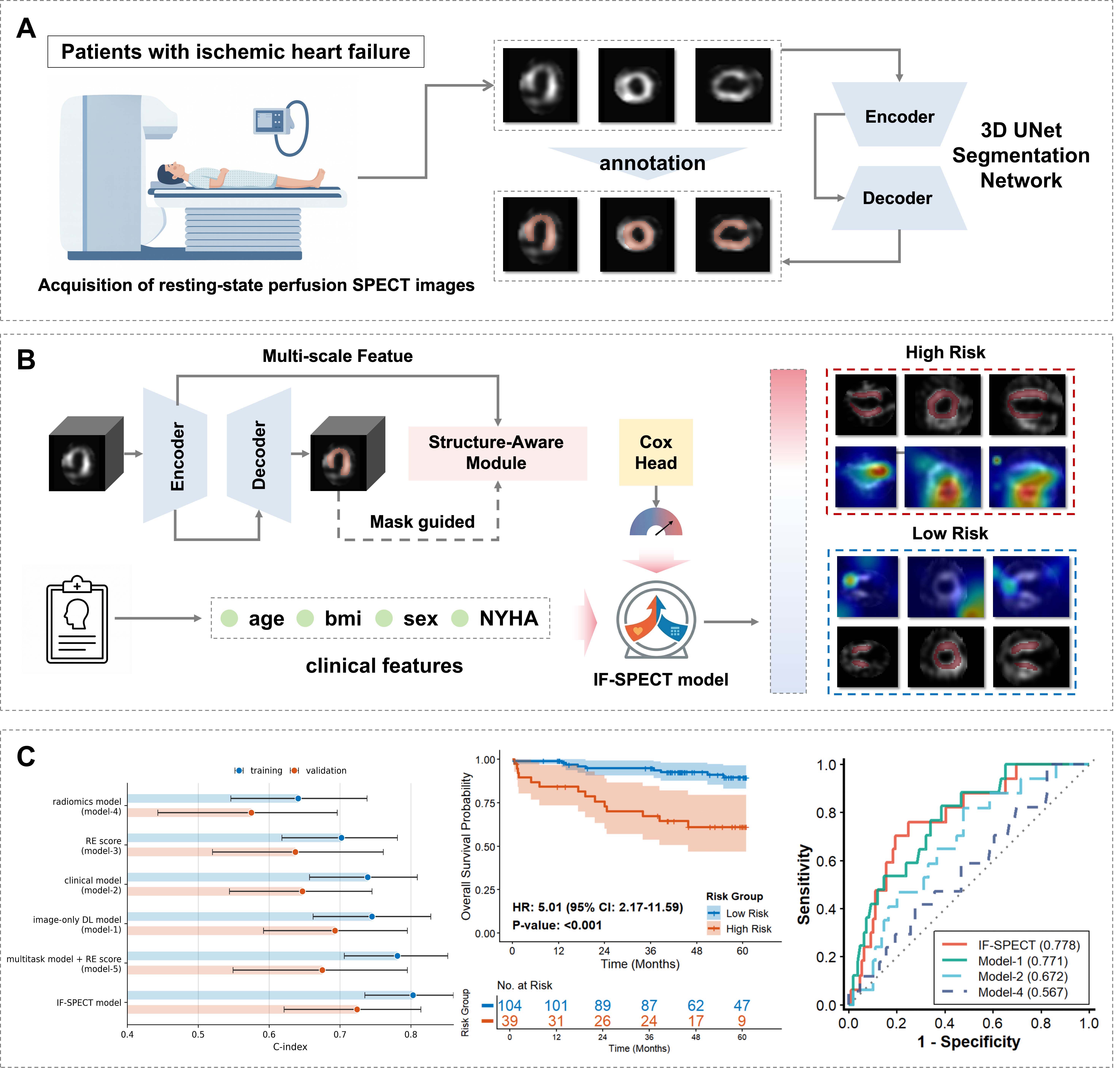
Figure 1. Overview of the study. (A) Acquisition and segmentation of resting-state perfusion SPECT images using a 3D U-Net. (B) Overall architecture of the multitask deep learning model. The neural network processes resting 3D SPECT images to generate a deep learning-derived imaging risk score, with left ventricular segmentation serving as an auxiliary task. The imaging risk score is subsequently combined with age, sex, BMI, and NYHA_3_4 in a multivariable Cox model to generate the final prognostic estimate. (C) Model performance evaluation using the C-index, Kaplan-Meier survival analysis, and time-dependent ROC curves.




