


fig7
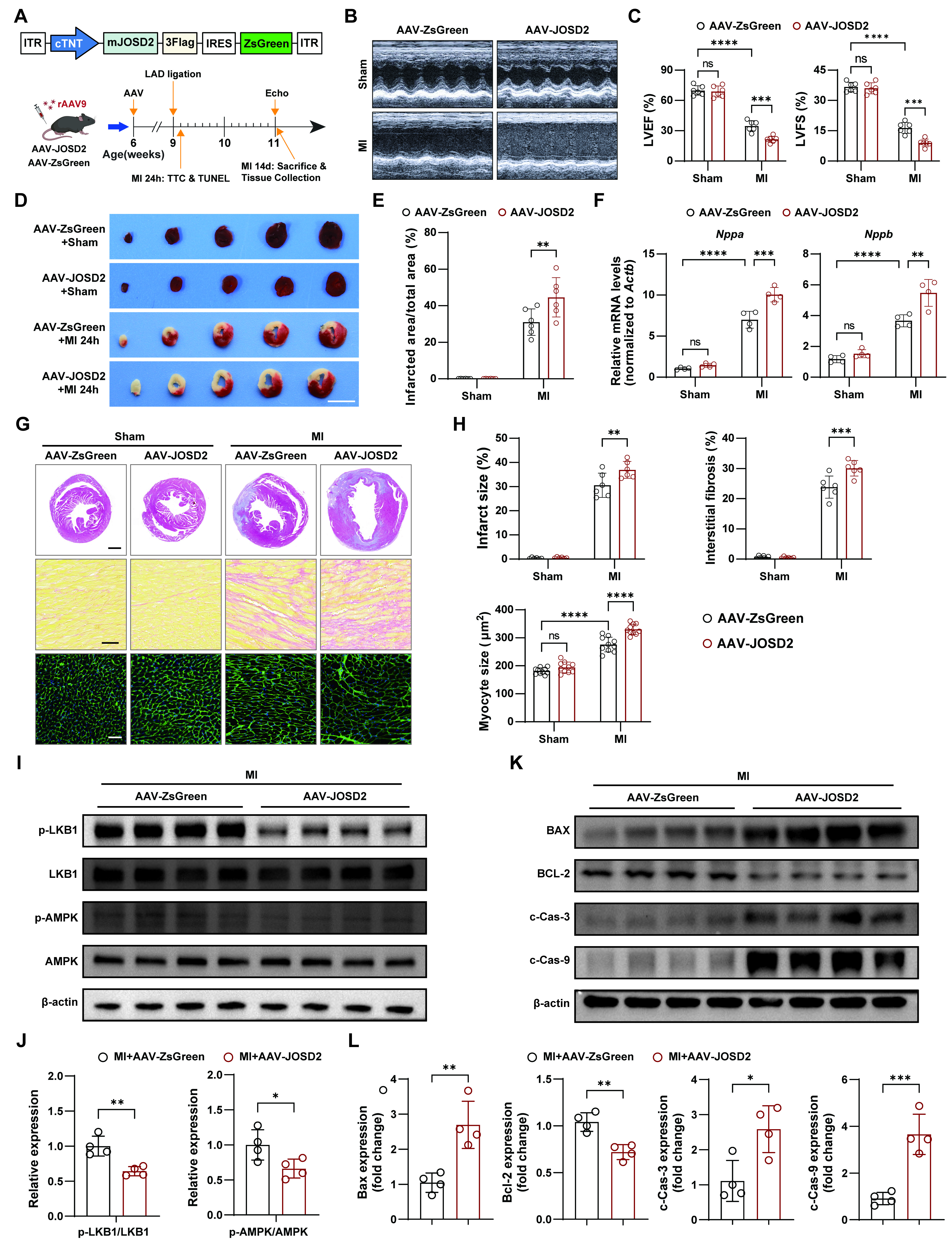
Figure 7. Aggravation of infarction and adverse remodeling after MI by cardiomyocyte-specific JOSD2 overexpression. (A) Schematic of AAV9-mediated cardiomyocyte-specific JOSD2 overexpression, MI/sham surgery, and tissue collection timelines; (B) Representative echocardiograms from C57BL/6J mice treated with AAV-ZsGreen or AAV-JOSD2 and subjected to MI/sham surgery; (C) Quantification of LVEF (%) and LVFS (%) at 14 days post-MI (n = 6 mice per group); (D) Representative TTC-stained heart sections at 1 day post-MI or sham. Scale bar, 5 mm; (E) Quantification of infarcted area/total area (%) from TTC staining (n = 6 mice per group); (F) RT-qPCR analysis of heart failure markers Nppa and Nppb in hearts at 14 days post-MI or sham (n = 4 mice per group); (G) Representative photomicrographs showing infarct size (scale bar, 1 mm), interstitial fibrosis (scale bar, 50 μm), and cardiomyocyte hypertrophy (scale bar, 50 μm) in AAV-ZsGreen- and AAV-JOSD2-treated mice subjected to sham or MI; (H) Quantitative evaluation of infarct size, interstitial fibrosis, and myocyte cross-sectional area (n = 6 mice per group); (I) Western blots of phosphorylated and total LKB1 and AMPK in hearts from the indicated groups; (J) Densitometric quantification of p-LKB1/LKB1 and p-AMPK/AMPK ratios (n = 4); (K) Western blots of apoptosis-associated proteins (BAX, BCL-2, c-Cas-3, and c-Cas-9); (L) Densitometric quantification of apoptosis-associated proteins in (K) (n = 4). Data are presented as mean ± SD. Data were analyzed by two-way ANOVA with Tukey’s post hoc test (C, E, F, and H) and unpaired two-tailed Student’s t-test (J and L). ns, not significant; *P < 0.05, **P < 0.01, ***P < 0.001, ****P < 0.0001. JOSD2: Josephin domain-containing protein 2; RT-qPCR: reverse transcription quantitative polymerase chain reaction; MI: myocardial infarction; OGD: oxygen-glucose deprivation; TUNEL: transferase dUTP nick-end labelling; SD: standard deviation; LVEF: left ventricular ejection fraction; LVFS: left ventricular fractional shortening; TTC: triphenyltetrazolium chloride; AAV: adeno-associated virus; AMPK: AMP-activated protein kinase; LKB1: liver kinase B1; ANOVA: analysis of variance; ITR: inverted terminal repeat; p-AMPK: phosphorylation of AMPK; p-LKB1: phosphorylation of LKB1; BAX: BCL-2-associated X protein; BCL-2: B-cell lymphoma 2.


