

fig4
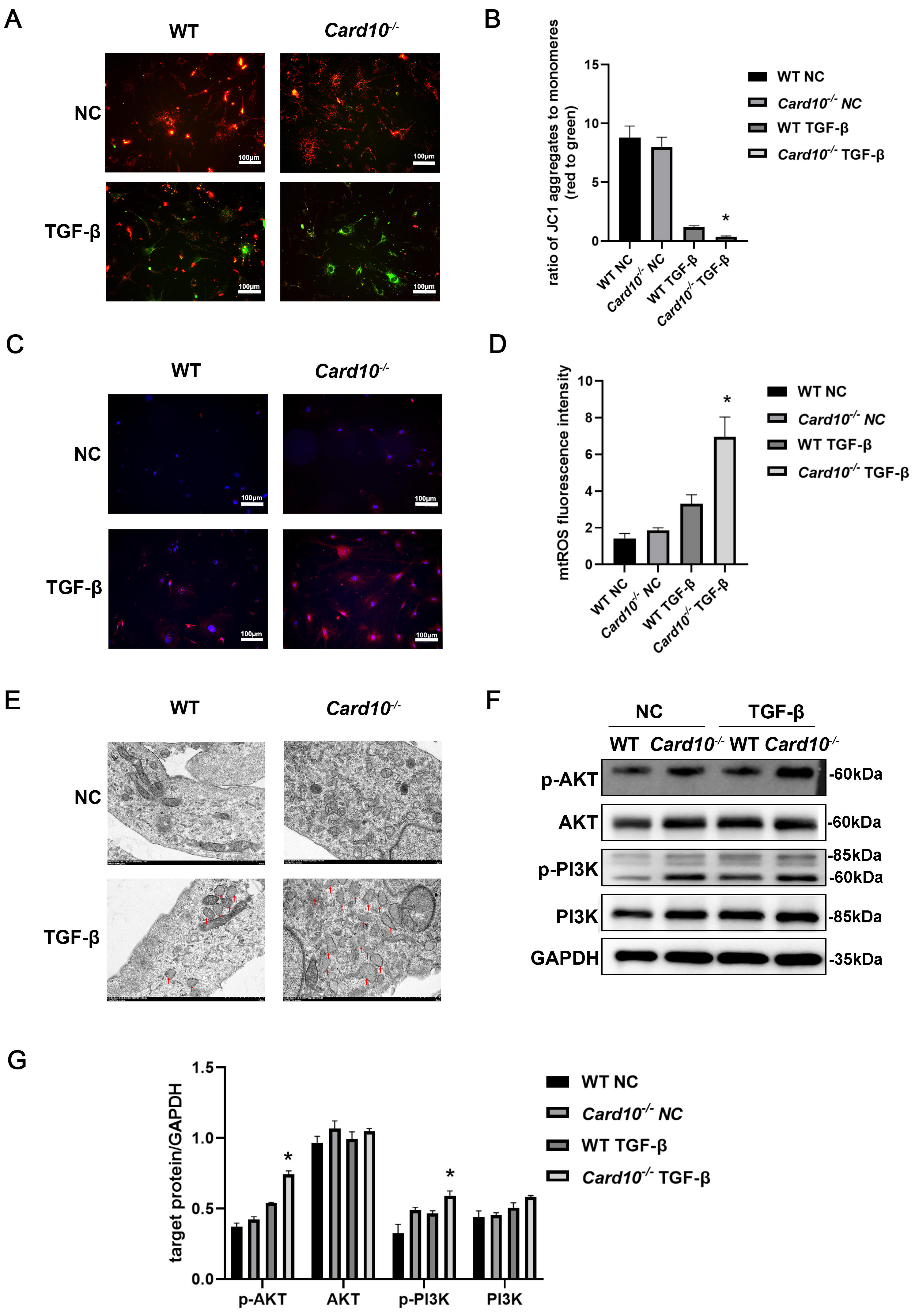
Figure 4. CARD10 deficiency aggravates mitochondrial dysfunction by activating the AKT/PI3K signaling pathway. The assays were conducted in CFs isolated from Card10-/- and WT adult mice and stimulated with TGF-β. Representative images of JC-1 staining (biological replicates n = 3, technical replicates 1) (A) and quantification of JC-1 levels (B); NC, negative control. Objective: 10×. Representative images (C) and quantification of MitoSOX Red fluorescence intensity (biological replicates n = 3, technical replicates 1) (D) for determination of mtROS levels. Objective: 10×; (E) Mitochondrial morphology was assessed by TEM (biological replicates n = 2, technical replicates 2). Scale bars: 1 µm. Western blot (p-PI3K&PI3K: biological replicates n = 3, technical replicates 2, p-AKT&AKT: biological replicates n = 3, technical replicates 2) (F) and densitometric analysis (G) showed increased levels of p-AKT and p-PI3K expressed in cell lysates of CFs isolated from Card10-/- adult mice and stimulated with TGF-β. Comparisons between different groups were performed using the two-way ANOVA followed by Šídák's multiple comparisons test. * P < 0.05 Card10-/- TGF-β vs. WT TGF-β. CARD10: Caspase recruitment domain-containing protein 10; AKT: protein kinase B; PI3K: phosphoinositide 3-kinase; CFs: cardiac fibroblasts; WT: wildtype; MI: myocardial infarction; TGF-β: transforming growth factor-beta; IF: immunofluorescence; mtROS: mitochondrial reactive oxidative species; TEM: transmission electron microscopy.




