

fig3
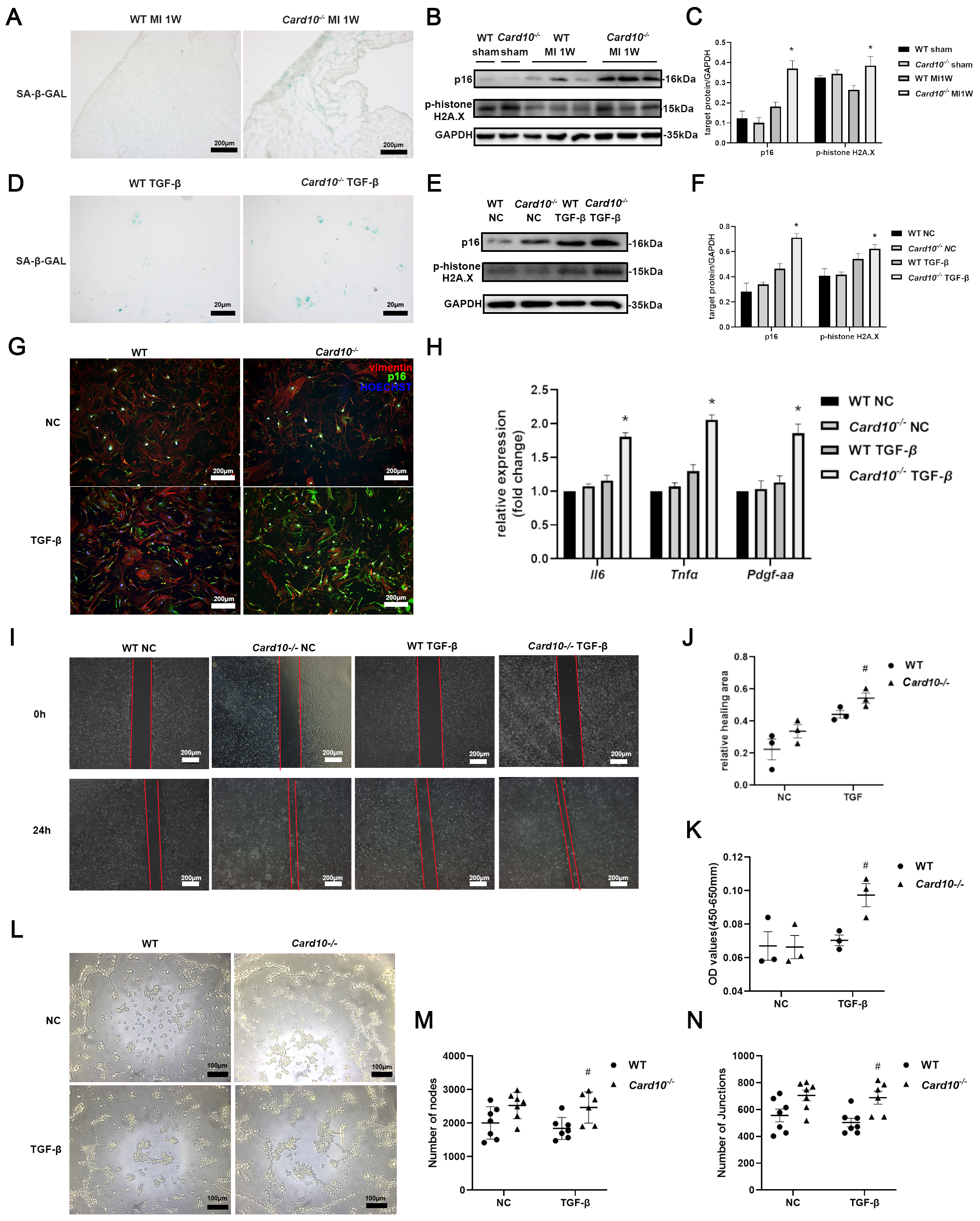
Figure 3. CARD10 deficiency induced CF senescence. (A) Representative images of SA-β-gal staining (biological replicates n = 3, technical replicates 1) showing increased SA-β-gal signal in cardiac tissue obtained from Card10-/- and WT mice one week (1 W) after MI surgery. Objective: 10×; (B) Western blot (biological replicates n = 3, technical replicates 2) and (C) densitometric analysis showing that senescence markers p16 and p-histone H2A.X were more abundant in Card10-/- MI mice heart compared to WT MI; (D) Representative images of SA-β-gal staining of CFs (biological replicates n = 3, technical replicates 1) obtained from Card10-/- and WT adult mice after TGF-β stimulation. Objective: 20×; (E) Western blot (p16: biological replicates n = 3, technical replicates 1, p-histone H2A.X: biological replicates n = 2, technical replicates 2) and (F) densitometric analysis showing that senescence markers p16 and p-histone H2A.X were more abundant in CFs obtained from Card10-/- and WT adult mice after TGF-β stimulation. NC, negative control; (G) Representative IF staining (biological replicates n = 3, technical replicates 1) showing colocalization of CF marker vimentin and p16 in primary CFs isolated from Card10-/- and WT adult mice after TGF-β stimulation. Objective: 10×; (H) RT-qPCR analysis (biological replicates n = 3, technical replicates 3) of senescence-associated secretory phenotype factors Il6, Tnfa, and Pdgfa in primary CFs isolated from Card10-/- and WT adult mice after TGF-β stimulation. Then HUVECs were cultured in conditioned medium derived from Card10-/- or WT cardiac fibroblasts treated with TGF-β for 4 days. Wound healing assay (biological replicates n = 3, technical replicates 1) (I) and quantitative analysis (J) were used to determine HUVEC migration. Objective: 10×. NC, negative control (no stimulation of CFs with TGF-β); CCK8 assay (biological replicates n = 3, technical replicates 3) (K) was used to determine HUVEC proliferation; Representative images of the tube formation assay (Card10-/- TGF-β group biological replicates n = 3, technical replicates 2, other group biological replicates n = 3, technical replicates 4) (L) and quantitative analysis of nodes (M) and junctions (N) showed more angiogenesis in HUVECs cultured with Card10-/- CF-derived conditioned medium compared to HUVECs cultured with WT CF-derived conditioned medium. Comparisons between different groups were performed using the two-way ANOVA followed by Šídák's multiple comparisons test. * P < 0.05 WT MI vs. Card10-/- MI, or WT TGF-β vs. Card10-/- TGF-β. # P < 0.05 WT conditioned medium vs. Card10-/- conditioned medium. CARD10: Caspase recruitment domain-containing protein 10; CFs: cardiac fibroblasts; WT: wildtype; MI: myocardial infarction; TGF -β: transforming growth factor-beta; IF: immunofluorescence; RT-qPCR: real-time quantitative polymerase chain reaction; HUVECs: human umbilical vein endothelial cells; ANOVA: analysis of variance; SA-β-gal: senescence-associated β-galactosidase.




