

fig1
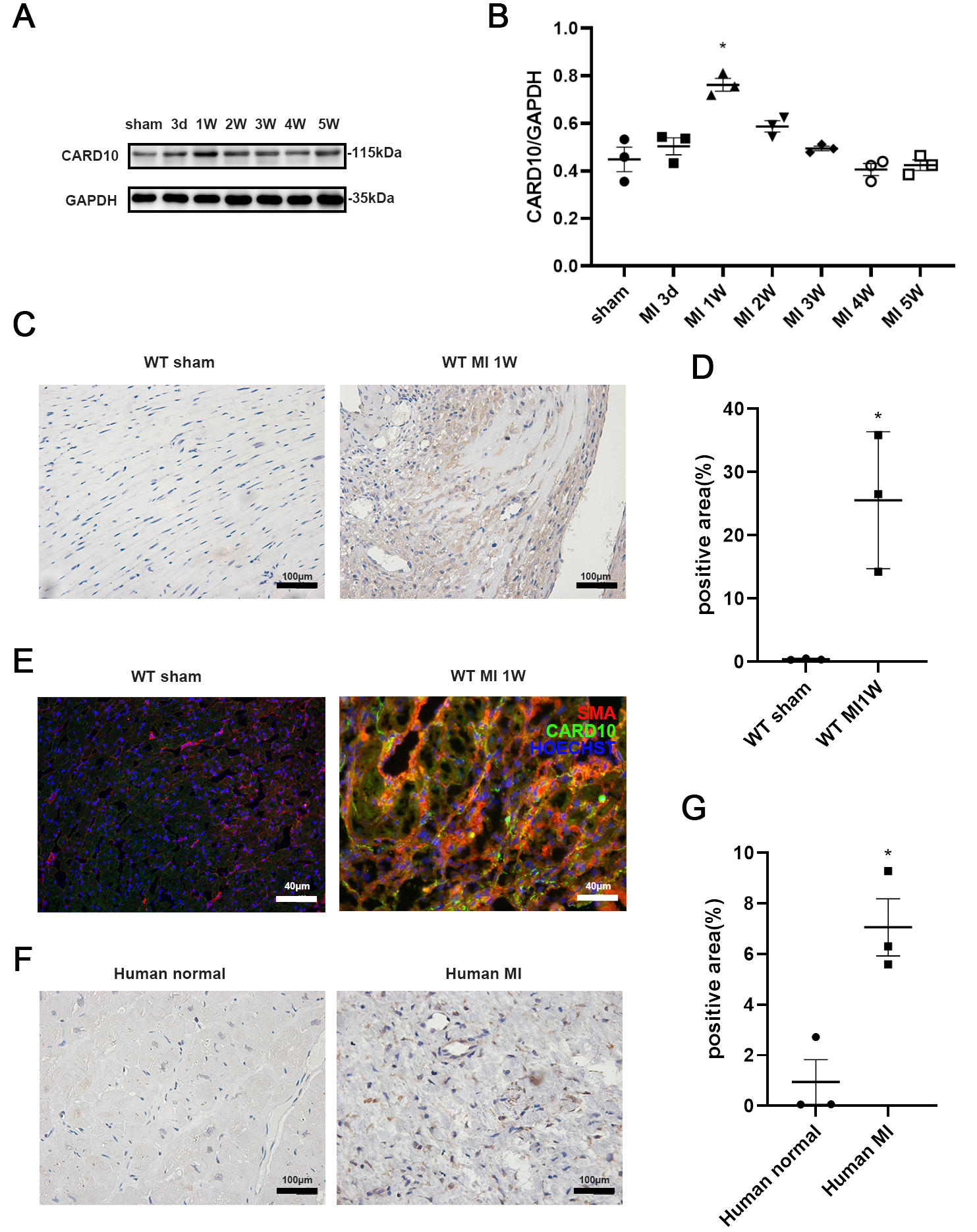
Figure 1. Increased CARD10 expression in heart tissues after MI induction. Western blot (A) (biological replicates n = 3, technical replicates 1) and densitometric analysis (B) in mouse whole heart lysates three days (3d) and one to five weeks (1 W to 5 W) after MI induction. CARD10 expression was highest one week after MI induction. (n = 3 per group); Representative images (C) and analysis (D) of IHC staining for CARD10 (biological replicates n = 3, technical replicates 1) in mouse heart one week after MI. Objective: 10×; (E) IF staining (biological replicates n = 3, technical replicates 1) showing scattered distribution of CARD10 and co-localization with a-SMA in infarcted heart tissue 1 week after MI, indicating predominant expression in CFs. Objective: 20×; Representative images (F) and analysis (G) of IHC staining for CARD10 (biological replicates n = 3, technical replicates 1) in human MI tissue. Objective: 10×. Differences in CARD10 expression among multiple groups were analyzed using one-way ANOVA. Comparisons between two groups were performed using the Mann-Whitney U test. * P < 0.05. CARD10: Caspase recruitment domain-containing protein 10; MI: myocardial infarction; IHC: Immunohistochemistry; IF: immunofluorescence; α-SMA: α-smooth muscle actin; CFs: cardiac fibroblasts; ANOVA: analysis of variance; WT: wildtype.




