

The Sub1-p53 axis in cardiac aging and HFpEF
 0



 Download PDF
Cardiovascular disease (CVD) remains a leading cause of morbidity and death worldwide. Aging is one of the major risk factors for overall CVD; advancing age is strongly associated with structural and functional decline of the heart and vasculature. Consequently, older adults bear a substantial burden of CVD, including but not limited to heart failure (HF) and atherosclerotic complications[1]. Increasingly aging populations around the world highlight challenges in prevention and treatment of CVD[2].
Among the various manifestations of CVD, HF is particularly relevant in older adults because its incidence and prevalence increase markedly with age. Based on left ventricular ejection fraction (LVEF), the 2026 European Society of Cardiology guidelines now classify HF into HF with reduced ejection fraction (HFrEF; LVEF < 50%) and HF with preserved ejection fraction (HFpEF; LVEF ≥ 50%), without retaining HF with mildly reduced ejection fraction as a separate phenotype[3].
HFpEF disproportionately affects older adults and remains heterogeneous despite therapeutic advances[3,4]. Most current pharmacological therapies proven effective for HFrEF failed to show consistent benefit in patients with HFpEF. This highlights the urgent need for novel therapeutic strategies for HFpEF. Targeting biological aging is one of the emerging approaches that have garnered increasing interest[3]. By targeting cellular senescence and other hallmarks of aging, such strategies may modify the underlying myocardial disease processes rather than merely addressing their downstream hemodynamic consequences[5]. However, based on the current state of research data, interventions that modify biological aging should still be viewed as complements to, and potentially synergistic partners with, established therapies rather than replacements.
Sub1 is a transcriptional coactivator, which has roles in transcription, stress responses, and genomic stability[6-8]. In a recent study, Zhao et al. investigated the possibility of targeting Sub1 as a regulator of cardiac aging and HFpEF-like disease in mice[9] [Figure 1]. The strongest evidence comes from reciprocal genetic models: cardiomyocyte-specific Sub1 overexpression accelerated cardiac senescence and produced an HFpEF-like phenotype, whereas partial global Sub1 deficiency mitigated age-related and two-hit HFpEF phenotypes[9]. Cardiomyocyte-specific Sub1 knockdown, mediated by late-life delivery of adeno-associated virus serotype 9 (AAV9) encoding cardiomyocyte-specific Sub1 shRNA, also improved diastolic function, exercise capacity, fibrosis, and aging markers in naturally aged mice[9].
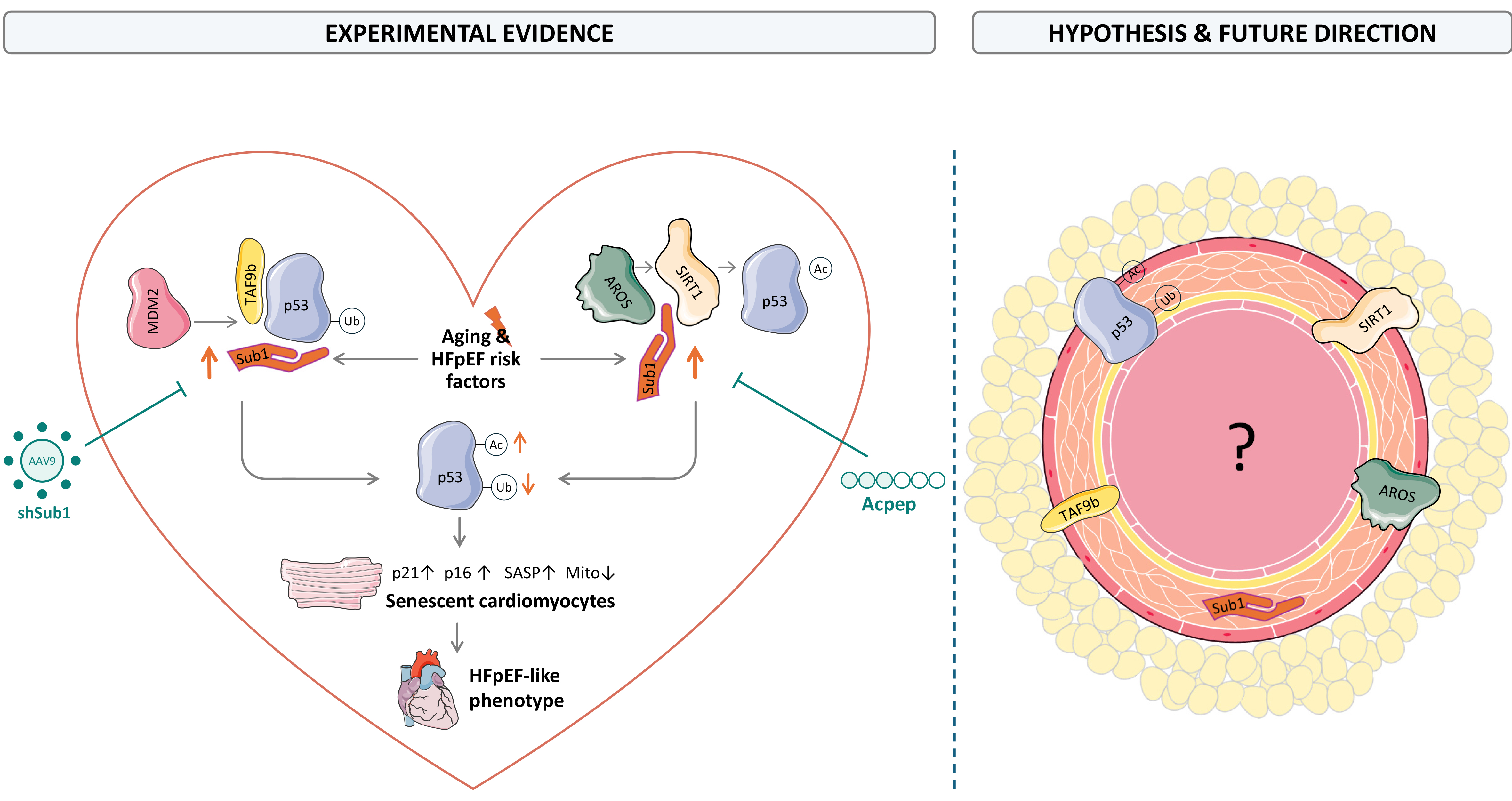
Figure 1. Sub1-p53 signaling links cardiac aging to HFpEF and raises unresolved questions about vascular aging. Age-related upregulation of Sub1 in cardiomyocytes enhances p53 stability and activity through two cooperating mechanisms (left). Acting as a “supporting hand”, Sub1 facilitates TAF9b-p53 association, potentially limiting MDM2 binding and thereby reducing MDM2-mediated p53 ubiquitination and proteasomal degradation. In parallel, acting as an “intercepting hand”, Sub1 binds AROS and reduces the AROS-SIRT1 association, thereby limiting AROS-dependent SIRT1 activation and preserving p53 acetylation. The resulting increases in p53 abundance and transcriptional activity promote cardiomyocyte senescence, characterized by increased p21, p16, and SASP markers together with mitochondrial dysfunction, thereby contributing to cardiac aging, remodeling, and HFpEF phenotypes in mice. Sub1 deficiency and cardiomyocyte-directed Sub1 knockdown ameliorated aging- and HFpEF-associated phenotypes, whereas Acpep-mediated disruption of the Sub1-p53 interaction attenuated senescence in cultured cardiomyocytes and cardiac organoids. Right: The extent to which cardiomyocyte-directed Sub1 suppression generalizes across experimental models and human HFpEF, whether its effects are sex-dependent, and whether Sub1-p53 signaling promotes vascular-cell senescence and contributes to hypertension or atherosclerosis remain unresolved. The pictures of the proteins, the cardiomyocyte, the heart and the artery are from Servier Medical Art (https://smart.servier.com/), licensed under CC BY 4.0. HFpEF: Heart failure with preserved ejection fraction; MDM2: mouse double minute 2 homolog; AROS: active regulator of SIRT1; SIRT1: Sirtuin 1; SASP: senescence-associated secretory phenotype; AAV9: adeno-associated virus serotype 9.
Importantly, cardiomyocyte-specific Sub1 overexpression was sufficient to induce premature cardiac aging and associated functional abnormalities, including diastolic dysfunction, pulmonary congestion, and reduced exercise capacity, without inducing comparable aging phenotypes in the liver, kidney, or skeletal muscle[9]. By contrast, partial global Sub1 deficiency reduced blood pressure in the two-hit HFpEF model and exerted modest extracardiac effects in naturally aged mice, including reduced senescence-associated β-galactosidase staining in the liver and kidney and increased skeletal muscle fiber cross-sectional area[9]. Thus, the cardiac benefits observed with global Sub1 deficiency may partly reflect reduced afterload and systemic anti-aging effects in addition to direct cardiac actions. The different organ profiles of these models are not necessarily contradictory. The cardiomyocyte-directed AAV9 experiment narrows this gap, although tissue selectivity and durability require validation. Together, these data support causality in murine cardiac aging, but not yet pharmacological efficacy in established HFpEF.
Mechanistically, Sub1 promoted the TAF9b-p53 interaction, reduced p53 ubiquitination and degradation by inhibiting mouse double minute 2 homolog (MDM2)-mediated ubiquitination and degradation, and competed with Sirtuin 1 (SIRT1) for active regulator of SIRT1 (AROS) binding, preserving p53 acetylation and senescence signaling[9]. Interaction and rescue experiments support this model, but displacement of MDM2 by the Sub1-TAF9b complex was not directly demonstrated and remains proposed [Figure 1]. Altered ATP-metabolism gene sets and mitochondrial ultrastructure likewise indicate mitochondrial abnormalities, not direct proof of impaired mitochondrial function. Acpep, a tri-acetylated peptide derived from the C-terminal region of p53, disrupted the Sub1-p53 interaction and reduced senescence in bleomycin-treated HL-1 cells and cardiac organoids, but has not been validated in vivo[6]. In vivo, doxorubicin treatment abolished Sub1-mediated protection in HFpEF mice. Bleomycin models cellular senescence and doxorubicin models cardiotoxic injury; neither reproduces natural physiological cardiac aging. Public myocardial datasets showed higher Sub1 transcripts in HFpEF and linked cardiac Sub1 expression to age in GTEx[9], but protein localization, association with severity or outcome, and therapeutic responsiveness in patients are unknown. The evidence hierarchy from the current study is therefore mechanistic validation, murine genetic proof-of-concept, early in vitro pharmacology, and preliminary human association. Subsequent studies must elucidate the dose-effect relationship, reversibility, non-cardiac effects, therapeutic window, and overall safety. Cardiomyocyte-selective delivery or interaction-specific inhibitors may be safer than chronic systemic suppression.
Despite the limitations and the necessity of conducting further investigation, the findings of Zhao et al. also raise a broader question regarding the potential role of Sub1 in cardiovascular aging beyond the myocardium[9]. Cardiac aging and HFpEF share several phenotypic features, including impaired functional reserve, myocardial fibrosis, and cellular senescence. Analogously, vascular aging and age-related vascular diseases, including hypertension and atherosclerosis, share common features such as endothelial dysfunction, arterial stiffness, vascular remodeling, and chronic inflammation[10-12]. Increasing evidence implicates cellular senescence as an important contributor to age-related CVD. Despite their distinct cellular and tissue contexts, cardiac and vascular senescence involve several common mechanisms, including activation of the p53/p21 and p16/Rb pathways, the senescence-associated secretory phenotype (SASP), and oxidative stress. Clinically, cardiac senescence is associated with age-related deterioration of cardiac function, whereas vascular senescence has been linked to hypertension, atherosclerosis, and increased cardiovascular risk[9].
The susceptibility of vascular cells to senescence increases with age, and p53 activation has been implicated in multiple vascular pathologies, including atherosclerosis and hypertension. Although diverse stressors can induce cellular senescence, many converge on a limited number of key signaling pathways. In particular, stress-induced premature senescence can be triggered by oxidative and other cellular stresses through activation of the p53/p21 and p16/Rb pathways and represents an important senescence phenotype in vascular disease. Regulation of p53 in vascular cells occurs at multiple levels, including phosphorylation, acetylation, and ubiquitination[6]. Zhao et al. demonstrated that Sub1 promotes cardiomyocyte senescence by interacting with TAF9b and AROS to regulate p53 stability and activity, thereby identifying the Sub1-p53 axis as a potential mechanistic link between cardiac aging and HFpEF[9].
A vascular role of Sub1 remains hypothesis-generating. The single-cell dataset included endothelial cells and pericytes, but did not establish causality in either compartment[9]. For the potential role of Sub1 in vasculature, endothelial cell- and vascular smooth muscle cell-specific deletion should be compared with cardiomyocyte manipulation using blood-pressure telemetry, endothelium-dependent relaxation, arterial stiffness, remodeling, and atherosclerosis models[10-12]. Temporal studies could test whether vascular Sub1 induction and dysfunction precede myocardial remodeling and explain the lower blood pressure of globally deficient mice.
In summary, the study by Zhao et al. strengthens the evidence for a mechanistic link between cardiac aging and HFpEF and identifies a previously unrecognized pathway through which Sub1 promotes cardiomyocyte senescence by regulating p53 via TAF9b and AROS[9]. Nevertheless, the therapeutic evidence remains at the preclinical proof-of-concept stage. Although the beneficial effects of Sub1 suppression were demonstrated in vivo using genetic approaches, including AAV9-mediated knockdown, Acpep was evaluated only in cultured cells and cardiac organoids. Sub1 therefore represents a promising therapeutic target, but further studies should follow a clearly defined translational pathway - from mechanistic target validation and preclinical proof of concept to in vivo pharmacological validation, safety assessment, and ultimately clinical translation. Beyond the heart, these findings raise the possibility that Sub1-dependent regulation of cellular senescence contributes to other manifestations of cardiovascular aging. Defining the cell- and tissue-specific functions of Sub1 and the mechanisms governing its age-dependent regulation will be important for establishing the broader relevance - and potential limitations - of this pathway across the cardiovascular system.
DECLARATIONS
Authors’ contributions
The author contributed solely to the article.
Availability of data and materials
Not applicable.
AI and AI-assisted tools statement
During the preparation of this manuscript, the AI tool ChatGPT (version 5.6, released 2026-07-09) was used solely for language editing. The tool did not influence the study design, data collection, analysis, interpretation, or the scientific content of the work. All authors take full responsibility for the accuracy, integrity, and final content of the manuscript.
Financial support and sponsorship
None.
Conflicts of interest
The author declared that there are no conflicts of interest.
Ethical approval and consent to participate
Not applicable.
Consent for publication
Not applicable.
Copyright
© The Author(s) 2026.
REFERENCES
1. Zheng P, Yan W, Ding Y, et al. Cardiovascular ageing: hallmarks, signaling pathways, diseases and therapeutic targets. Signal Transduct Target Ther. 2026;11:142.
2. Zhao D, Wang Y, Wong ND, Wang J. Impact of aging on cardiovascular diseases: from chronological observation to biological insights: JACC family series. JACC Asia. 2024;4:345-58.
3. Køber L, Adamo M, Ruwald AC, et al.; ESC Guidelines Advisory Group. 2026 ESC Guidelines for the management of heart failure. Eur Heart J. 2026;47:5346-457.
4. Redfield MM, Borlaug BA. Heart failure with preserved ejection fraction: a review. JAMA. 2023;329:827-38.
5. Abdellatif M, Rainer PP, Sedej S, Kroemer G. Hallmarks of cardiovascular ageing. Nat Rev Cardiol. 2023;20:754-77.
6. Ge H, Roeder RG. Purification, cloning, and characterization of a human coactivator, PC4, that mediates transcriptional activation of class II genes. Cell. 1994;78:513-23.
8. Sikder S, Kumari S, Mustafi P, et al. Nonhistone human chromatin protein PC4 is critical for genomic integrity and negatively regulates autophagy. FEBS J. 2019;286:4422-42.
9. Zhao D, Lin L, Zhou Y, et al. Sub1 contributes to heart failure with preserved ejection fraction driven by aging in mice. Nat Commun. 2026;17:8332.
10. Ungvari Z, Tarantini S, Donato AJ, Galvan V, Csiszar A. Mechanisms of vascular aging. Circ Res. 2018;123:849-67.
11. Picos A, Seoane N, Campos-Toimil M, Viña D. Vascular senescence and aging: mechanisms, clinical implications, and therapeutic prospects. Biogerontology. 2025;26:118.
Cite This Article
 Open Access
How to Cite
Xia N. The Sub1-p53 axis in cardiac aging and HFpEF. J Cardiovasc Aging. 2026;6:50. https://dx.doi.org/10.20517/jca.2026.127
Download Citation
If you have the appropriate software installed, you can download article citation data to the citation manager of your choice. Simply select your manager software from the list below and click on download.
Export Citation File
Type of Import
Tips on Downloading Citation
Citation Manager File Format
Type of Import
Direct Import: When the Direct Import option is selected (the default state), a dialogue box will give you the option to Save or Open the downloaded citation data. Choosing Open will either launch your citation manager or give you a choice of applications with which to use the metadata. The Save option saves the file locally for later use.
Indirect Import: When the Indirect Import option is selected, the metadata is displayed and may be copied and pasted as needed.
About This Article
Copyright

Data & Comments
Data
0










Comments
Comments must be written in English. Spam, offensive content, impersonation, and private information will not be permitted. If any comment is reported and identified as inappropriate content by OAE staff, the comment will be removed without notice. If you have any queries or need any help, please contact us at [email protected].