

fig5

Fulminant myocarditis complicated with left ventricular thrombus: a case report with complete functional recovery

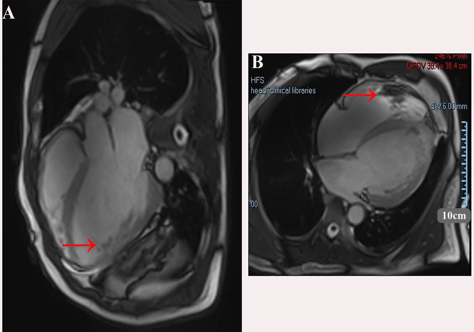
Figure 5. Showing the Cardiac magnetic resonance imaging, balanced steady-state free precession (bSSFP) cine sequence. (A) four-chamber view; (B) short-axis view. A large thrombus is seen adjacent to the apical/anterior wall of the left ventricle (red arrows). The left ventricle is markedly dilated.


The Journal of Cardiovascular Aging
ISSN 2768-5993 (Online)

Portico

All published articles are preserved here permanently:

https://www.portico.org/publishers/oae/

Portico

All published articles are preserved here permanently:

https://www.portico.org/publishers/oae/


