

fig3
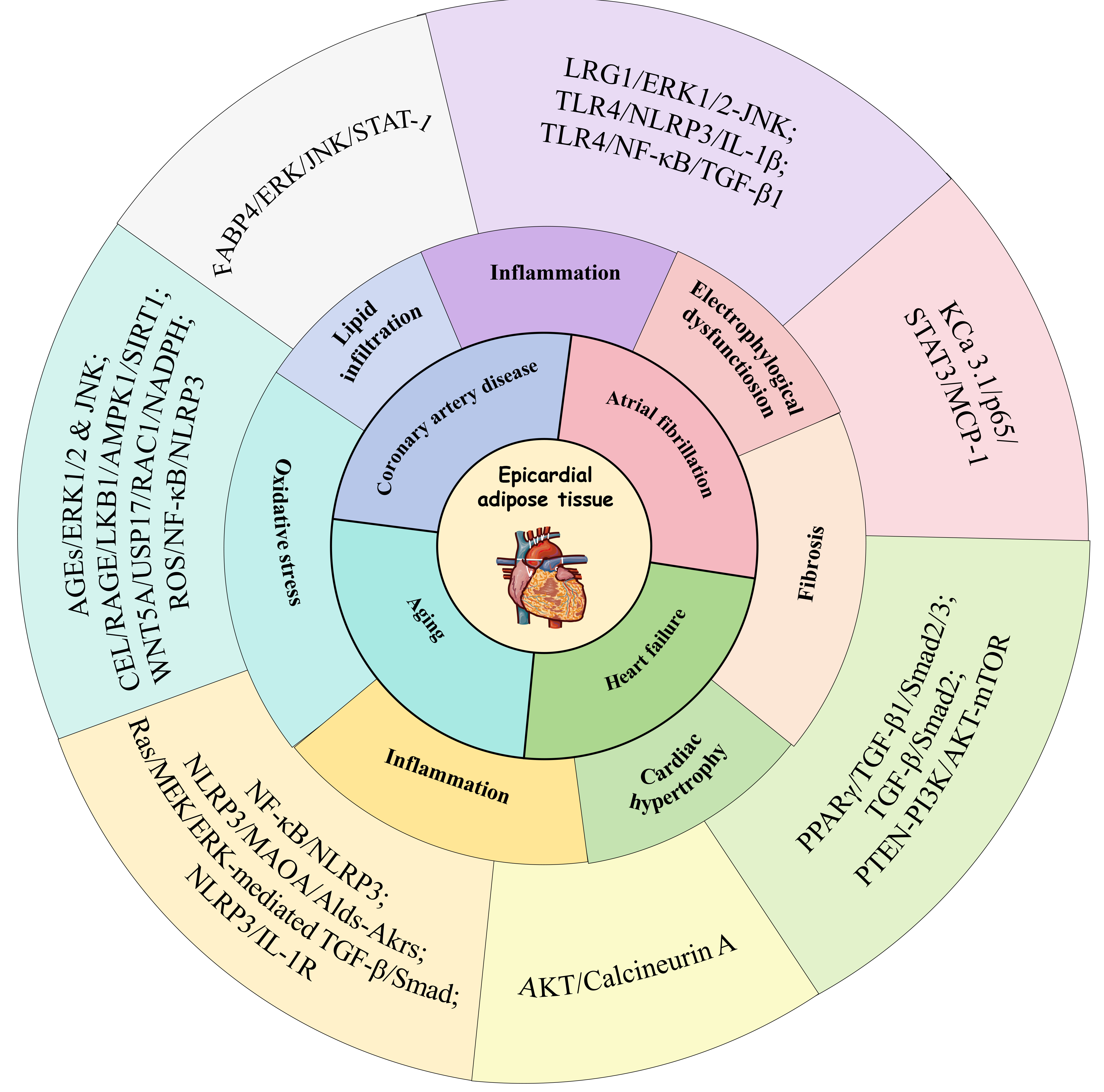
Figure 3. Mechanistic pathways linking EAT dysfunction to CVD and aging. The pathological expansion and dysfunction of EAT promote inflammatory responses, lipid infiltration, and fibrosis in the cardiac microenvironment through complex signaling cascade networks. These pathways contribute to the development of significant clinical outcomes, including CAD, AF, and HF. AF: Atrial fibrillation; CAD: coronary artery disease; CVD: cardiovascular disease; EAT: epicardial adipose tissue; HF: heart failure; FABP4: fatty acid-binding protein 4; NF-κB: nuclear factor κB; TGF-β: transforming growth factor-β; AGEs: advanced glycation end products; JNK: Jun N-terminal kinase; ROS: reactive oxygen species; mTOR: mammalian target of rapamycin; RAGE: receptor for advanced glycation end products; LKB1: liver kinase B1; AMPK1: AMP-activated protein kinase 1; SIRT1: sirtuin 1; WNT5A: wingless-related integration site 5A; USP17: ubiquitin-specific protease 17; NADPH: nicotinamide adenine dinucleotide phosphate; NLRP3: NOD-like receptor family pyrin domain-containing 3; PPARγ: peroxisome proliferator-activated receptor γ; PTEN: phosphatase and tensin homolog; PI3K: phosphatidylinositol 3-kinase; ERK: extracellular signal-regulated kinase; CEL: Nε-carboxyethyl-lysin; STAT1: signal transducer and activator of transcription 1; LRG1: leucine-rich alpha-2 glycoprotein 1; TLR4: toll like receptor 4; IL: interleukin; MCP: monocyte chemoattractant protein; MAOA: monoamine oxidase A; Alds: aldehyde dehydrogenases; Akrs: aldo-keto reductases; Ras: rat sarcoma; MEK: mitogen-activated protein kinase kinase.




