

fig1
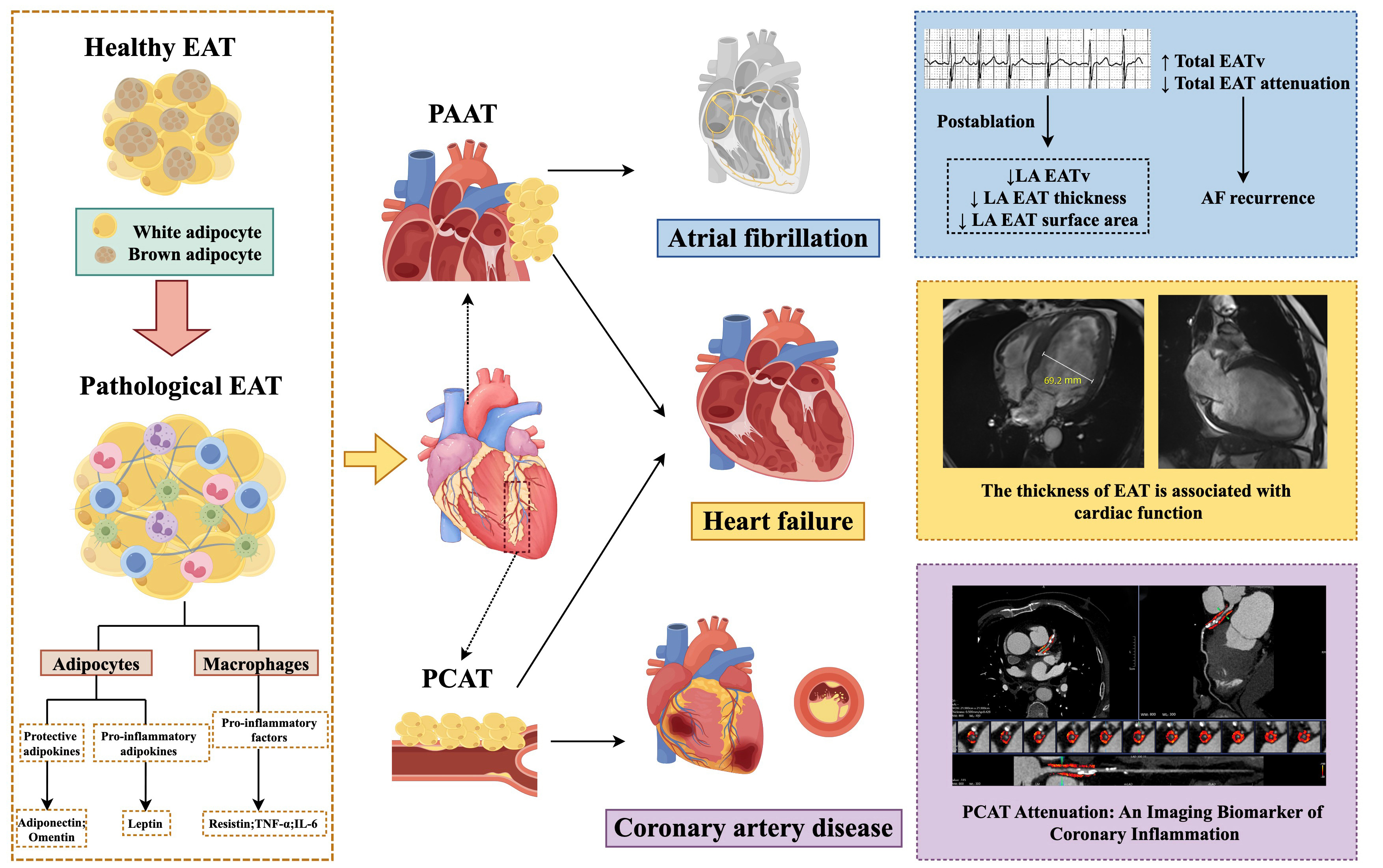
Figure 1. Regional distribution of EAT and its differential impact on CVD. Under pathological conditions, EAT undergoes a phenotypic transition from a protective phenotype to an inflammatory substrate, becoming a source of pro-inflammatory, pro-atherogenic, and pro-arrhythmogenic mediators (bottom-left). Following AF ablation, LA EATv decreases. Patients experiencing AF recurrence exhibit higher pre-ablation total EATv, lower EAT attenuation values, and progressive EATv enlargement during follow-up (top-right). EAT thickness is associated with cardiac function (middle-right). Elevated PCAT attenuation/density correlates with CAD. Compared to patients with stable CAD, those with ACS exhibit a stronger inflammatory response around coronary lesions (bottom-right). The top-right, middle-right and bottom-right panels show patients from Beijing Anzhen Hospital, Capital Medical University, Beijing, China. Patient consent was obtained for the use of this image, and all personally identifiable information has been removed. The image was created on the website FigDraw.com (ID: OTUPWf87ff). ACS: Acute coronary syndrome; AF: atrial fibrillation; CAD: coronary artery disease; CCTA: coronary computed tomography angiography; CVD: cardiovascular disease; EAT: epicardial adipose tissue; IL-6: interleukin-6; PCAT: peri-coronary adipose tissue; TNF-α: tumor necrosis factor-α; LA: left atrium; EATv: epicardial adipose tissue volume; PAAT: periatrial adipose tissue.




