


fig5
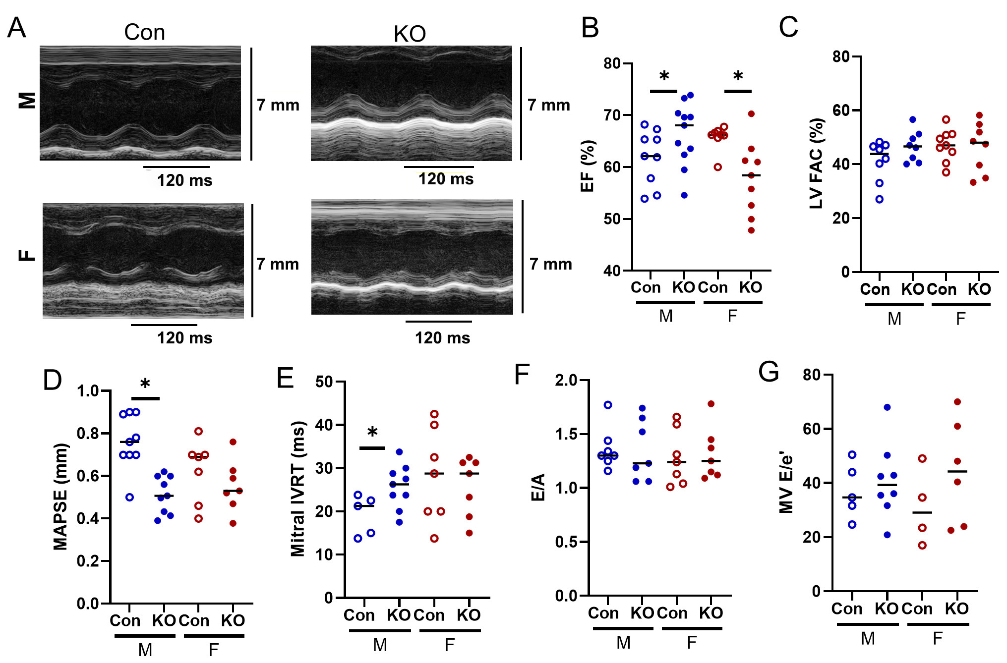
Figure 5. LV echocardiography in Kv1.5 KO and Con mice. (A) Representative LV Ejection Fraction (EF) M-mode images. (B) LV EF was higher in KO male but mildly lower in KO female. (C) LV fractional area change (FAC) was unchanged. (D) Mitral annual plane systolic excursion (MAPSE) decreased in KO males and was unchanged in females. (E) Mitral isovolumic relaxation time (IVRT) increased in KO males and was unchanged in females. (F) Early filling divided by late filling (E/A) and G) E/e’ did not differ by genotype. *P < 0.05 as assessed by Student’s t-test to compare within-sex differences between KO and Con. Data are expressed as mean ± SEM. Blue: male (M), red: female (F).


