


fig2
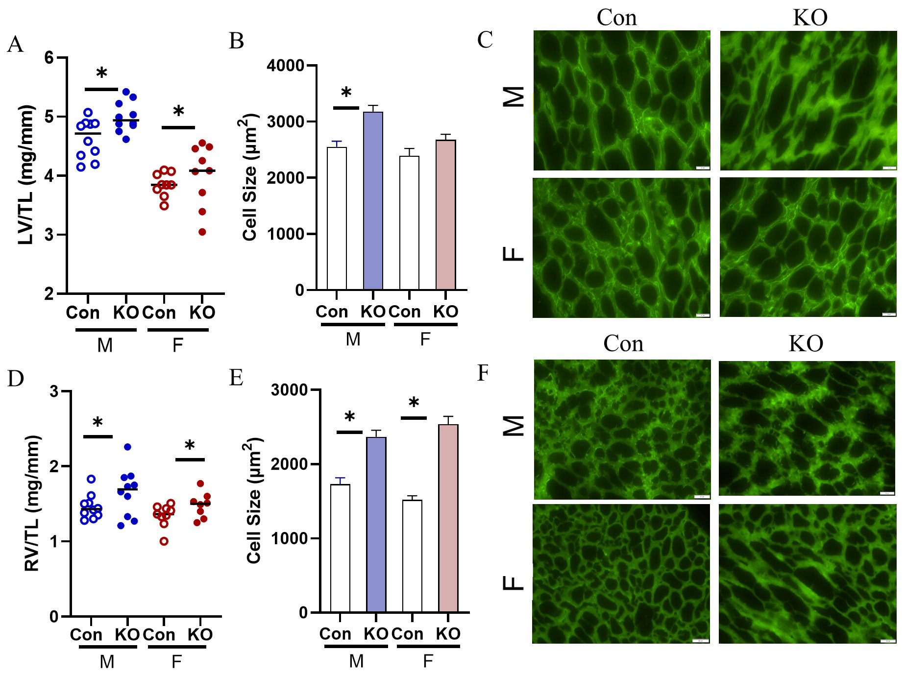
Figure 2. Cardiac remodeling in response to Kv1.5 KO. (A) LV/TL was elevated in both male and female KO mice. (B) LV myocyte cell size was elevated with Kv1.5 deletion in male mice. (C) Representative images of LV lectin-stained cardiac myocytes. Scale bar: 20 mm. (D) RV/TL was elevated in male and female KO mice. (E) RV myocyte cell size was elevated in male and female KO mice. (F) Representative images of RV lectin-stained cardiac myocytes. Scale bar: 20 mm. n = 3 mice with technical triplicate for histology. Data are expressed as mean ± SEM. *P < 0.05 within sex by Student’s t-test. Blue: male (M), red: female (F).


