fig5
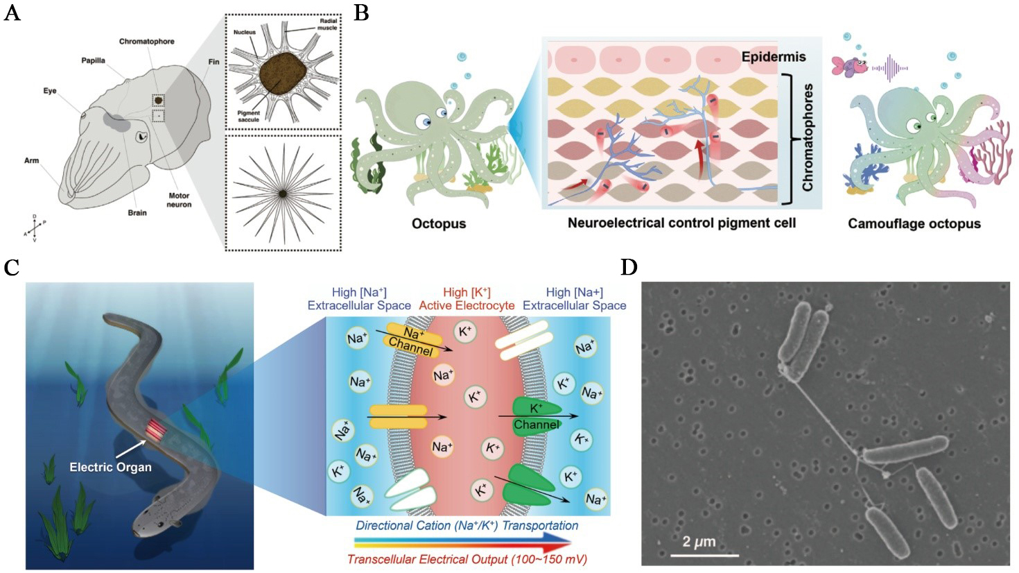
Figure 5. Biological ionic processes at organismal and collective scales. (A) Cephalopod skin is covered in thousands to millions of chromatophores - expandable neuromuscular organs that are controlled by motor neurons projecting from the brain. Upper box: expanded chromatophore; lower box: relaxed chromatophore. This figure is quoted with permission from Shook et al.[95]; (B) Schematic of the cephalopod reflex arc illustrating how sensory inputs are converted into neural signals that drive chromatophore muscle contraction, enabling rapid, reversible color change. This figure is quoted with permission from Yu et al.[96]; (C) Electrophorus electricus and a schematic of its electric organ. The right image depicts ion fluxes during a discharge, with the innervated posterior membrane becoming Na+-permeable and the anterior side K+-selective, so that many electrocytes in series collectively produce a high-voltage output. This figure is quoted with permission from Zhang et al.[27]; (D) Scanning electron micrograph of S. oneidensis MR-1 grown under electron-acceptor-limited conditions, showing bundles of pili-like, electrically conductive nanowires that bridge cells. Scale bar, 2 µm. This figure is quoted with permission from Gorby et al.[97]. MR: Manganese reducer.

