

Arc is a novel regulator of Tau packaging into extracellular vesicles and promotes Tau spreading
 0



 Download PDF
MAIN TEXT
The microtubule-associated protein Tau plays a central role in stabilizing axonal microtubules in neurons, where Tau is highly expressed[1]. However, with aging, Tau becomes increasingly susceptible to misfolding and aggregation. The accumulation of these pathological Tau species underlies a group of neurodegenerative disorders collectively known as tauopathies[1], including Alzheimer’s disease (AD), the most common cause of aging dementia. A key pathological feature of misfolded Tau is its ability to act as a proteopathic “seed” that templates the conformational conversion of native Tau into oligomeric and fibrillar aggregates. Seeding-competent Tau exists both as “free” aggregates[2] or encapsulated within extracellular vesicles (EVs)[3,4]. Although both forms are capable of propagating Tau pathology, the mechanisms governing Tau incorporation into EVs have remained poorly understood. A major advance in this area has now been reported by Tyagi et al., whose findings are discussed in this Research Highlight[5].
Most studies of EV-mediated Tau transmission have focused on small EVs
Arc is a key regulator of synaptic plasticity and memory consolidation[10]. In the brain, Arc expression is largely restricted to calcium/calmodulin-dependent protein kinase II (CaMKII)-positive glutamatergic neurons in the hippocampus and neocortex, while no expression has been reported in glial cells[11]. Furthermore, Arc protein is predominantly localized to dendrites and appears largely absent from axons and presynaptic terminals[12]. Another remarkable feature of Arc is its ability to spontaneously assemble into virus-like capsids that are subsequently packaged into EVs, a process facilitated by the recruitment of the inverse BAR (I-BAR) domain-containing protein insulin receptor substrate protein of 53 kDa (IRSp53)[13]. To investigate the role of Arc in Tau secretion, wild-type and Arc-knockout neurons were transduced with a lentiviral construct expressing enhanced green fluorescent protein (eGFP)-P2A-hTau(P301L) under the control of the human synapsin 1 (hSyn) promoter. When EVs isolated from the culture medium were compared with neuronal lysates, the authors found that mutant hTau(P301L) levels were markedly reduced in Arc-knockout-derived EVs, whereas neuronal lysates contained comparable amounts of hTau in both wild-type and Arc-knockout neurons. These findings demonstrate that the packaging of monomeric mutant hTau into EVs is strongly dependent on Arc expression.
The study then evaluated EVs isolated from 4-month-old rTg4510 mice, which also express mutant hTau(P301L)[14]. For comparison, rTg4510 mice were crossed with Arc-knockout animals to generate rTg4510ArcKO mice. Consistent with the neuronal culture experiments, hTau was readily detected in EVs derived from rTg4510 brains but was markedly reduced in EVs from rTg4510ArcKO mice. Importantly, neither the size distribution nor the abundance of EVs differed between genotypes, indicating that Arc specifically regulates Tau loading rather than EV biogenesis. Immunogold electron microscopy further revealed that approximately 12% of all EVs were separately positive for each protein, whereas only 3.5% were double-positive for both Arc and hTau. This unexpected and relatively low degree of colocalization may reflect the heterogeneous cellular origin of brain-derived EVs, which are released by neurons, glia, and other brain cell types. Moreover, EVs isolated from rTg4510 brains robustly induced Tau aggregation in human embryonic kidney 293 (HEK293) Tau biosensor cells[2], whereas EVs from rTg4510ArcKO mice exhibited minimal seeding activity. Together, these findings support a role for Arc in the packaging of seed-competent Tau into EVs, and suggest that the reduced seeding activity observed in EVs derived from rTg4510ArcKO mice may result from the markedly lower levels of total hTau, including both monomeric and aggregated species, present within these vesicles, as determined by Western blot analysis.
The authors also examined EVs isolated from age-matched individuals with AD and non-demented controls and observed findings broadly consistent with those observed in Tau-transgenic mice. EV size distribution and the patterns of Arc and hTau association with vesicles were similar between the human and mouse datasets. Notably, EVs isolated from brains with late-stage (Braak VI) AD exhibited only a modest, non-significant increase in phosphorylated hTau levels compared with control samples, despite the marked accumulation of phosphorylated hTau observed in AD brain homogenates. However, Arc and phosphorylated hTau levels in EVs showed a positive correlation, such that brain-derived EVs enriched in Arc consistently displayed higher levels of phosphorylated hTau in AD cases than in control samples. Furthermore, co-immunoprecipitation experiments using postmortem prefrontal cortex tissue demonstrated that Arc and IRSp53 associated with hTau in both control and AD brains. These observations were consistent with glutathione S-transferase (GST) pull-down and immunoprecipitation studies showing that purified Arc binds phosphorylated hTau with substantially higher affinity than its non-phosphorylated counterpart.
Using adeno-associated virus (AAV)-mediated expression of hSyn-eGFP-P2A-hTau(P301L) in primary neuronal cultures and in the brains of wild-type and Arc-knockout mice, the authors demonstrated that intercellular transmission of hTau(P301L) between connected neurons and across brain regions was substantially reduced in the absence of Arc. To investigate the consequences of impaired EV-mediated Tau packaging in vivo, hippocampal tissue from rTg4510ArcKO mice was examined. Compared with age-matched rTg4510 controls, 4-month-old rTg4510ArcKO mice exhibited markedly increased hTau accumulation within neuronal somata and dendrites. This intracellular accumulation of hTau was accompanied by increased neuronal cell death, as assessed by terminal deoxynucleotidyl transferase-mediated dUTP nick-end labeling (TUNEL) staining. Based on these findings, one might predict that the enhanced accumulation of hTau at 4 months of age would accelerate Tau pathology in rTg4510ArcKO mice. Surprisingly, however, analysis of 8-month-old animals using thioflavin S staining revealed no significant increase in fibrillar Tau pathology relative to rTg4510 controls. Thus, despite an earlier accumulation of hTau and increased neurotoxicity, the absence of Arc did not appear to accelerate the development of overt Tau pathology in aged rTg4510ArcKO mice. This unexpected result raises several important and unresolved questions. Firstly, do EVs isolated from 8-month-old rTg4510ArcKO mice still exhibit the pronounced reduction in EV-associated hTau observed at an earlier age? Secondly, do hippocampal lysates from 4-month-old rTg4510ArcKO mice contain higher levels of seed-competent Tau, as measured using Tau biosensor cells[2], thereby reflecting an accumulation of pathogenic Tau species rather than merely increased levels of monomeric or phosphorylated hTau? Addressing this question could help explain why the increased intracellular Tau burden observed at 4 months did not ultimately translate into more severe Tau pathology at 8 months. Thirdly, does Arc deletion specifically impair the incorporation of Tau into EVs, or does it also affect the release of non-vesicular Tau species, including free extracellular aggregates? Finally, it remains unclear whether the loss of Arc induces compensatory mechanisms that may attenuate Tau propagation. Such mechanisms may include downregulation of low-density lipoprotein receptor-related protein 1 (LRP1)[15], a key receptor mediating the uptake of extracellular hTau (P301L), including that expressed by the Syn-eGFP-P2A-hTau(P301L) construct[15]. Additional mechanisms may involve impaired endolysosomal escape[16,17] or reduced constitutive neuronal leakiness[18], both of which could limit Tau dissemination.
Based on these findings, the authors proposed a model by which Arc facilitates the packaging of hTau into EVs through direct protein-protein interaction [Figure 1]. Because Arc and IRSp53 are predominantly localized to dendrites and promote the formation of small EVs that bud directly from the dendritic plasma membrane[13], the authors suggested that EV-mediated Tau transmission probably occur between dendrites (i.e., from postsynapse to postsynapse). However, Arc is also released in small EVs that arise via the classical exosome pathway involving multivesicular bodies[19]. Therefore, axon-to-dendrite (presynaptic-to-postsynaptic) transmission cannot be excluded. Nevertheless, the authors argue that EV-mediated Tau transfer is more likely to occur involving postsynaptic compartments, either via the postsynaptic-to-postsynaptic or postsynaptic-to-presynaptic route [Figure 1].
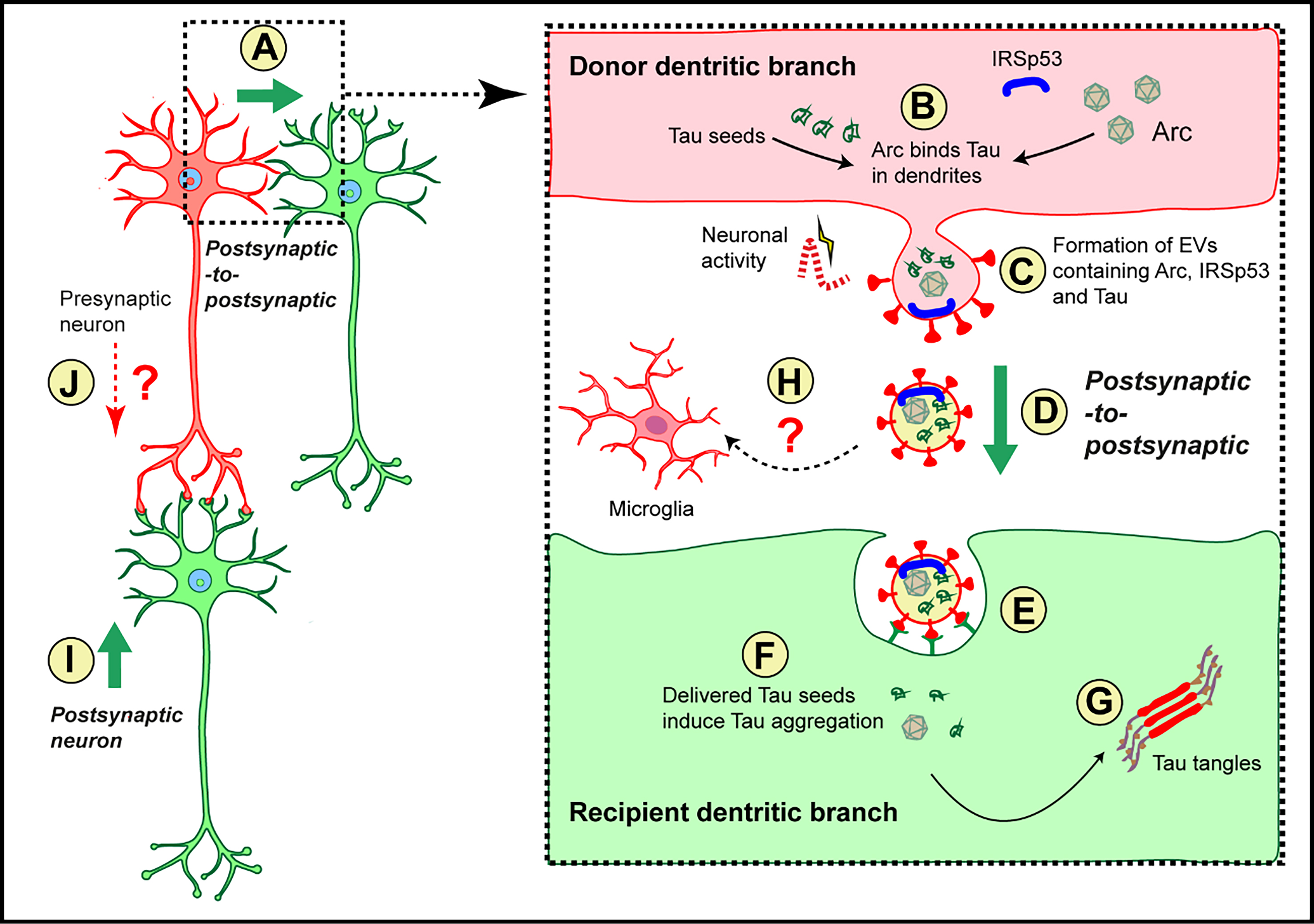
Figure 1. Potential model of Arc-facilitated Tau packaging into EVs and their propagation in the initiation of Tau pathology. (A) The model of dendrite-to-dendrite (postsynaptic-to-postsynaptic) transmission of EVs containing Tau. This mode of transmission is illustrated in greater detail in the right panel; (B) Under pathological conditions, Tau becomes enriched in dendrites, where it interacts with Arc, a protein that is predominantly localized in this compartment[12]; (C) In dendrites, neuronal activity can potentiate the interaction between Arc and IRSp53 with the plasma membrane within actin-rich extrasynaptic microdomains, promoting the formation of EVs that bud from the cell surface[13]; (D) Tau-containing EVs are released and transferred to neighboring dendrites, thereby enabling postsynaptic-to-postsynaptic transmission; (E) These Arc-IRSp53 EVs are subsequently internalized by dendrites of neighboring neurons; (F) Following internalization, Tau and Arc should be delivered to the cytosol; (G) Tau seeds then template the conversion of endogenous soluble Tau into oligomeric species and, ultimately, into fibrillar Tau aggregates characteristic of Tau pathology[20]; (H) Potential uptake of Arc-IRSp53-Tau-containing EVs by microglia (dotted arrow) may further promote Tau propagation and disease progression, consistent with the established EV-mediated role of microglia in driving tauopathy progression in other mouse models[21]; (I) An alternative model of EV-mediated Tau transmission from dendrites to axons (postsynaptic-to-presynaptic) is also depicted; (J) Because Arc can also be released in small EVs generated via the classical exosome pathway involving multivesicular bodies[19], axon-to-dendrite (presynaptic-to-postsynaptic) transmission cannot be ruled out. EVs: Extracellular vesicles; IRSp53: insulin receptor substrate protein of 53 kDa; Arc: activity-regulated cytoskeleton-associated protein.
The discovery that Arc functions as a critical regulator of Tau packaging into EVs opens an entirely new avenue for therapeutic intervention in AD and related tauopathies. Rather than disrupting the intracellular handling of toxic Tau by Arc, future therapies could focus on blocking the dissemination of Tau-containing EVs or their uptake by recipient cells. By restricting the neuron-to-neuron spread of pathogenic Tau, such strategies may slow disease progression while preserving the beneficial intracellular functions of Arc.
DECLARATIONS
Acknowledgments
The author thanks Götz J, Cruz E, both from the Clem Jones Centre for Ageing Dementia Research, for their critical review of the manuscript and valuable feedback.
Authors’ contributions
The author contributed solely to the article.
Availability of data and materials
Not applicable.
AI and AI-assisted tools statement
During the preparation of this manuscript, the AI tool Microsoft Copilot (version 157.0.4341.60, released 2026-11-13) was used solely for language editing and improving the readability of selected portions of the author’s original text. All AI-generated content was subsequently reviewed and edited by the author. The tool did not influence the study design, data collection, analysis, interpretation, or the scientific content of the work. The author takes full responsibility for the accuracy, integrity, and final content of the manuscript.
Financial support and sponsorship
This work was supported by a grant from the National Health and Medical Research Council of Australia (GNT2018919) to Polanco JC.
Conflicts of interest
The author declared that there are no conflicts of interest.
Ethical approval and consent to participate
Not applicable.
Consent for publication
Not applicable.
Copyright
© The Author(s) 2026.
REFERENCES
1. Chung DC, Roemer S, Petrucelli L, Dickson DW. Cellular and pathological heterogeneity of primary tauopathies. Mol Neurodegener. 2021;16:57.
2. Holmes BB, Furman JL, Mahan TE, et al. Proteopathic tau seeding predicts tauopathy in vivo. Proc Natl Acad Sci U S A. 2014;111:E4376-85.
3. Oosterlynck M, Leroux E, Namasivayam B, et al. Stratification of brain-derived extracellular vesicles of Alzheimer’s disease patients indicates a unique proteomic content and a higher seeding capacity of small extracellular vesicles. Transl Neurodegener. 2025;14:63.
4. Polanco JC, Scicluna BJ, Hill AF, Götz J. Extracellular vesicles isolated from the brains of rTg4510 mice seed Tau protein aggregation in a threshold-dependent manner. J Biol Chem. 2016;291:12445-66.
5. Tyagi M, de Hoog E, Grega M, et al. Arc mediates intercellular tau transmission via extracellular vesicles. Cell. 2026;189:4706-23.e13.
6. Wang Y, Balaji V, Kaniyappan S, et al. The release and trans-synaptic transmission of Tau via exosomes. Mol Neurodegener. 2017;12:5.
7. Dujardin S, Bégard S, Caillierez R, et al. Ectosomes: a new mechanism for non-exosomal secretion of tau protein. PLoS One. 2014;9:e100760.
8. Fowler SL, Behr TS, Turkes E, et al. Tau filaments are tethered within brain extracellular vesicles in Alzheimer’s disease. Nat Neurosci. 2025;28:40-8.
9. Pooler AM, Phillips EC, Lau DH, Noble W, Hanger DP. Physiological release of endogenous tau is stimulated by neuronal activity. EMBO Rep. 2013;14:389-94.
10. Das S, Lituma PJ, Castillo PE, Singer RH. Maintenance of a short-lived protein required for long-term memory involves cycles of transcription and local translation. Neuron. 2023;111:2051-64.e6.
11. Vazdarjanova A, Ramirez-Amaya V, Insel N, et al. Spatial exploration induces ARC, a plasticity-related immediate-early gene, only in calcium/calmodulin-dependent protein kinase II-positive principal excitatory and inhibitory neurons of the rat forebrain. J Comp Neurol. 2006;498:317-29.
12. Steward O, Worley PF. Selective targeting of newly synthesized Arc mRNA to active synapses requires NMDA receptor activation. Neuron. 2001;30:227-40.
13. Sullivan K, Ravens A, de Hoog E, et al. Arc mediates intercellular synaptic plasticity via IRSp53-dependent extracellular vesicle biogenesis. bioRxiv. 2026;bioRxiv:2024.01.30.578027.
14. Santacruz K, Lewis J, Spires T, et al. Tau suppression in a neurodegenerative mouse model improves memory function. Science. 2005;309:476-81.
15. Rauch JN, Luna G, Guzman E, et al. LRP1 is a master regulator of tau uptake and spread. Nature. 2020;580:381-5.
16. Joshi BS, de Beer MA, Giepmans BNG, Zuhorn IS. Endocytosis of extracellular vesicles and release of their cargo from endosomes. ACS Nano. 2020;14:4444-55.
17. Polanco JC, Hand GR, Briner A, Li C, Götz J. Exosomes induce endolysosomal permeabilization as a gateway by which exosomal tau seeds escape into the cytosol. Acta Neuropathol. 2021;141:235-56.
18. Sanyal A, Scanavachi G, Somerville E, et al. Neuronal constitutive endolysosomal perforations enable α-synuclein aggregation by internalized PFFs. J Cell Biol. 2025;224:e202401136.
19. Mehta K, Yentsch H, Lee J, et al. Phosphatidylinositol-3-phosphate mediates Arc capsid secretion through the multivesicular body pathway. Proc Natl Acad Sci U S A. 2024;121:e2322422121.
20. Ruan Z, Pathak D, Venkatesan Kalavai S, et al. Alzheimer’s disease brain-derived extracellular vesicles spread tau pathology in interneurons. Brain. 2021;144:288-309.
Cite This Article
 Open Access
How to Cite
Polanco JC. Arc is a novel regulator of Tau packaging into extracellular vesicles and promotes Tau spreading. Extracell Vesicles Circ Nucleic Acids. 2026;7:2108-13. https://dx.doi.org/10.20517/evcna.234
Download Citation
If you have the appropriate software installed, you can download article citation data to the citation manager of your choice. Simply select your manager software from the list below and click on download.
Export Citation File
Type of Import
Tips on Downloading Citation
Citation Manager File Format
Type of Import
Direct Import: When the Direct Import option is selected (the default state), a dialogue box will give you the option to Save or Open the downloaded citation data. Choosing Open will either launch your citation manager or give you a choice of applications with which to use the metadata. The Save option saves the file locally for later use.
Indirect Import: When the Indirect Import option is selected, the metadata is displayed and may be copied and pasted as needed.
About This Article
Copyright

Data & Comments
Data
0










Comments
Comments must be written in English. Spam, offensive content, impersonation, and private information will not be permitted. If any comment is reported and identified as inappropriate content by OAE staff, the comment will be removed without notice. If you have any queries or need any help, please contact us at [email protected].