

fig1
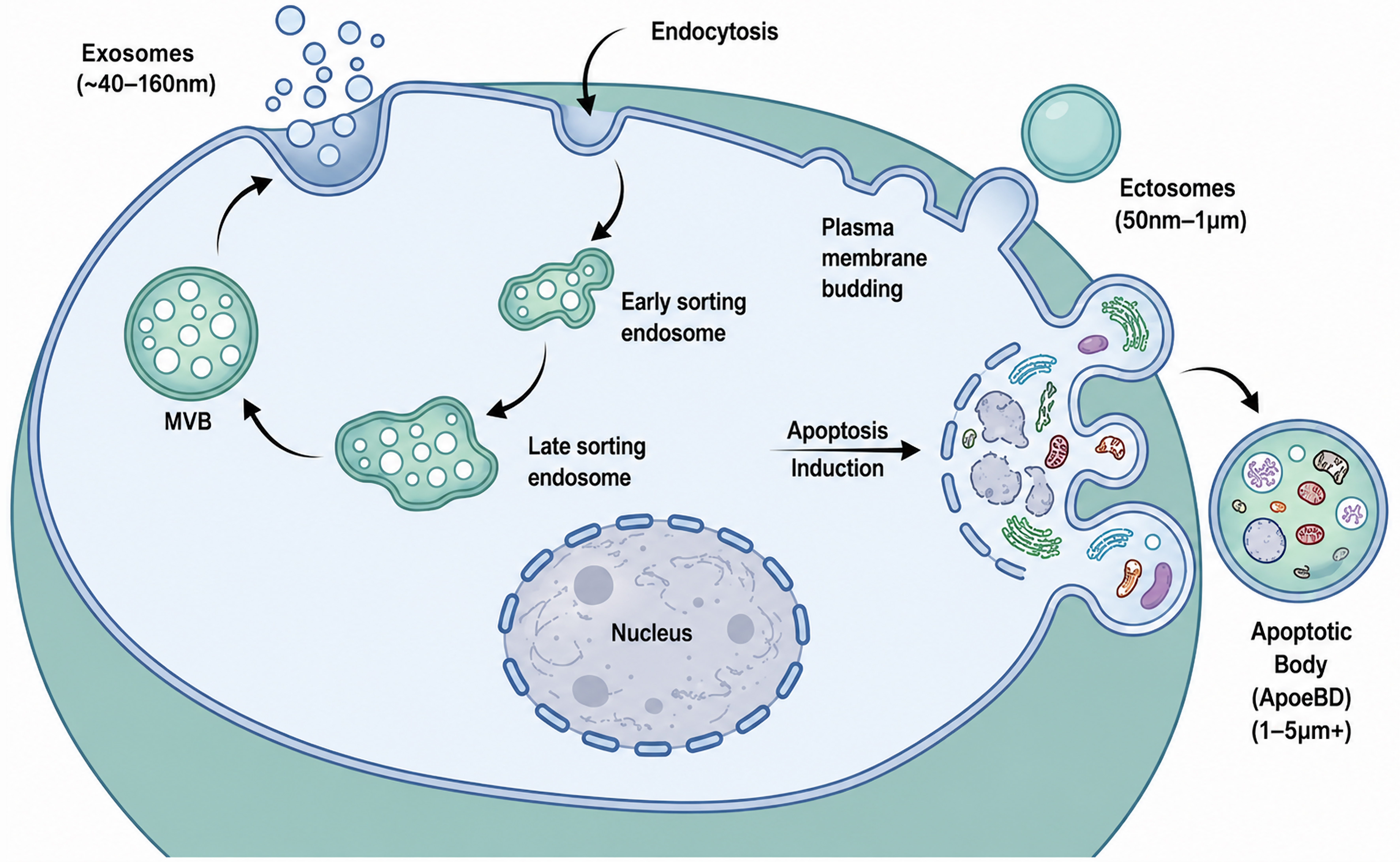
Figure 1. Biogenesis and classification of extracellular vesicle subtypes. Exosomes (approximately 40-160 nm) arise through the endosomal pathway, where early and late sorting endosomes mature into MVBs that fuse with the plasma membrane to release intraluminal vesicles[6]. Ectosomes or microvesicles (approximately 50 nm-1 μm) are generated by outward budding of the plasma membrane[8,9]. During apoptosis, membrane blebbing and cellular fragmentation generate ApoBDs (typically 1-5 μm or larger) that can enclose cytoplasmic components, organelles, and nuclear material. Substantial overlap in size and composition limits strict assignment of individua l extracellular particles to a single biogenetic pathway[10,11]. References[8-11] support the scientific content depicted and were not used as sources for adaptation or reproduction. Created with BioRender. MVB: Multivesicular body; ApoBDs: apoptotic bodies.




