

fig6
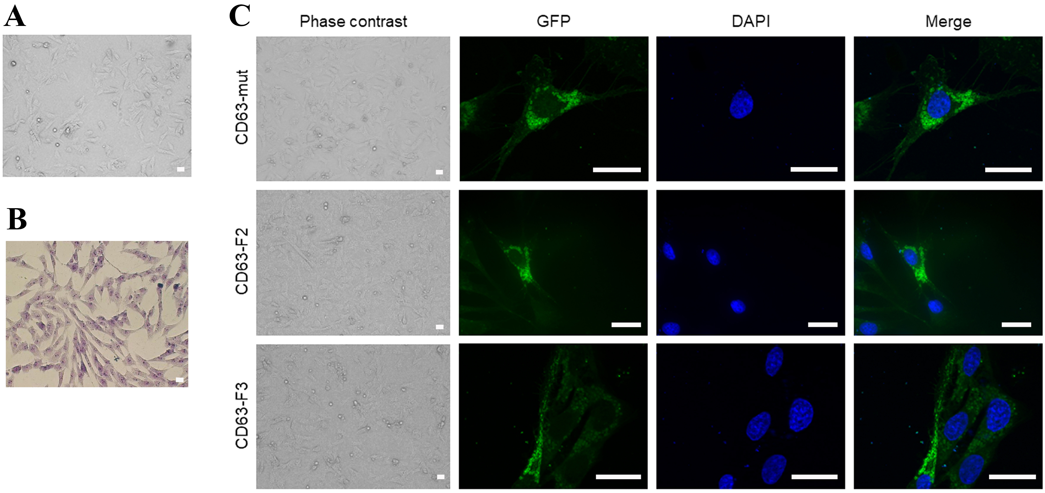
Figure 6. Canine MSC morphology and expression of CD63-based constructs. (A) Phase-contrast image; (B) representative Giemsa-stained image of untransfected canine MSCs in culture, both images showing a typical fibroblast-like morphology and strong adherence to plastic. Scale bar: 20 micrometer; (C) Distribution and localization of CD63-GFP, CD63-F2-GFP, and CD63-F3-GFP in transfected MSCs. Phase-contrast images; GFP, DAPI, and merged images. Scale bar: 20 micrometer. CD63: Cluster of differentiation 63; DAPI: 4’,6-diamidino-2-phenylindole; GFP: green fluorescent protein; MSC: mesenchymal stromal cell.




