

fig3
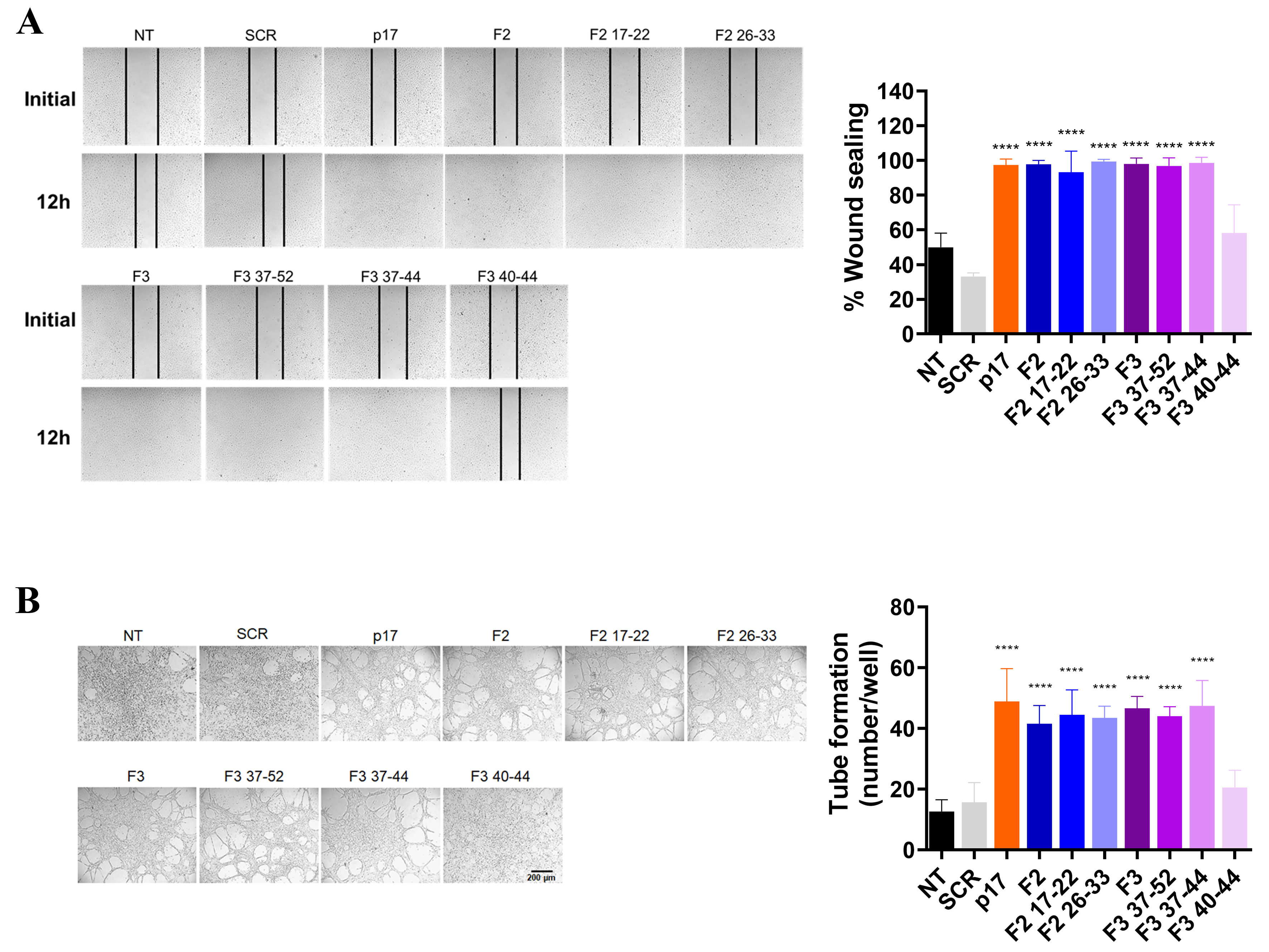
Figure 3. Effects of different p17 F2- and F3-derived peptides on migration and tube formation of HUVECs under stress conditions. (A) Pro-migratory activity evaluated by a wound-healing assay in serum-starved HUVECs treated with NT, SCR, full-length p17, or the indicated p17-derived peptides; (B) angiogenic activity evaluated by tube formation assay in HUVECs, treated as indicated. Scale bar: 200 µm. (A and B) Images are representative of three independent experiments with similar results (original magnification, ×10). Data are expressed as mean ± SD of three independent experiments performed in triplicate. Statistical analysis was performed by one-way ANOVA followed by Bonferroni’s post hoc test (****P < 0.0001). ANOVA: Analysis of variance; HUVEC: human umbilical vein endothelial cell; NT: no treatment; SCR: scrambled peptide; SD: standard deviation.




