

fig2
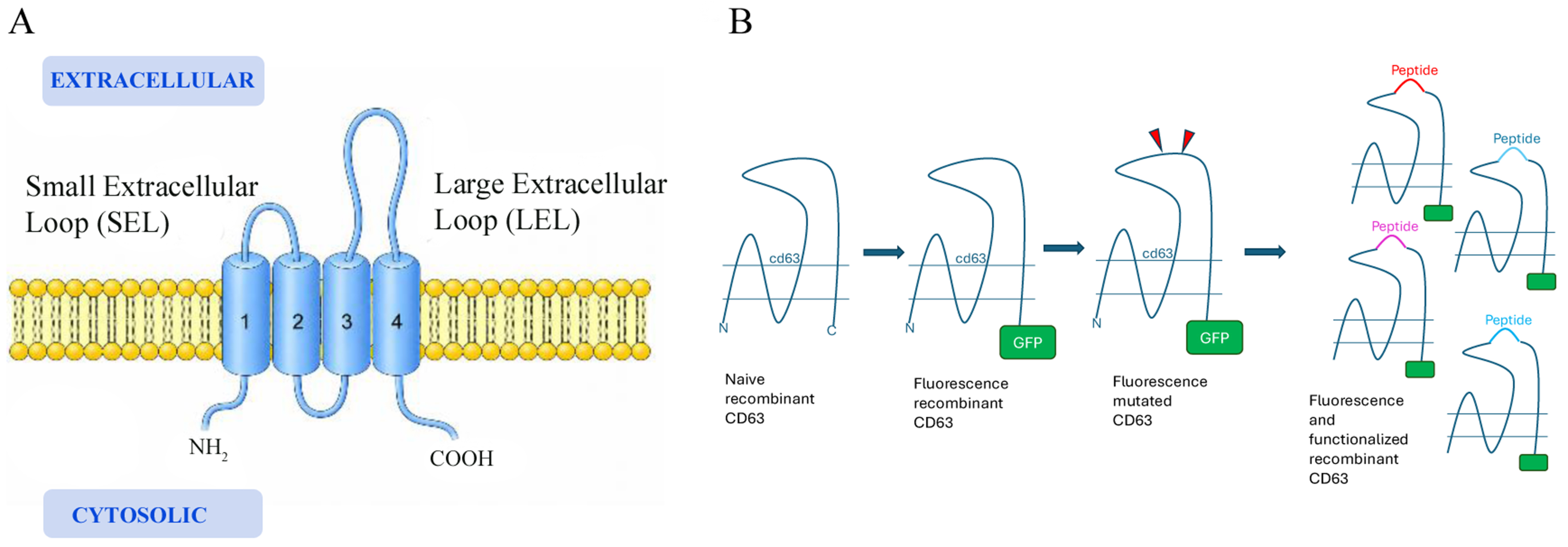
Figure 2. (A) Schematic representation of CD63 tetraspanin topology. Tetraspanins consist of four transmembrane domains (TM1-TM4) connected by a SEL and a LEL. The N- and C-terminal regions are located on the cytosolic head; (B) schematic representation of the stepwise engineering strategy used to evaluate different peptide sequences on the same scaffold protein (CD63). Native CD63 was first fused to GFP and then modified to introduce a peptide insertion site. Different peptides were subsequently displayed on the extracellular domain, enabling comparative functional analysis while maintaining a constant structural backbone. CD63: Cluster of differentiation 63; GFP: green fluorescent protein; LEL: large extracellular loop; SEL: small extracellular loop; TM: transmembrane domain.




