

fig1
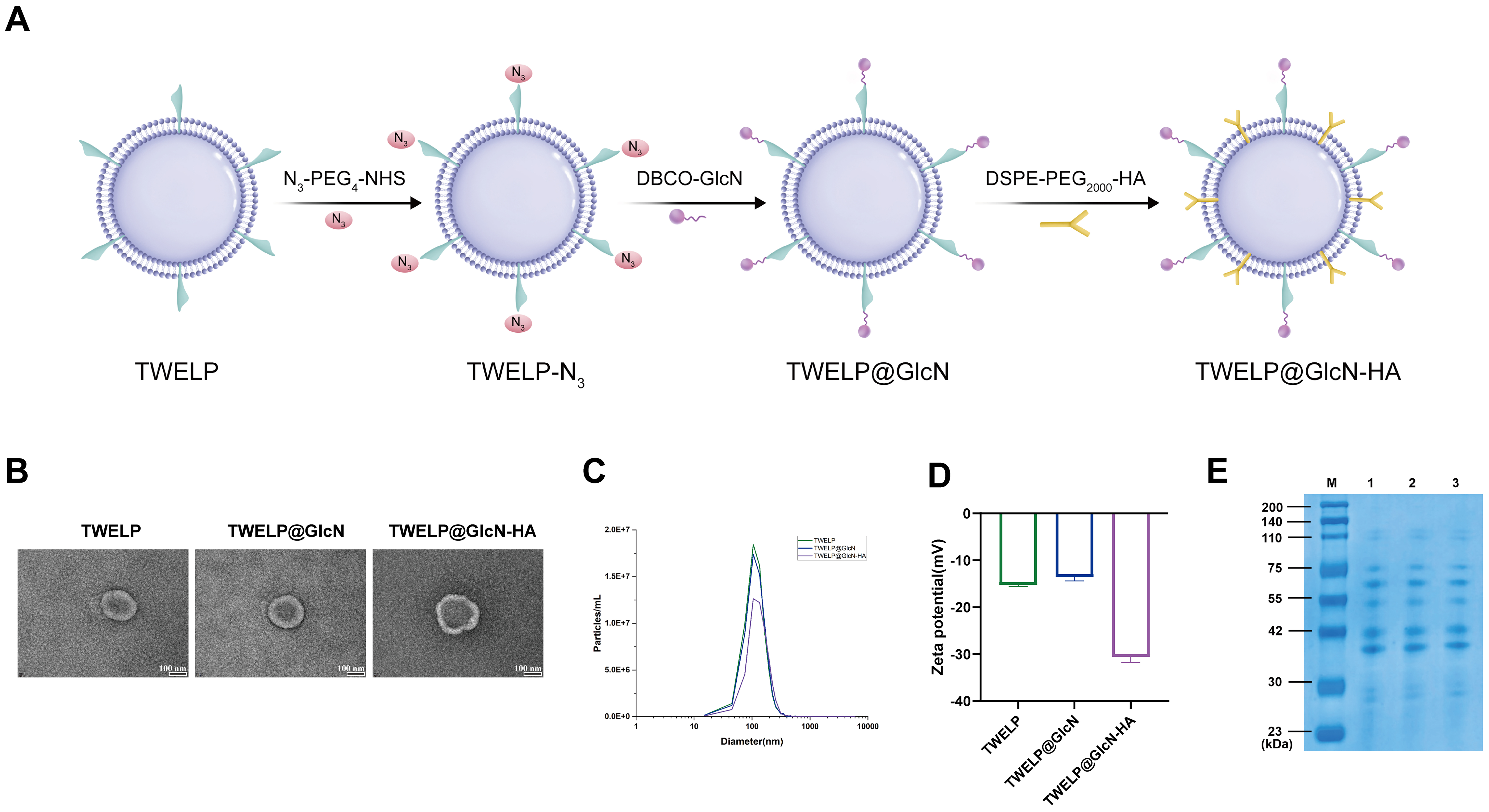
Figure 1. Preparation and characterization of TWELP@GlcN-HA. (A) Schematic of TWELP surface modification to generate TWELP@GlcN-HA; (B) TEM images of TWELP, TWELP@GlcN, and TWELP@GlcN-HA (scale bar = 100 nm); (C) NTA particle size distribution; (D) Zeta potential measured by NTA; (E) SDS-PAGE protein profiles (M: marker; lanes 1-3: TWELP, TWELP@GlcN, TWELP@GlcN-HA). DBCO-GlcN: Dibenzocyclooctyne-conjugated glucosamine; DSPE-PEG2000-HA: 1,2-distearoyl-sn-glycero-3-phosphoethanolamine-poly(ethylene glycol) 2000-hyaluronic acid conjugate; GlcN: glucosamine; HA: hyaluronic acid; N3-PEG4-NHS: azide-poly(ethylene glycol)4-N-hydroxysuccinimide ester; NTA: nanoparticle tracking analysis; SDS-PAGE: sodium dodecyl sulfate-polyacrylamide gel electrophoresis; TEM: transmission electron microscopy; TWELP: Tripterygium wilfordii-derived exosome-like nanoparticles; TWELP-N3: azide-modified TWELP; TWELP@GlcN: glucosamine-modified TWELP; TWELP@GlcN-HA: engineered Tripterygium wilfordii-derived exosome-like nanoparticle system.




