

fig3
From: Region-specific human cardiac extracellular vesicles differentially regulate mitochondrial function
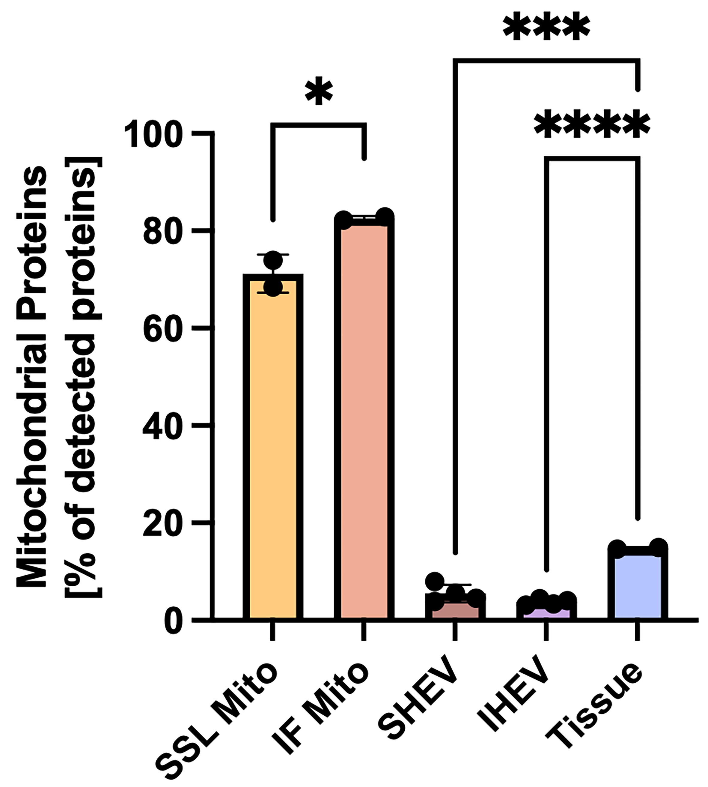
Figure 3. Percentage of detected proteins that are mitochondrial in SSL or IF mitochondria, or SSL- or IF-associated heart EVs (SHEV, IHEV, respectively), or left ventricular tissue. For clarity, statistical comparisons between mitochondria and vesicle or tissue samples are not shown. *P < 0.05, ***P < 0.001, ****P < 0.0001; N = 2-4 donor hearts. Error bars show SEM. Analysis was performed using ordinary one-way ANOVA with multiple comparisons corrected using Tukey’s test. SSL: Subsarcolemmal; IF: interfibrillar; EV: extracellular vesicle; SHEV: subsarcolemmal heart extracellular vesicle; IHEV: interfibrillar heart extracellular vesicle; SEM: standard error of the mean; ANOVA: analysis of variance.




