

fig2
From: Region-specific human cardiac extracellular vesicles differentially regulate mitochondrial function
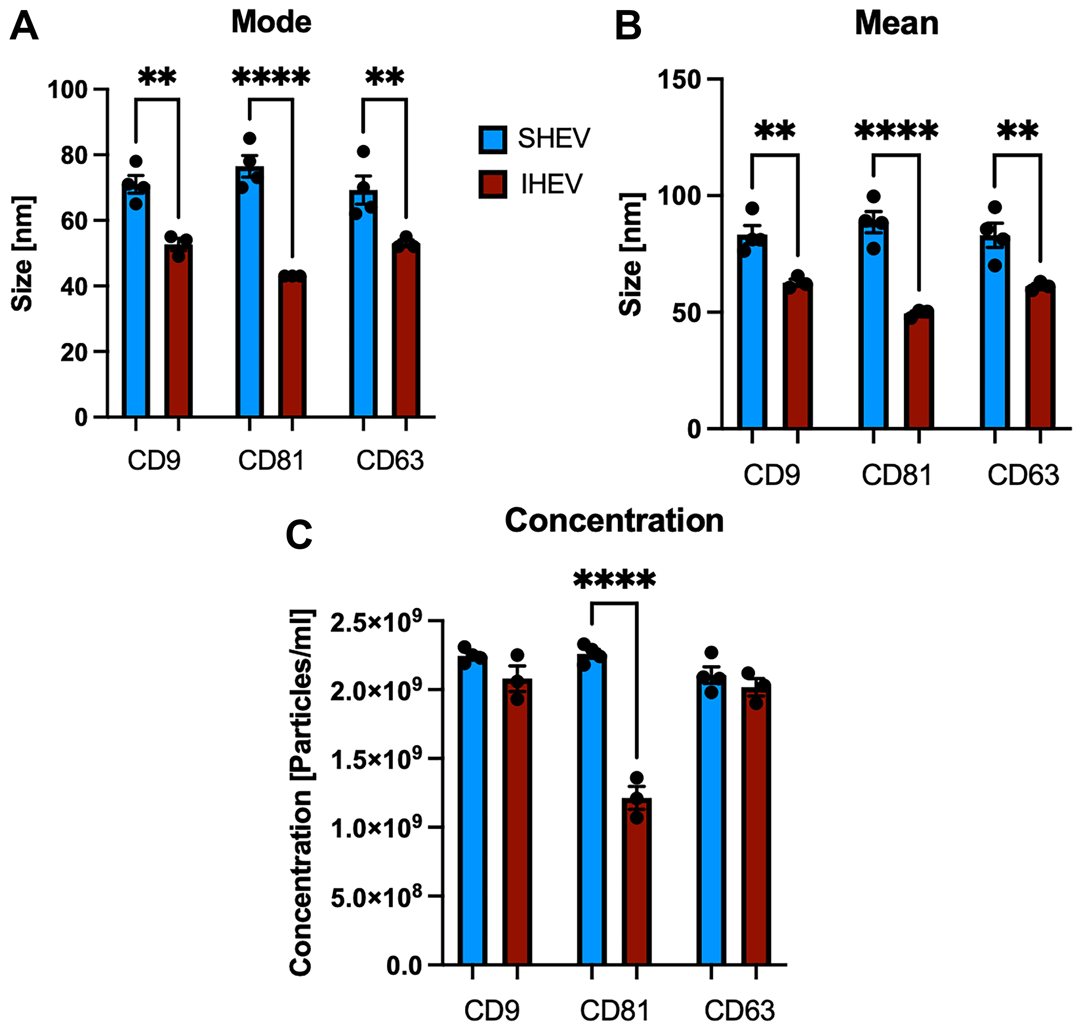
Figure 2. Leprechaun analysis: (A) mode, (B) mean size, and (C) concentration of SHEV and IHEV particles containing CD9, CD81, and CD63. Comparisons were performed only between SHEV and IHEV for each CD protein. **P < 0.01, ****P < 0.0001; N = 4 SHEV or 3 IHEV donor hearts. Error bars show SEM. Analysis was performed using ordinary one-way ANOVA with multiple comparisons corrected using Tukey’s test. SHEV: Subsarcolemmal heart extracellular vesicle; IHEV: interfibrillar heart extracellular vesicle; SEM: standard error of the mean; ANOVA: analysis of variance.




