

fig8
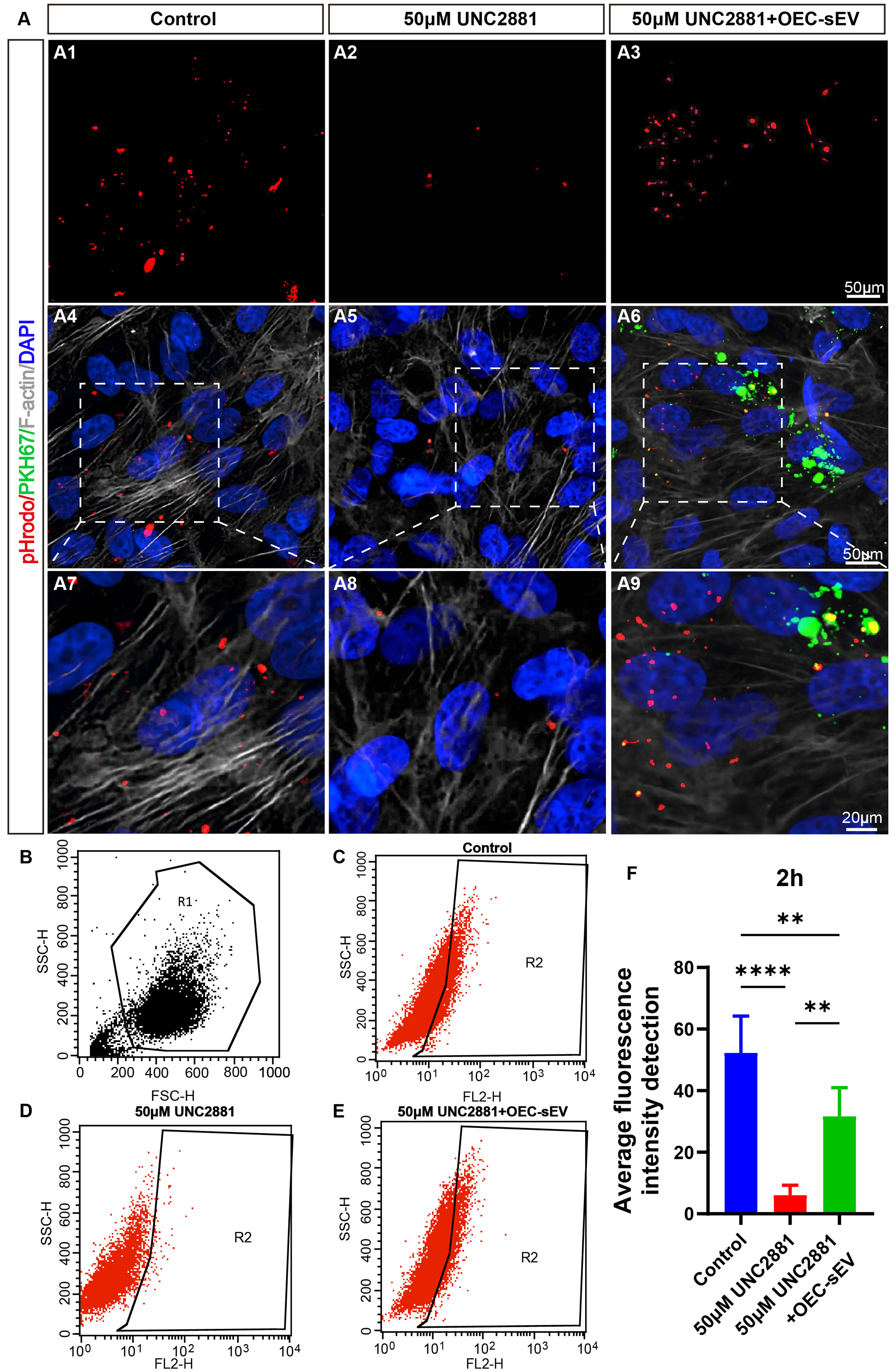
Figure 8. OEC-sEVs rescue UNC2881-induced phagocytic suppression in ARPE-19 cells. (A) Visualization of pHrodoTM BioParticlesTM conjugates and PKH67, and fluorescent staining of F-actin in the control group, 50 μM UNC2881 group, and 50 μM UNC2881 + OEC-sEV group. Scale bar = 50 μm (original images); scale bar = 20 μm (enlarged images). The dashed boxes denote the regions selected for magnification in the subsequent panels; (B-E) Flow cytometry analysis of pHrodoTM dyes in all three groups. R1 gate: Initial gating on FSC-H vs. SSC-H plot to identify the target cell population based on size and granularity, excluding debris and cell aggregates. R2 gate: pH-sensitive pHrodoTM dye-positive population within the viable cell gate; (F) Average fluorescence intensity detection of pHrodoTM dyes. n = 3 biologically independent cell cultures per group. Data are represented as mean ± SD. **P < 0.01, ****P < 0.0001. One-way ANOVA, followed by Tukey’s multiple comparisons test, was conducted for multiple comparisons. ANOVA: Analysis of variance; ARPE-19: adult retinal pigment epithelial cell line-19; F-actin: filamentous actin; FSC-H: forward scatter height; OEC-sEVs: olfactory ensheathing cell-derived small extracellular vesicles; SD: standard deviation; SSC-H: side scatter height.




