

fig2
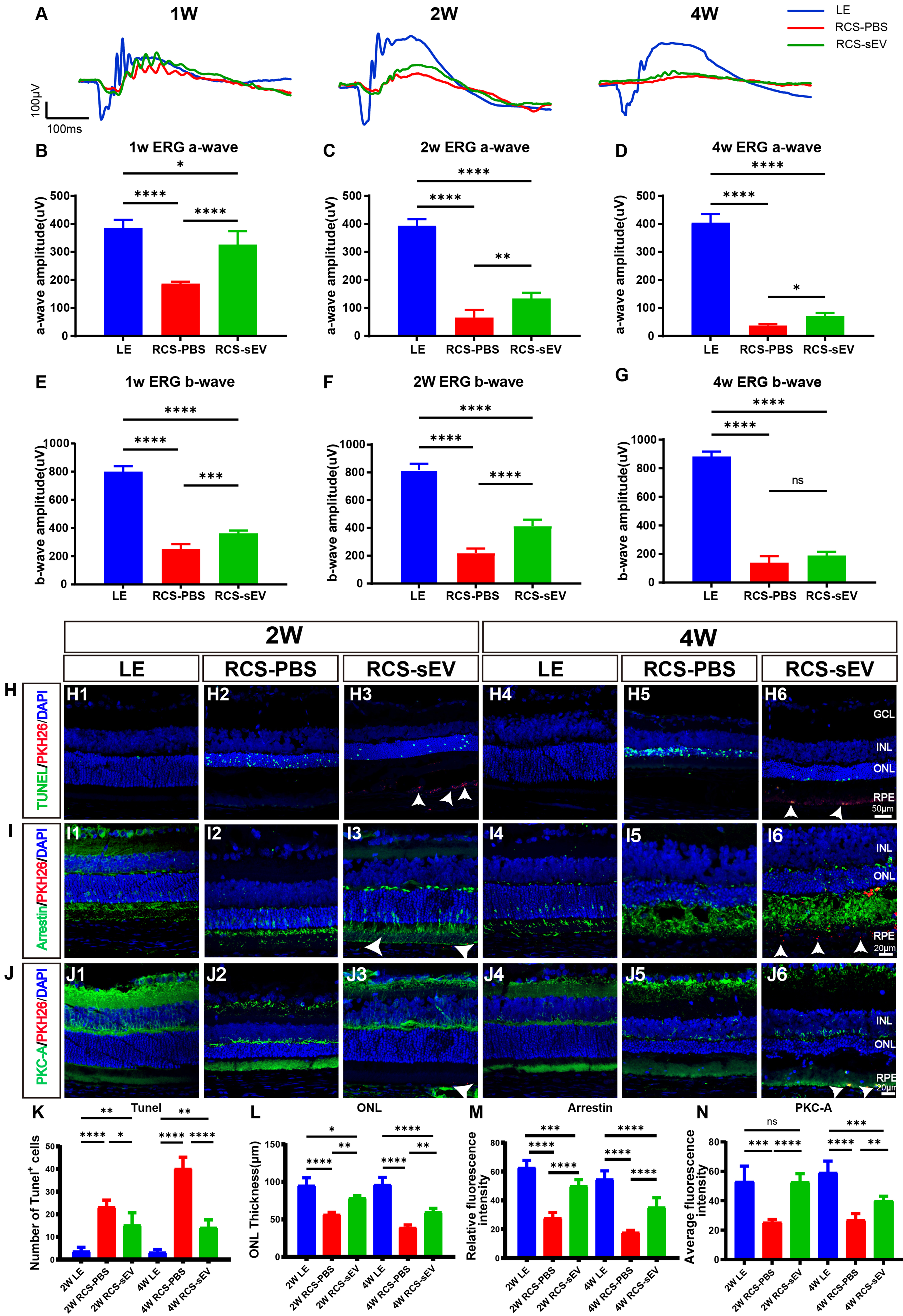
Figure 2. Protective effects of OEC-sEVs on photoreceptor and bipolar cell degeneration in RCS rats. (A) ERG graph of LE, RCS-PBS, and RCS-sEV groups at 1, 2 and 4 weeks post-injection; (B-D) Quantitative analysis of a-wave amplitude; (E-G) Quantitative analysis of b-wave amplitude; (H) PKH26-labeled OEC-sEV internalization (red) and TUNEL staining (green) in retinal sections of LE, RCS-PBS, and RCS-sEV groups at 2 and 4 weeks post-injection. Nuclei were stained with DAPI (blue). Scale bars = 50 μm; (I) PKH26-labeled OEC-sEVs internalization (red) and immunofluorescence staining of photoreceptors (Arrestin, green). Scale bars = 20 μm. (Since OEC-sEVs may fuse with the membrane or enter the intracellular space after uptake by the cell, PKH26 signaling may reflect the distribution or residue of sEV membrane components rather than the presence of intact sEVs.); (J) PKH26-labeled OEC-sEVs internalization (red) and immunofluorescence staining of bipolar cells (PKC-A, green). Scale bars = 20 μm; (K) Quantification of TUNEL-positive cells in LE, RCS-PBS, and RCS-sEV groups; (L) ONL thickness in LE retinas, PBS- and sEV-treated retinas; (M) Relative fluorescence intensity of Arrestin in LE, RCS-PBS, and RCS-sEV groups; (N) Relative fluorescence intensity of PKC-A in LE, RCS-PBS, and RCS-sEV groups. n = 4 biologically independent rats per group. Arrows show the presence of PKH26-labeled OEC-sEVs within the RPE layer. Data are represented as mean ± SD. *P < 0.05, **P < 0.01, ***P < 0.001, ****P < 0.0001, ns, no statistical difference. One-way ANOVA, followed by Tukey’s multiple comparisons test, was conducted for multiple comparisons. ANOVA: Analysis of variance; DAPI: 4’,6-diamidino-2-phenylindole; ERG: electroretinogram; GCL: ganglion cell layer; INL: inner nuclear layer; LE: Long Evans; OEC-sEVs: olfactory ensheathing cell-derived small extracellular vesicles; ONL: outer nuclear layer; PBS: phosphate-buffered saline; PKC-A: protein kinase C alpha; RCS: Royal College of Surgeons; RCS-PBS: Royal College of Surgeons rats treated with phosphate-buffered saline; RCS-sEV: Royal College of Surgeons rats treated with small extracellular vesicles; RPE: retinal pigment epithelium; SD: standard deviation; sEV: small extracellular vesicle; sEVs: small extracellular vesicles; TUNEL: terminal deoxynucleotidyl transferase dUTP nick end labeling.




