

fig6
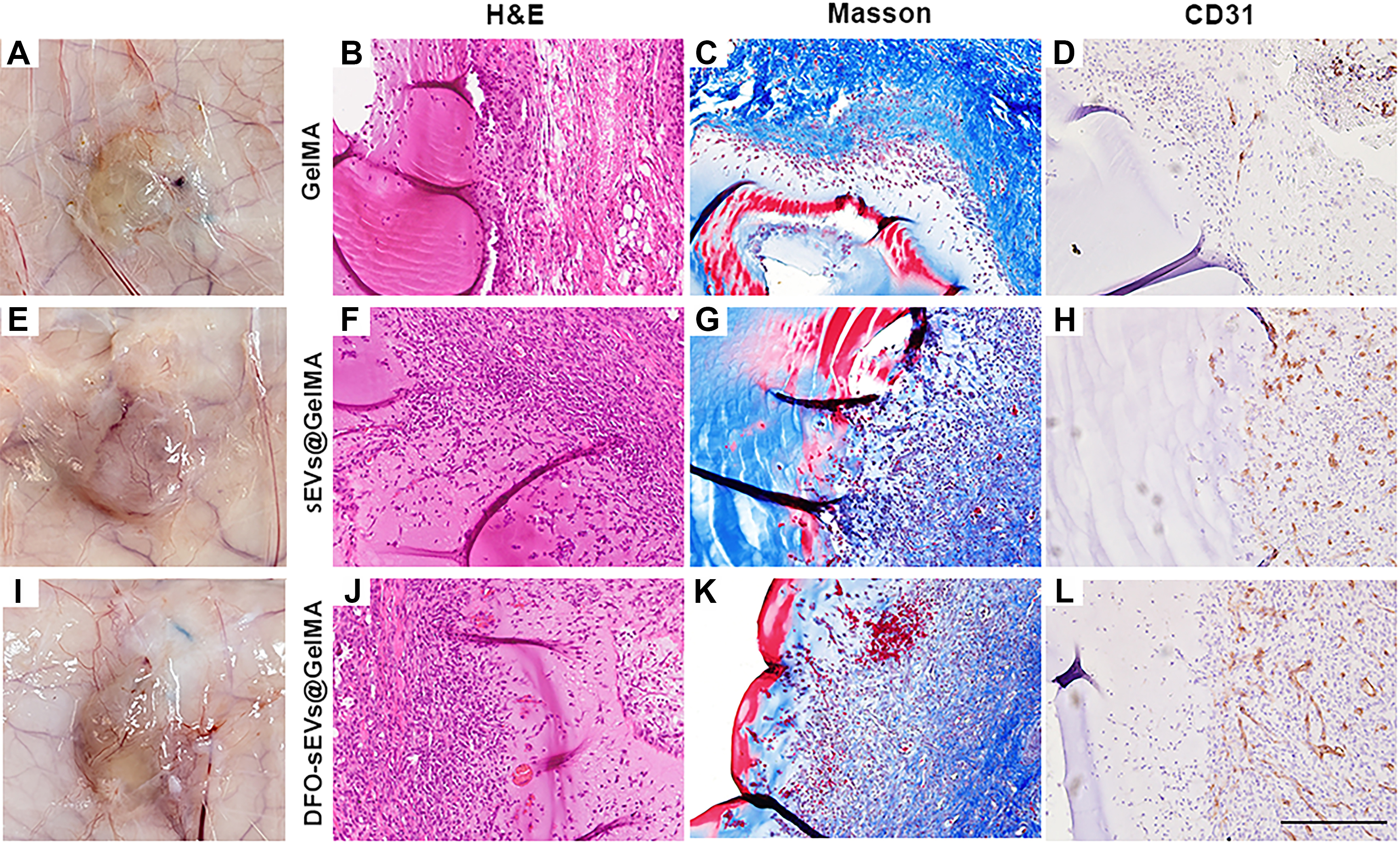
Figure 6. Subcutaneous implantation of DFO-sEVs-loaded GelMA hydrogel in rats. (A, E, and I) Gross views of the implants one week after implantation; (B, F, and J) Histological evaluation (H&E staining); (C, G, and K) Masson’s trichrome; (D, H, and L) CD31 immunohistochemistry. S represents the location of the GelMA hydrogel scaffold; the black arrows indicate the newly formed vessels, and red stars indicate the CD31-positive sites. Each scaffold group contained four independent implantation sites (n = 4). Representative histological images are shown for each scaffold group; Scale bar = 200 μm. CD31: Cluster of differentiation 31; DFO: deferoxamine; GelMA: gelatin methacryloyl; H&E: hematoxylin and eosin; sEVs: small extracellular vesicles.




