

fig5
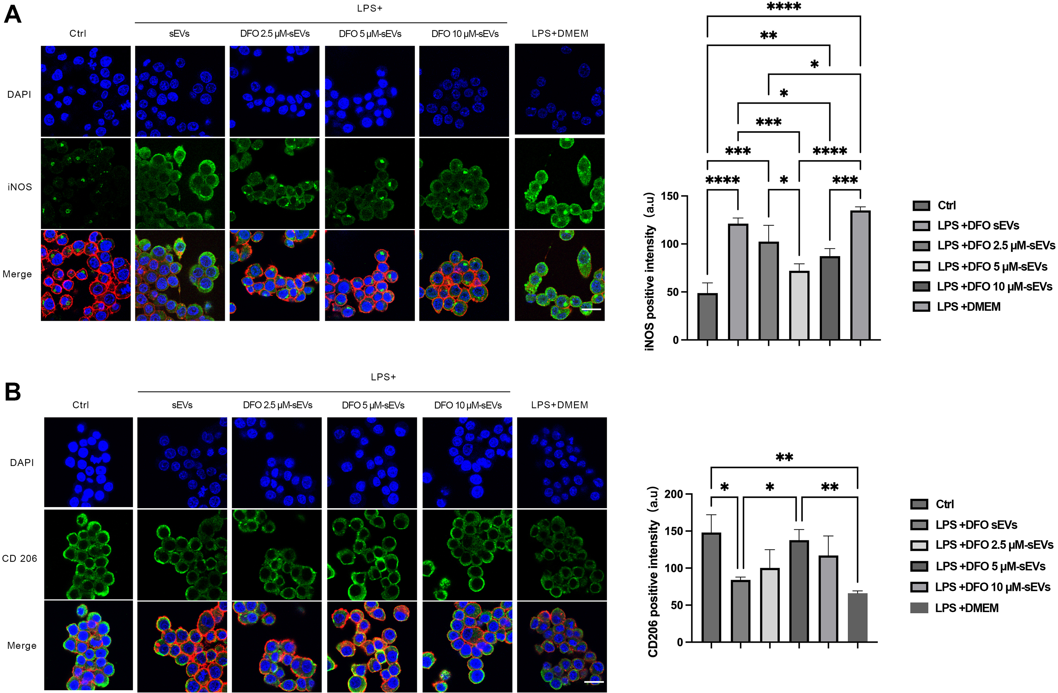
Figure 5. Modulatory effects of DFO-sEVs on RAW264.7 cell polarization. (A) iNOS expression on day 3; Scale bar = 20 μm, with quantification of immunofluorescence intensity; (B) CD206 expression on day 3; Scale bar = 20 μm, with quantification of immunofluorescence intensity. Values are expressed as mean ± SD. Data were analyzed by one-way ANOVA with Tukey’s HSD post hoc comparisons; three independent biological replicates were included. *P < 0.05; **P < 0.01; ***P < 0.001; ****P < 0.0001. ANOVA: Analysis of variance; a.u.: arbitrary units; CD206: cluster of differentiation 206; Ctrl: control; DAPI: 4′,6-diamidino-2-phenylindole; DFO: deferoxamine; DMEM: Dulbecco’s modified Eagle medium; HSD: honestly significant difference; iNOS: inducible nitric oxide synthase; LPS: lipopolysaccharide; RAW264.7: murine macrophage cell line; SD: standard deviation; sEVs: small extracellular vesicles.




